Cited by 143Angiogenesis and VEGF in CancerNitric Oxide and Endothelin EffectsRedox biology and oxidative stress

The DOI matches its Crossref record; 72 of the 73 references it cites were found in a registry or an index.

Targeting Vascular NADPH Oxidase 1 Blocks Tumor Angiogenesis through a PPARα Mediated Mechanism

Open access
Garrido-Urbani, Sarah, Jemelin, Stephane, Deffert, Christine, Carnesecchi, Stéphanie, Basset, Olivier, Szyndralewiez, Cédric, Heitz, Freddy, Page, Patrick, Montet, Xavier, Michalik, Liliane, Arbiser, Jack, Rüegg, Curzio, Krause, Karl Heinz, Imhof, Beat·2011·PLoS ONE

Abstract

Reactive oxygen species, ROS, are regulators of endothelial cell migration, proliferation and survival, events critically involved in angiogenesis. Different isoforms of ROS-generating NOX enzymes are expressed in the vasculature and provide distinct signaling cues through differential localization and activation. We show that mice deficient in NOX1, but not NOX2 or NOX4, have impaired angiogenesis. NOX1 expression and activity is increased in primary mouse and human endothelial cells upon angiogenic stimulation. NOX1 silencing decreases endothelial cell migration and tube-like structure formation, through the inhibition of PPARα, a regulator of NF-κB. Administration of a novel NOX-specific inhibitor reduced angiogenesis and tumor growth in vivo in a PPARα dependent manner. In conclusion, vascular NOX1 is a critical mediator of angiogenesis and an attractive target for anti-angiogenic therapies.

Overview

The claims and statistics of this paper haven’t been checked yet.

Run the check — free, no account needed. It reads the open-access text and checks the sources it cites.

A few free checks a day · papers already verified open instantly · results are public

Integrity signals

Registry

Crossref · 10.1371/journal.pone.0014665

Cites

72 of 72 references verified0 citation errors · 0 not checkable

Statistics

Figures

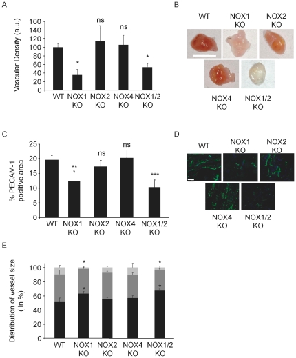
Figure 1 NOX1 deficient mice exhibit reduced angiogenic capacity. Matrigel was loaded with 500 ng/ml bFGF and implanted subcutaneously into NOX deficient mice. After one week, iodinated liposomes were injected i.v. and the plugs analyzed by X-ray tomography. (a) Quantification of matrigel plug vascularization. Graph shows mean of grey density ± s.e.m. For WT, NOX1 KO and NOX1/2 KO n = 8 and for NOX2 KO and NOX4 KO n = 6. (b) Photographs of excised plugs, scale bars represent 1 cm. c. Blood vessel density in plugs from the experiment in (a). Graph shows percentage of PECAM-1 positive area ± s.e.m. (d). Photographs of PECAM-1 immunostaining, PECAM-1 in green, nuclei in blue, scale bars represent 20 µm. Images were acquired with a 20x/0.5 numeric aperture lens and analyzed using LSM510 Meta confocal microscope (Carl Zeiss). e. Vessel size analysis; vessel with lumen under 20 µm are considered as small (black), from 20 to 50 medium (dark grey) and over 50 µm as large (light grey) ± s.e.m. Anova p<0.01; * p<0.05, ** p<0.01, *** p<0.001.
Figure 1 NOX1 deficient mice exhibit reduced angiogenic capacity. Matrigel was loaded with 500 ng/ml bFGF and implanted subcutaneously into NOX deficient mice. After one week, iodinated liposomes were injected i.v. and the plugs analyzed by X-ray tomography. (a) Quantification of matrigel plug vascularization. Graph shows mean of grey density ± s.e.m. For WT, NOX1 KO and NOX1/2 KO n = 8 and for NOX2 KO and NOX4 KO n = 6. (b) Photographs of excised plugs, scale bars represent 1 cm. c. Blood vessel density in plugs from the experiment in (a). Graph shows percentage of PECAM-1 positive area ± s.e.m. (d). Photographs of PECAM-1 immunostaining, PECAM-1 in green, nuclei in blue, scale bars represent 20 µm. Images were acquired with a 20x/0.5 numeric aperture lens and analyzed using LSM510 Meta confocal microscope (Carl Zeiss). e. Vessel size analysis; vessel with lumen under 20 µm are considered as small (black), from 20 to 50 medium (dark grey) and over 50 µm as large (light grey) ± s.e.m. Anova p<0.01; * p<0.05, ** p<0.01, *** p<0.001.CC BY · PubMed Central
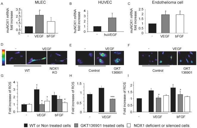
Figure 2 NOX1 expression and activity is up-regulated by angiogenic stimuli. NOX1 expression was measured by quantitative real-time PCR in mouse primary lung endothelial cells (MLEC, a), HUVEC (b), mouse endothelioma cell line (c) after stimulation with VEGF or b-FGF at 20 ng/ml for 3 hours. The quantity of NOX1 mRNA was normalized to the quantity of a housekeeping gene, tubulin for mouse cells and β2-microglobulin for human cells, ± s.e.m, n = 3. Activity of NOX1 was evaluated by ROS measurement using DHE substrate on MLEC (d,g), HUVEC (e, h) and mouse endothelioma (f, i). ROS production is up-regulated by VEGF and b-FGF stimulation in a NOX1-dependent manner. g, h, i. Graphs show quantification of ROS level in endothelial cells stimulated in presence or absence of VEGF or b-FGF. g. MLEC WT (black bar), MLEC WT + GKT136901 (grey bar), MLEC NOX1KO (white bar). h. HUVEC untreated (black bar) or treated with GKT136901 at 10 µM (grey bar). i. Mouse endothelioma cell line untreated (black bar), treated with GKT136901 at 10 µM (grey bar), treated with NOX1 siRNA (white bar). Results are expressed in fold increase ± s.e.m, n = 3. *p<0.05, ***<0.001 using Student's t-test.
Figure 2 NOX1 expression and activity is up-regulated by angiogenic stimuli. NOX1 expression was measured by quantitative real-time PCR in mouse primary lung endothelial cells (MLEC, a), HUVEC (b), mouse endothelioma cell line (c) after stimulation with VEGF or b-FGF at 20 ng/ml for 3 hours. The quantity of NOX1 mRNA was normalized to the quantity of a housekeeping gene, tubulin for mouse cells and β2-microglobulin for human cells, ± s.e.m, n = 3. Activity of NOX1 was evaluated by ROS measurement using DHE substrate on MLEC (d,g), HUVEC (e, h) and mouse endothelioma (f, i). ROS production is up-regulated by VEGF and b-FGF stimulation in a NOX1-dependent manner. g, h, i. Graphs show quantification of ROS level in endothelial cells stimulated in presence or absence of VEGF or b-FGF. g. MLEC WT (black bar), MLEC WT + GKT136901 (grey bar), MLEC NOX1KO (white bar). h. HUVEC untreated (black bar) or treated with GKT136901 at 10 µM (grey bar). i. Mouse endothelioma cell line untreated (black bar), treated with GKT136901 at 10 µM (grey bar), treated with NOX1 siRNA (white bar). Results are expressed in fold increase ± s.e.m, n = 3. *p<0.05, ***<0.001 using Student's t-test.CC BY · PubMed Central
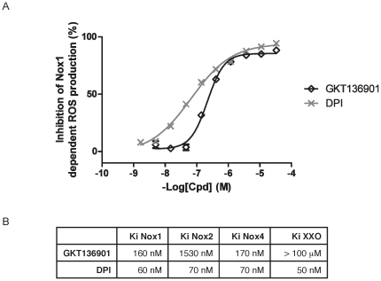
Figure 3 Inhibition of NOX1-dependent ROS production by GKT136901. (a) Concentration-response curve of GKT136901 (◊) and DPI (x) on membranes prepared from NOX1 over-expressing cells. Results represent one out of four experiments performed in triplicate. Values are presented as means ± s.e.m. (b) Affinities of GKT136901 and DPI on NOX1, NOX2, NOX4 and Xanthine Oxidase. GKT136901 is a NADPH-oxidase specific inhibitor, with selectivity on NOX1 and NOX4 over NOX2, whereas DPI inhibits all the NADPH oxidases tested with the same potency.
Figure 3 Inhibition of NOX1-dependent ROS production by GKT136901. (a) Concentration-response curve of GKT136901 (◊) and DPI (x) on membranes prepared from NOX1 over-expressing cells. Results represent one out of four experiments performed in triplicate. Values are presented as means ± s.e.m. (b) Affinities of GKT136901 and DPI on NOX1, NOX2, NOX4 and Xanthine Oxidase. GKT136901 is a NADPH-oxidase specific inhibitor, with selectivity on NOX1 and NOX4 over NOX2, whereas DPI inhibits all the NADPH oxidases tested with the same potency.CC BY · PubMed Central
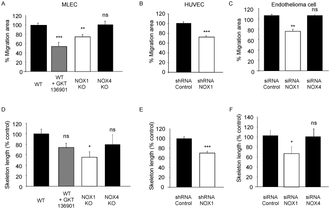
Figure 4 NOX1 inhibition blocks endothelial cell migration and tube-like structure formation. In vitro migration was analyzed by wound-healing assay on mouse primary endothelial cells (a), human primary endothelial cells (b) and endothelioma cell lines (c). Tubular structure formation was measured by 3D culture of mouse primary endothelial cells (d), human primary endothelial cells (e) or endothelioma cell lines (f). Results are expressed in % of control ± s.e.m, n = 3. *p<0.05, **p<0.01, ***<0.001 using Student's t-test. GKT136901 was used at 10 µM.
Figure 4 NOX1 inhibition blocks endothelial cell migration and tube-like structure formation. In vitro migration was analyzed by wound-healing assay on mouse primary endothelial cells (a), human primary endothelial cells (b) and endothelioma cell lines (c). Tubular structure formation was measured by 3D culture of mouse primary endothelial cells (d), human primary endothelial cells (e) or endothelioma cell lines (f). Results are expressed in % of control ± s.e.m, n = 3. *p<0.05, **p<0.01, ***<0.001 using Student's t-test. GKT136901 was used at 10 µM.CC BY · PubMed Central
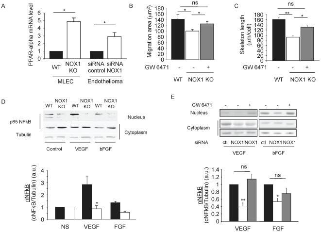
Figure 5 NOX1 negatively regulates PPARα expression and activity. NOX1 deficiency induces an up-regulation of PPARα expression and inhibition of pro-angiogenic signaling in endothelial cells. (a). Expression level of PPARα in MLEC and endothelioma cell lines by quantitative real-time PCR, normalized to the tubulin expression. (b–c) PPARα antagonist, GW6471 compensated NOX1 deficiency in MLEC. (b). Migration of NOX1-deficient MLEC in presence or absence of PPARα antagonist (10 µM). (c). Tube-like structure formation of NOX1-deficient MLEC in presence or absence of PPARα antagonist (10 µM). VEGF and b-FGF induced-NFκB nuclear translocation is inhibited in absence of NOX1 and is dependent on PPARα activity. (d). Western blot analysis of p65 NFκB in nuclear and cytoplasmic fractions of WT and NOX1-deficient cells stimulated 30 min with VEGF and bFGF (20 ng/ml). (e). Western blot analysis of p65 NFkB in nuclear and cytoplasmic fractions of NOX1-silenced cells treated or not with 10 µM of GW6471 before stimulation with VEGF and bFGF. The graphs show the abundance of nuclear p65 NF-κB relative to cytoplasmic p65 NF-κB ± s.e.m as determined by densitometry. n = 3. * p<0.05, ** p<0.01 (Student's t-test).
Figure 5 NOX1 negatively regulates PPARα expression and activity. NOX1 deficiency induces an up-regulation of PPARα expression and inhibition of pro-angiogenic signaling in endothelial cells. (a). Expression level of PPARα in MLEC and endothelioma cell lines by quantitative real-time PCR, normalized to the tubulin expression. (b–c) PPARα antagonist, GW6471 compensated NOX1 deficiency in MLEC. (b). Migration of NOX1-deficient MLEC in presence or absence of PPARα antagonist (10 µM). (c). Tube-like structure formation of NOX1-deficient MLEC in presence or absence of PPARα antagonist (10 µM). VEGF and b-FGF induced-NFκB nuclear translocation is inhibited in absence of NOX1 and is dependent on PPARα activity. (d). Western blot analysis of p65 NFκB in nuclear and cytoplasmic fractions of WT and NOX1-deficient cells stimulated 30 min with VEGF and bFGF (20 ng/ml). (e). Western blot analysis of p65 NFkB in nuclear and cytoplasmic fractions of NOX1-silenced cells treated or not with 10 µM of GW6471 before stimulation with VEGF and bFGF. The graphs show the abundance of nuclear p65 NF-κB relative to cytoplasmic p65 NF-κB ± s.e.m as determined by densitometry. n = 3. * p<0.05, ** p<0.01 (Student's t-test).CC BY · PubMed Central
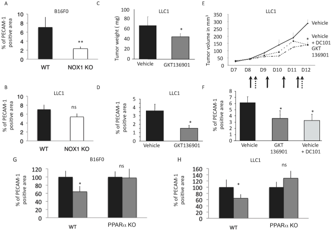
Figure 6 NOX1 inhibition reduces tumor growth and angiogenesis. B16F0 (a) or LLC1 (b) tumor cells were injected subcutaneously into WT or NOX1 KO mice. Tumors were allowed to develop for 10 days and the density of tumor vasculature was identified using PECAM-1 staining by measuring the positive (vascular) and total (tumor) area ± s.e.m expressed as percentage. n = 8; WT mice injected with LLC1 tumor cells were then treated with the NOX inhibitor GKT136901 at 40 mg/kg/day by oral administration for 8 days; (c). Graph represents tumor weight in mg ± s.e.m n = 8 per group. (d). Tumor vessel density in tumors from the experiment in (c). Graph shows tumor vascularization expressed in percentage of PECAM-1 positive area ± s.e.m.; (e). Changes in tumor volume in mm3 after therapeutic treatment starting 8 days post tumor cell injection with GKT136901 (black arrows) or anti-VEGFR2 (DC101) (pointed arrows). Tumor volume is measured using a caliper and the formula V = 4/3π(L/2*l/2*w/2). n = 8; (f). Blood vessel density in tumors from the experiment in (e). at day 12. Graph shows tumor vascularization expressed in percentage of PECAM-1 positive area ± s.e.m. WT or PPARα KO mice injected with B16F0 (g) or LLC1 (h) were then treated with the NOX inhibitor GKT136901 at 40 mg/kg/day by oral administration for 8 days. Graph shows tumor vascularization expressed in percentage of PECAM-1 positive area ± s.e.m. n = 6. * p<0.05, ** p<0.01 (Student's t-test).
Figure 6 NOX1 inhibition reduces tumor growth and angiogenesis. B16F0 (a) or LLC1 (b) tumor cells were injected subcutaneously into WT or NOX1 KO mice. Tumors were allowed to develop for 10 days and the density of tumor vasculature was identified using PECAM-1 staining by measuring the positive (vascular) and total (tumor) area ± s.e.m expressed as percentage. n = 8; WT mice injected with LLC1 tumor cells were then treated with the NOX inhibitor GKT136901 at 40 mg/kg/day by oral administration for 8 days; (c). Graph represents tumor weight in mg ± s.e.m n = 8 per group. (d). Tumor vessel density in tumors from the experiment in (c). Graph shows tumor vascularization expressed in percentage of PECAM-1 positive area ± s.e.m.; (e). Changes in tumor volume in mm3 after therapeutic treatment starting 8 days post tumor cell injection with GKT136901 (black arrows) or anti-VEGFR2 (DC101) (pointed arrows). Tumor volume is measured using a caliper and the formula V = 4/3π(L/2*l/2*w/2). n = 8; (f). Blood vessel density in tumors from the experiment in (e). at day 12. Graph shows tumor vascularization expressed in percentage of PECAM-1 positive area ± s.e.m. WT or PPARα KO mice injected with B16F0 (g) or LLC1 (h) were then treated with the NOX inhibitor GKT136901 at 40 mg/kg/day by oral administration for 8 days. Graph shows tumor vascularization expressed in percentage of PECAM-1 positive area ± s.e.m. n = 6. * p<0.05, ** p<0.01 (Student's t-test).CC BY · PubMed Central
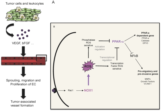
Figure 7 Schematic representation of the role of NOX1 in tumor angiogenesis. (a) Tumor cells and tumor-infiltrating leukocytes during tumor development produce proangiogenic factors such as VEGF and bFGF. These factors activate the preexisting vessels to form neovessels by sprouting, migration and proliferation. (b) Endothelial cells receive the angiogenic stimulus by fixation of the angiogenic factor to the surface receptor. This interaction initiates a signaling cascade, which leads to NOX1 activation through Rac1. NOX1 dependent ROS inhibits the nuclear hormone receptor PPARα by post-translational modification and transcriptional regulation. This inhibition leads to NF-κB activation and transcription of angiogenic factors such as MMPs, growth factors or promigratory molecules.
Figure 7 Schematic representation of the role of NOX1 in tumor angiogenesis. (a) Tumor cells and tumor-infiltrating leukocytes during tumor development produce proangiogenic factors such as VEGF and bFGF. These factors activate the preexisting vessels to form neovessels by sprouting, migration and proliferation. (b) Endothelial cells receive the angiogenic stimulus by fixation of the angiogenic factor to the surface receptor. This interaction initiates a signaling cascade, which leads to NOX1 activation through Rac1. NOX1 dependent ROS inhibits the nuclear hormone receptor PPARα by post-translational modification and transcriptional regulation. This inhibition leads to NF-κB activation and transcription of angiogenic factors such as MMPs, growth factors or promigratory molecules.CC BY · PubMed Central

Verification

Claims the paper makes

The claims and statistics of this paper haven’t been checked yet.

Run the check — free, no account needed. It reads the open-access text and checks the sources it cites.

A few free checks a day · papers already verified open instantly · results are public

Claims other papers make about this one

Each is checked against this paper’s own text: does it say what the citing paper says it does?

Self-check

Percentages, sums and reported differences re-computed from the paper’s own numbers.

The self-check needs the paper’s full text.

Citations

Cites

1 retracted

Show all 73 references

Cited by

Sorted by citations

See all 143

Metadata

Authors
Garrido-Urbani, S., Jemelin, S., Deffert, C., Carnesecchi, S., Basset, O., Szyndralewiez, C., Heitz, F., Page, P., Montet, X., Michalik, L., Arbiser, J., Rüegg, C., Krause, K. H., & Imhof, B.
Affiliations
  • University of Geneva (CH)
  • Genkyotex (Switzerland) (CH)
  • University of Lausanne (CH)
  • Emory University (US)
  • University of Fribourg (CH)
Corresponding author
Imhof, Beat
Venue
PLoS ONE
Published
2011-02-07
Volume
6
Issue
2
Pages
e14665
ISSN
1932-6203 (ISSN-L)
OpenAlex
W1977185778
Cited by
143
References
73
Citations by year
2025: 3 · 2024: 3 · 2023: 6 · 2022: 7 · 2021: 7 · 2020: 7 · 2019: 8 · 2018: 5 · 2017: 11 · 2016: 7 · 2015: 14 · 2014: 21 · 2013: 17 · 2012: 19
FWCI
8.43
Type
journal article
Language
en
Open access
Locations
  • PLoS ONE (publishedVersion, cc-by)
  • PubMed Central (publishedVersion, cc-by)
  • PubMed (publishedVersion)
  • Archive ouverte UNIGE (University of Geneva) (publishedVersion, other-oa)
  • reroDoc Digital Library (submittedVersion)
  • IRIS (submittedVersion)
  • DOAJ (DOAJ: Directory of Open Access Journals) (submittedVersion, cc-by-sa)
  • Figshare (submittedVersion, cc-by)
  • SERVAL (Université de Lausanne) (submittedVersion, other-oa)
  • Archive ouverte UNIGE (University of Geneva) (submittedVersion, cc-by)
Funders
  • National Science Foundation (310030)
  • Schweizerischer Nationalfonds zur Förderung der Wissenschaftlichen Forschung (120184, 310030)
  • Université de Lausanne
Topics
  • Life Sciences › Biochemistry, Genetics and Molecular Biology › Molecular Biology › Angiogenesis and VEGF in Cancer
  • Health Sciences › Medicine › Physiology (medical) › Nitric Oxide and Endothelin Effects
  • Life Sciences › Biochemistry, Genetics and Molecular Biology › Molecular Biology › Redox biology and oxidative stress
Keywords
NOX1, tumor angiogenesis, PPARα, reactive oxygen species, endothelial cells, endothelial cell migration, tube formation, NF-κB, NOX inhibitors, anti-angiogenic therapy, NOX2, NOX4
MeSH terms
NADPH Oxidase 1, Animals, Cells, Cultured, Female, Humans, Mice, Inbred C57BL, Models, Biological, NADH, NADPH Oxidoreductases, Neoplasms, Neovascularization, Pathologic, Pyrazoles, Pyridones, Signal Transduction, Mice, Knockout, Gene Targeting, Angiogenesis Inhibitors, RNA, Small Interfering, Endothelial Cells, PPAR alpha, Mice, Gene Knockdown Techniques, Molecular Targeted Therapy

About these checks

Registry, retraction, Cites and statistics results are computed, not judged, with one exception: a printed reference that no index lists is read once by a model before it counts as a citation error. Claims are extracted by a model and kept only when their quote appears verbatim in the text; each cited source is then checked, with a model’s help, for whether it says what the claim relies on. Those verdicts can be wrong — read the claim’s quote and the cited source before relying on one.

Statistics and arithmetic are recomputed, not judged.

Confidence bands are shown only for fields calibrated on our annotated test set. None is calibrated yet, so no band is shown.

What counts as a claim ›How we measure confidence ›How we check this ›

Are you an author, or do you see a problem with this page? Request a review