

The DOI matches its Crossref record; 37 of the 37 references it cites were found in a registry or an index.
Layer-Specific fMRI Reflects Different Neuronal Computations at Different Depths in Human V1
Open accessAbstract
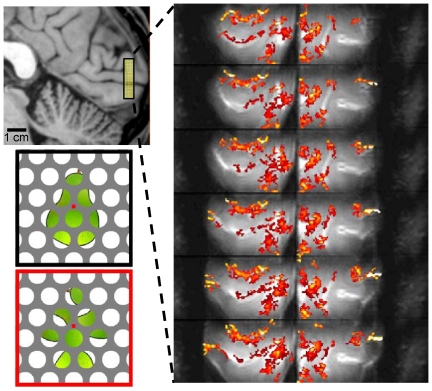
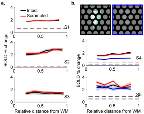
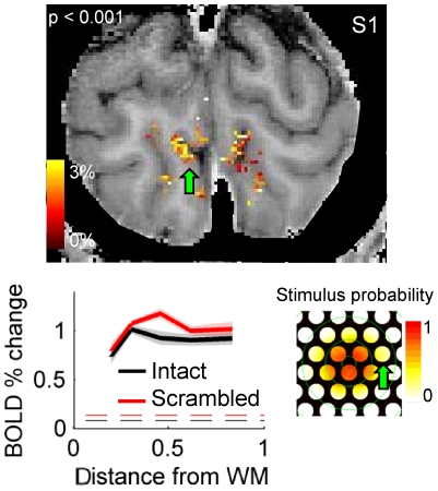
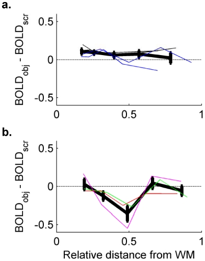
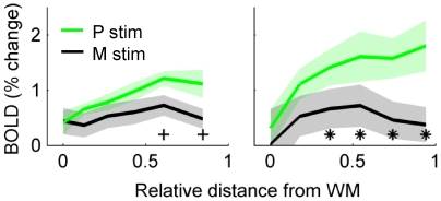
Recent work has established that cerebral blood flow is regulated at a spatial scale that can be resolved by high field fMRI to show cortical columns in humans. While cortical columns represent a cluster of neurons with similar response properties (spanning from the pial surface to the white matter), important information regarding neuronal interactions and computational processes is also contained within a single column, distributed across the six cortical lamina. A basic understanding of underlying neuronal circuitry or computations may be revealed through investigations of the distribution of neural responses at different cortical depths. In this study, we used T(2)-weighted imaging with 0.7 mm (isotropic) resolution to measure fMRI responses at different depths in the gray matter while human subjects observed images with either recognizable or scrambled (physically impossible) objects. Intact and scrambled images were partially occluded, resulting in clusters of activity distributed across primary visual cortex. A subset of the identified clusters of voxels showed a preference for scrambled objects over intact; in these clusters, the fMRI response in middle layers was stronger during the presentation of scrambled objects than during the presentation of intact objects. A second experiment, using stimuli targeted at either the magnocellular or the parvocellular visual pathway, shows that laminar profiles in response to parvocellular-targeted stimuli peak in more superficial layers. These findings provide new evidence for the differential sensitivity of high-field fMRI to modulations of the neural responses at different cortical depths.
Overview
The claims and statistics of this paper haven’t been checked yet.
Run the check — free, no account needed. It reads the open-access text and checks the sources it cites.
Integrity signals
Registry
Cites
Statistics
Figures
Verification
Claims the paper makes
The claims and statistics of this paper haven’t been checked yet.
Run the check — free, no account needed. It reads the open-access text and checks the sources it cites.
Claims other papers make about this one
Each is checked against this paper’s own text: does it say what the citing paper says it does?
The cited source says what the claim attributes to it.
In the citing paper
“With the advent of high-field MRI, it is now possible, in principle, to obtain sufficiently high resolution that separate cortical layers can be imaged in humans (e.g., Koopmans et al., 2010; Olman et al., 2012).”
From Allostatic Self-efficacy: A Metacognitive Theory of Dyshomeostasis-Induced Fatigue and Depression (2016) · cites this paper as Olman et al., 2012
Self-check
Percentages, sums and reported differences re-computed from the paper’s own numbers.
The self-check needs the paper’s full text.
Citations
Cites
Brainard, D. H. (1997) · Spatial Vision · doi:10.1163/156856897x00357 · Cited by 20,012
The VideoToolbox software for visual psychophysics: transforming numbers into movies
Pelli, D. G. (1997) · Spatial Vision · doi:10.1163/156856897x00366 · Cited by 11,110
Rossion, B., & Pourtois, G. (2004) · Perception · doi:10.1068/p5117 · Cited by 978
A Quantitative Map of the Circuit of Cat Primary Visual Cortex
Binzegger, T., Douglas, R. J., & Martin, K. A. C. (2004) · Journal of Neuroscience · doi:10.1523/jneurosci.1400-04.2004 · Cited by 893
Cortical blood vessels of the human brain
Duvernoy, H. M., Delon, S., & Vannson, J. L. (1981) · Brain Research Bulletin · doi:10.1016/0361-9230(81)90007-1 · Cited by 849
Edge co-occurrence in natural images predicts contour grouping performance
Geisler, W. S., Perry, J. S., Super, B. J., & Gallogly, D. P. (2001) · Vision Research · doi:10.1016/s0042-6989(00)00277-7 · Cited by 678
High-field fMRI unveils orientation columns in humans
Yacoub, E. S., Harel, N., & Uǧurbil, K. (2008) · Proceedings of the National Academy of Sciences · doi:10.1073/pnas.0804110105 · Cited by 564
The thalamus is more than just a relay
Sherman, S. M. (2007) · Current Opinion in Neurobiology · doi:10.1016/j.conb.2007.07.003 · Cited by 559
Shape perception reduces activity in human primary visual cortex
Murray, S. O., Kersten, D., Olshausen, B. A., Schrater, P., & Woods, D. L. (2002) · Proceedings of the National Academy of Sciences · doi:10.1073/pnas.192579399 · Cited by 496 · cited 4×
“This stronger response to scrambled objects is consistent with previous reports of reduced BOLD response during perception of coordinated, rather than disjoint or unconnected, image features [5].”
BackgroundResultInfluentialThe representation of perceived angular size in human primary visual cortex
Murray, S. O., Boyacı, H., & Kersten, D. (2006) · Nature Neuroscience · doi:10.1038/nn1641 · Cited by 483
Research works printed but not linked: 2
- G Adriany, 2001, A half-volume transmit/receive coil combination for 7 Tesla applications., Glasgow, 1097
- DA Feinberg, 2008, Sub-millimeter single-shot 3D GRASE with inner volume selection for T2-weighted fMRI applications at 7 Tesla., Toronto
Cited by
Sorted by citations
A multi-modal parcellation of human cerebral cortex
Glasser, M. F., Coalson, T. S., Robinson, E. C., Hacker, C. D., Harwell, J., Yacoub, E., Ugurbil, K., Andersson, J., Beckmann, C. F., Jenkinson, M., et al. (2016) · Nature · doi:10.1038/nature18933 · Cited by 5,111
Uğurbil, K., Xu, J., Auerbach, E. J., Moeller, S., Vu, A. T., Duarte-Carvajalino, J. M., Lenglet, C., Wu, X., Schmitter, S., Van de Moortele, P. F., et al. (2013) · NeuroImage · doi:10.1016/j.neuroimage.2013.05.012 · Cited by 812
Michalareas, G., Vezoli, J., van Pelt, S., Schoffelen, J.-M., Kennedy, H., & Fries, P. (2016) · Neuron · doi:10.1016/j.neuron.2015.12.018 · Cited by 696
Pros and cons of ultra-high-field MRI/MRS for human application
Ladd, M. E., Bachert, P., Meyerspeer, M., Moser, E., Nagel, A. M., Norris, D. G., Schmitter, S., Speck, O., Straub, S., & Zaiß, M. (2018) · Progress in Nuclear Magnetic Resonance Spectroscopy · doi:10.1016/j.pnmrs.2018.06.001 · Cited by 586
Contextual Feedback to Superficial Layers of V1
Muckli, L., De Martino, F., Vizioli, L., Petro, L. S., Smith, F. W., Ugurbil, K., Goebel, R., & Yacoub, E. (2015) · Current Biology · doi:10.1016/j.cub.2015.08.057 · Cited by 423
Metadata
- Authors
- Olman, C. A., Harel, N., Feinberg, D. A., He, S., Zhang, P., Ugurbil, K., & Yacoub, E.
- Affiliations
- University of Minnesota (US)
- Advanced MRI Technologies (United States) (US)
- Corresponding author
- Yacoub, Essa
- Venue
- PLoS ONE
- Publisher
- Public Library of Science (PLoS)
- Published
- 2012-03-20
- Volume
- 7
- Issue
- 3
- Pages
- e32536
- ISSN
- 1932-6203 (ISSN-L)
- PMC
- PMC3308958
- OpenAlex
- W2062819254
- Cited by
- 194
- References
- 37
- Citations by year
- 2026: 4 · 2025: 14 · 2024: 13 · 2023: 8 · 2022: 9 · 2021: 21 · 2020: 18 · 2019: 12 · 2018: 11 · 2017: 25 · 2016: 22 · 2015: 12 · 2014: 8 · 2013: 13 · 2012: 3
- FWCI
- 5.89
- Type
- journal article
- Language
- en
- Open access
- gold
- doi.org
- Locations
- PLoS ONE (publishedVersion, cc-by)
- Europe PMC (PubMed Central) (publishedVersion, cc-by)
- PubMed Central (publishedVersion, cc-by)
- PubMed (publishedVersion)
- RePEc: Research Papers in Economics (submittedVersion)
- DOAJ (DOAJ: Directory of Open Access Journals) (submittedVersion, cc-by-sa)
- Figshare (submittedVersion, cc-by)
- Funders
- National Science Foundation (P30 NS057091, 9907842)
- W. M. Keck Foundation
- National Institutes of Health (S10 RR1395, RR08079)
- National Institute of Mental Health (R01MH070800)
- National Eye Institute (R01EY015261)
- National Institute of Neurological Disorders and Stroke (R44NS063537, P30NS057091)
- National Institute of Biomedical Imaging and Bioengineering (R01EB008645, R01EB000331)
- National Center for Research Resources (S10-RR1395, P41RR001395, P41RR008079, P41RR08079)
- Topics
- Life Sciences › Neuroscience › Cognitive Neuroscience › Visual perception and processing mechanisms
- Health Sciences › Medicine › Radiology, Nuclear Medicine and Imaging › Advanced MRI Techniques and Applications
- Health Sciences › Medicine › Radiology, Nuclear Medicine and Imaging › Advanced Neuroimaging Techniques and Applications
- Keywords
- primary visual cortex, cortical depth, high-field fMRI, visual object recognition, occlusion, magnocellular pathway, parvocellular pathway, neural circuits
- MeSH terms
- Adult, Brain Mapping, Cerebral Cortex, Cerebrovascular Circulation, Data Interpretation, Statistical, Female, Humans, Image Processing, Computer-Assisted, Magnetic Resonance Imaging, Male, Neurons, Oxygen
About these checks
Registry, retraction, Cites and statistics results are computed, not judged, with one exception: a printed reference that no index lists is read once by a model before it counts as a citation error. Claims are extracted by a model and kept only when their quote appears verbatim in the text; each cited source is then checked, with a model’s help, for whether it says what the claim relies on. Those verdicts can be wrong — read the claim’s quote and the cited source before relying on one.
Statistics and arithmetic are recomputed, not judged.
Confidence bands are shown only for fields calibrated on our annotated test set. None is calibrated yet, so no band is shown.
What counts as a claim ›How we measure confidence ›How we check this ›
Are you an author, or do you see a problem with this page? Request a review
