

The DOI matches its Crossref record; 33 of the 34 references it cites were found in a registry or an index.
Multicolour Single Molecule Imaging in Cells with Near Infra-Red Dyes
Open accessAbstract
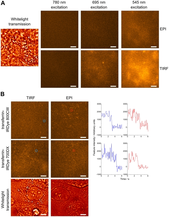
BACKGROUND: The autofluorescence background of biological samples impedes the detection of single molecules when imaging. The most common method of reducing the background is to use evanescent field excitation, which is incompatible with imaging beyond the surface of biological samples. An alternative would be to use probes that can be excited in the near infra-red region of the spectrum, where autofluorescence is low. Such probes could also increase the number of labels that can be imaged in multicolour single molecule microscopes. Despite being widely used in ensemble imaging, there is a currently a shortage of information available for selecting appropriate commercial near infra-red dyes for single molecule work. It is therefore important to characterise available near infra-red dyes relevant to multicolour single molecule imaging. METHODOLOGY/PRINCIPAL FINDINGS: A range of commercially available near infra-red dyes compatible with multi-colour imaging was screened to find the brightest and most photostable candidates. Image series of immobilised samples of the brightest dyes (Alexa 700, IRDye 700DX, Alexa 790 and IRDye 800CW) were analysed to obtain the mean intensity of single dye molecules, their photobleaching rates and long period blinking kinetics. Using the optimum dye pair, we have demonstrated for the first time widefield, multi-colour, near infra-red single molecule imaging using a supercontinuum light source in MCF-7 cells. CONCLUSIONS/SIGNIFICANCE: We have demonstrated that near infra-red dyes can be used to avoid autofluorescence background in samples where restricting the illumination volume of visible light fails or is inappropriate. We have also shown that supercontinuum sources are suited to single molecule multicolour imaging throughout the 470-1000 nm range. Our measurements of near infra-red dye properties will enable others to select optimal dyes for single molecule imaging.
Overview
The claims and statistics of this paper haven’t been checked yet.
Run the check — free, no account needed. It reads the open-access text and checks the sources it cites.
Integrity signals
Registry
Cites
Statistics
Figures
Verification
Claims the paper makes
The claims and statistics of this paper haven’t been checked yet.
Run the check — free, no account needed. It reads the open-access text and checks the sources it cites.
Claims other papers make about this one
Each is checked against this paper’s own text: does it say what the citing paper says it does?
The source backs part of the claim, not all of it — or backs it for a different population, condition or comparison.
In the citing paper
“The first single-molecule imaging with NIR emitting dyes was conducted using NIR emitting dyes IRdye 700DX and IRDye 800CW.”
From The Pursuit of Shortwave Infrared-Emitting Nanoparticles with Bright Fluorescence through Molecular Design and Excited-State Engineering of Molecular Aggregates (2022) · cites this paper as [213]
Experimental — accuracy not yet measuredThe cited source says what the claim attributes to it.
In the citing paper
“Single-molecule images of MCF-7 cells labeled with these NIR dyes were captured by using epi-fluorescence microscopy and total internal reflection fluorescence microscopy.213”
From The Pursuit of Shortwave Infrared-Emitting Nanoparticles with Bright Fluorescence through Molecular Design and Excited-State Engineering of Molecular Aggregates (2022) · cites this paper as [213]
Self-check
Percentages, sums and reported differences re-computed from the paper’s own numbers.
The self-check needs the paper’s full text.
Citations
Cites
A Human Cell Line From a Pleural Effusion Derived From a Breast Carcinoma 2
Soule, H. D., Vázquez, J., Long, A., Albert, S. M., & BRENNAN, M. D. (1973) · JNCI Journal of the National Cancer Institute · doi:10.1093/jnci/51.5.1409 · Cited by 2,452 · cited 1×
“MCF-7 cells ([32], European Collection of Cell Cultures) were cultured on 35 mm glass bottomed dishes (MatTek) in RPMI 1640 media supplemented with glutamine and fetal calf serum, at 37uC in the presence of 5% CO2.”
MethodMulticolor Super-Resolution Imaging with Photo-Switchable Fluorescent Probes
Bates, M., Huang, B., Dempsey, G. T., & Zhuang, X. (2007) · Science · doi:10.1126/science.1146598 · Cited by 1,507 · cited 1×
“Alexa 680, Cy7 and Alexa 750 have been used in stochastic optical reconstruction microscopy (STORM) of fixed cells and tissue, in which one step of image formation is the localisation of single molecules in acquired data [10,11].”
MethodEvaluation of fluorophores for optimal performance in localization-based super-resolution imaging
Dempsey, G. T., Vaughan, J. C., Chen, K. H., Bates, M., & Zhuang, X. (2011) · Nature Methods · doi:10.1038/nmeth.1768 · Cited by 1,405 · cited 1×
“(Selected data for certain dyes specifically for the related technique of STORM were given in a recent study [17]).”
MethodBright Ideas for Chemical Biology
Lavis, L. D., & Raines, R. T. (2008) · ACS Chemical Biology · doi:10.1021/cb700248m · Cited by 1,357
Pavani, S. R. P., Thompson, M. A., Biteen, J. S., Lord, S. J., Liu, N., Twieg, R. J., Piestun, R., & Moerner, W. E. (2009) · Proceedings of the National Academy of Sciences · doi:10.1073/pnas.0900245106 · Cited by 1,051
Near-infrared fluorescence: application to in vivo molecular imaging
Hilderbrand, S. A., & Weissleder, R. (2009) · Current Opinion in Chemical Biology · doi:10.1016/j.cbpa.2009.09.029 · Cited by 1,036
Total Internal Reflection Fluorescence Microscopy in Cell Biology
Axelrod, D. (2001) · Traffic · doi:10.1034/j.1600-0854.2001.21104.x · Cited by 968
Single-molecule imaging of EGFR signalling on the surface of living cells
Sako, Y., Minoghchi, S., & Yanagida, T. (2000) · Nature Cell Biology · doi:10.1038/35004044 · Cited by 889 · cited 1×
“This has been used to study proteins in both the apical and basolateral membranes of live cells [3], but TIRF is unable to follow these molecules as they internalise or to image organelles within the cell.”
MethodAlexa Dyes, a Series of New Fluorescent Dyes that Yield Exceptionally Bright, Photostable Conjugates
Panchuk-Voloshina, N., Haugland, R. P., Bishop-Stewart, J., Bhalgat, M. K., Millard, P. J., Mao, F., Leung, W., & Haugland, R. P. (1999) · Journal of Histochemistry & Cytochemistry · doi:10.1177/002215549904700910 · Cited by 883
Crichton, R. R., & CHARLOTEAUX‐WAUTERS, M. (1987) · European Journal of Biochemistry · doi:10.1111/j.1432-1033.1987.tb11155.x · Cited by 682
Fluorescence quantum yields of a series of red and near-infrared dyes emitting at 600-1000 nm.
Rurack, K., & Spieles, M. (2011) · Analytical Chemistry · doi:10.1021/ac101329h · Cited by 632
Nanometric three-dimensional tracking of individual quantum dots in cells
Holtzer, L., Meckel, T., & Schmidt, T. (2007) · Applied Physics Letters · doi:10.1063/1.2437066 · Cited by 263 · cited 1×
“In conjunction with methods for resolving the axial position of single molecules, which include astigmatism [26], multifocal imaging [27] and double-helix point spread functions [28], epi-illumination with NIR probes should enable 3D imaging of individual proteins in cells and tissue.”
MethodMitigating Unwanted Photophysical Processes for Improved Single-Molecule Fluorescence Imaging
Dave, R., Terry, D. S., Munro, J. B., & Blanchard, S. C. (2009) · Biophysical Journal · doi:10.1016/j.bpj.2008.11.061 · Cited by 210
Near-infrared quantum dots for deep tissue imaging
Girija, A. R., Yoshida, Y., Maekawa, T., & Kumar, D. S. (2010) · Analytical and Bioanalytical Chemistry · doi:10.1007/s00216-010-3643-6 · Cited by 187 · cited 1×
“Other NIR fluorescent tags – quantum dots [24] and expressible proteins [25] – also exist, with the same advantages and disadvantages as their visible versions, but have not been considered here.”
BackgroundMultimodal Nanoprobes for Radionuclide and Five-Color Near-Infrared Optical Lymphatic Imaging
Kobayashi, H., Koyama, Y., Barrett, T., Hama, Y., Regino, C. A. S., Shin, I. S., Jang, B., Le, N., Paik, C. H., Choyke, P. L., et al. (2007) · ACS Nano · doi:10.1021/nn700062z · Cited by 186 · cited 1×
“It would be desirable to image more than two fluorophores within the NIR spectrum, but this has only been achieved by spectral unmixing in software [30], for which the SNRs required are not found in single molecule microscopy.”
BackgroundBaddeley, D., Crossman, D. J., Roßberger, S., Cheyne, J. E., Montgomery, J. M., Jayasinghe, I., Cremer, C. G., Cannell, M. B., & Soeller, C. (2011) · PLoS ONE · doi:10.1371/journal.pone.0020645 · Cited by 167 · cited 1×
“Alexa 680, Cy7 and Alexa 750 have been used in stochastic optical reconstruction microscopy (STORM) of fixed cells and tissue, in which one step of image formation is the localisation of single molecules in acquired data [10,11].”
MethodSingle‐Molecule Four‐Color FRET
Lee, J., Lee, S.-H., Ragunathan, K., Joo, C., Ha, T., & Hohng, S. (2010) · Angewandte Chemie International Edition · doi:10.1002/anie.201005402 · Cited by 164
Kapanidis, A. N., & Weiss, S. (2002) · The Journal of Chemical Physics · doi:10.1063/1.1521158 · Cited by 159
Light Sheet Microscopy for Single Molecule Tracking in Living Tissue
Ritter, J., Veith, R., Veenendaal, A., Siebrasse, J. P., & Kubitscheck, U. (2010) · PLoS ONE · doi:10.1371/journal.pone.0011639 · Cited by 158 · cited 1×
“Light sheet microscopy is an alternative method of limiting fluorescence excitation and has been used to image deeper planes [4], but requires complex setups.”
MethodEggeling, C., Widengren, J., Brand, L., Schaffer, J., Felekyan, S., & Seidel, C. A. M. (2006) · The Journal of Physical Chemistry A · doi:10.1021/jp054581w · Cited by 152
A nonfluorescent, broad-range quencher dye for Förster resonance energy transfer assays
Peng, X., Chen, H., Draney, D. R., Volcheck, W. M., Schutz-Geschwender, A. R., & Olive, D. M. (2009) · Analytical Biochemistry · doi:10.1016/j.ab.2009.02.024 · Cited by 116
Single-Molecule Three-Color FRET with Both Negligible Spectral Overlap and Long Observation Time
Lee, S.-H., Lee, J., & Hohng, S. (2010) · PLoS ONE · doi:10.1371/journal.pone.0012270 · Cited by 103
Selection of Fluorophore and Quencher Pairs for Fluorescent Nucleic Acid Hybridization Probes
Marras, S. A. E. (2006) · Humana Press eBooks · doi:10.1385/1-59745-069-3:3 · Cited by 99 · cited 1×
“This variation may be due to the strong temperature dependence of Cy5’s quantum yield [20].”
BackgroundSabanayagam, C. R., Eid, J. S., & MELLER, A. (2005) · The Journal of Chemical Physics · doi:10.1063/1.2136157 · Cited by 84
Single Molecule Dynamics in Life Science
(2008) · Wiley eBooks · doi:10.1002/9783527626137 · Cited by 66 · cited 1×
“Molecular diffusion, interactions, the formation of complexes, molecular structure and conformational changes can all be studied through fluorescence microscopy at the single molecule level [1].”
BackgroundAutomated multidimensional single molecule fluorescence microscopy feature detection and tracking
Rolfe, D. J., McLachlan, C., Hirsch, M., Needham, S. R., Tynan, C. J., Webb, S. E. D., Martin-Fernandez, M. L., & Hobson, M. (2011) · European Biophysics Journal · doi:10.1007/s00249-011-0747-7 · Cited by 46 · cited 1×
“Analysis of single molecule datasets The detection and tracking of single molecules contained within multichannel image series was achieved by software that has been described in detail elsewhere [33].”
MethodSelf-citationPhthalocyanine dye as an extremely photostable and highly fluorescent near-infrared labeling reagent
Peng, X., Draney, D. R., Volcheck, W. M., Bashford, G. R., Lamb, D. T., Grone, D. L., Zhang, Y., & Johnson, C. M. (2006) · Proceedings of SPIE, the International Society for Optical Engineering/Proceedings of SPIE · doi:10.1117/12.669173 · Cited by 45 · cited 1×
“We attribute the different trends regarding the IRDyes reported elsewhere [23] to the use of different conditions, such as temperature, which were not included.”
DeRocco, V., Anderson, T., Piehler, J., Erie, D. A., & Weninger, K. (2010) · BioTechniques · doi:10.2144/000113551 · Cited by 38
Nanometric depth resolution from multi-focal images in microscopy
Dalgarno, H. I. C., Dalgarno, P. A., Dada, A. C., Towers, C. E., Gibson, G. J., Parton, R. M., Davis, I., Warburton, R. J., & GREENAWAY, A. H. (2011) · Journal of The Royal Society Interface · doi:10.1098/rsif.2010.0508 · Cited by 17 · cited 1×
“In conjunction with methods for resolving the axial position of single molecules, which include astigmatism [26], multifocal imaging [27] and double-helix point spread functions [28], epi-illumination with NIR probes should enable 3D imaging of individual proteins in cells and tissue.”
MethodMulticolour single molecule imaging on cells using a supercontinuum source
Webb, S. E. D., Zanetti-Domingues, L. C., Coles, B. C., Rolfe, D. J., Wareham, R. J., & Martin-Fernandez, M. L. (2012) · Biomedical Optics Express · doi:10.1364/boe.3.000400 · Cited by 15 · cited 2×
“This work extends the use of a supercontinuum source for imaging single molecules from the visible spectrum [29] to the NIR.”
BackgroundMethodSelf-citationBeyond the rainbow: new fluorescent proteins brighten the infrared scene
Lin, M. Z. (2011) · Nature Methods · doi:10.1038/nmeth.1678 · Cited by 14 · cited 1×
“Other NIR fluorescent tags – quantum dots [24] and expressible proteins [25] – also exist, with the same advantages and disadvantages as their visible versions, but have not been considered here.”
BackgroundDetection of single fluorescent proteins inside eukaryotic cells using two-photon fluorescence
Hou, X., & Cheng, W. (2012) · Biomedical Optics Express · doi:10.1364/boe.3.000340 · Cited by 9 · cited 1×
“This could be achieved either by two-photon excitation of visible fluorophores, as has recently been demonstrated with EGFP in fixed cells [5], or by using NIR probes.”
BackgroundSingle-Molecule Imaging of Cellular Signaling
de Keijzer, S., Snaar-Jagalska, E. B., Spaink, H. P., & Schmidt, T. (2007) · Springer series in biophysics · doi:10.1007/978-3-540-73924-1_5 · Cited by 4 · cited 1×
“The histograms could be fit by summed Gaussian distributions, each corresponding to a different number of molecules within the feature [21].”
Background
Cited by
Oliveira, E., Bértolo, E., Núñez, C., Pilla, V., Santos, H. M., Fernández‐Lodeiro, J., Fernández‐Lodeiro, A., Djafari, J., Capelo, J. L., & Lodeiro, C. (2017) · ChemistryOpen · doi:10.1002/open.201700135 · Cited by 143
Li, X., Wei, L., Pan, L., Yi, Z., Wang, X., Ye, Z., Xiao, L., Li, H., & Wang, J. (2018) · Analytical Chemistry · doi:10.1021/acs.analchem.8b00251 · Cited by 118
Ylidenemalononitrile Enamines as Fluorescent “Turn-On” Indicators for Primary Amines
Longstreet, A. R., Jo, M., Chandler, R. R., Hanson, K., Zhan, N., Hrudka, J. J., Mattoussi, H. M., Shatruk, M., & McQuade, D. T. (2014) · Journal of the American Chemical Society · doi:10.1021/ja509058u · Cited by 95
PSMA-targeting agents for radio- and fluorescence-guided prostate cancer surgery
Derks, Y. H. W., Löwik, D. W. P. M., Sedelaar, J. P. M., Gotthardt, M., Boerman, O. C., Rijpkema, M., Lütje, S., & Heskamp, S. (2019) · Theranostics · doi:10.7150/thno.36739 · Cited by 91
“suitable for fluorescence imaging due to their brightness and photostability [80].”
BackgroundFLIM-FRET for Cancer Applications
Rajoria, S., Zhao, L., Intes, X., & Barroso, M. (2014) · Current molecular imaging · doi:10.2174/2211555203666141117221111 · Cited by 68
Metadata
- Authors
- Tynan, C. J., Clarke, D. T., Coles, B. C., Rolfe, D. J., Martin-Fernandez, M. L., & Webb, S. E. D.
- Affiliations
- Rutherford Appleton Laboratory (GB)
- Corresponding author
- Webb, Stephen E. D.
- ORCID
- Venue
- PLoS ONE
- Publisher
- Public Library of Science (PLoS)
- Published
- 2012-04-25
- Volume
- 7
- Issue
- 4
- Pages
- e36265
- ISSN
- 1932-6203 (ISSN-L)
- PMC
- PMC3338497
- OpenAlex
- W2042537014
- Cited by
- 40
- References
- 34
- Citations by year
- 2025: 2 · 2024: 2 · 2023: 2 · 2022: 4 · 2021: 3 · 2020: 5 · 2019: 3 · 2018: 3 · 2017: 4 · 2016: 2 · 2015: 2 · 2014: 4 · 2013: 2 · 2012: 2
- FWCI
- 1.46
- Type
- journal article
- Language
- en
- Open access
- gold
- doi.org
- Locations
- PLoS ONE (publishedVersion, cc-by)
- PubMed Central (publishedVersion, cc-by)
- PubMed (publishedVersion)
- DOAJ (DOAJ: Directory of Open Access Journals) (submittedVersion)
- Figshare (submittedVersion, cc-by)
- Science and Technology Facilities Council (submittedVersion)
- Funders
- Directorate for Biological Sciences
- Biotechnology and Biological Sciences Research Council (BB/G007160/1, BB/G006911/1)
- Topics
- Life Sciences › Biochemistry, Genetics and Molecular Biology › Biophysics › Advanced Fluorescence Microscopy Techniques
- Physical Sciences › Materials Science › Materials Chemistry › Luminescence and Fluorescent Materials
- Health Sciences › Medicine › Radiology, Nuclear Medicine and Imaging › Optical Imaging and Spectroscopy Techniques
- Keywords
- single-molecule imaging, multicolor imaging, near-infrared dyes, photostability, photobleaching, supercontinuum light source, MCF-7 cells, dye selection
- MeSH terms
- Color, Fluorescent Dyes, Humans, Infrared Rays, Spectrometry, Fluorescence, Cell Line, Tumor, Molecular Imaging
About these checks
Registry, retraction, Cites and statistics results are computed, not judged, with one exception: a printed reference that no index lists is read once by a model before it counts as a citation error. Claims are extracted by a model and kept only when their quote appears verbatim in the text; each cited source is then checked, with a model’s help, for whether it says what the claim relies on. Those verdicts can be wrong — read the claim’s quote and the cited source before relying on one.
Statistics and arithmetic are recomputed, not judged.
Confidence bands are shown only for fields calibrated on our annotated test set. None is calibrated yet, so no band is shown.
What counts as a claim ›How we measure confidence ›How we check this ›
Are you an author, or do you see a problem with this page? Request a review
