

The DOI matches its Crossref record; 47 of the 47 references it cites were found in a registry or an index.
Sensitivity to Lysosome-Dependent Cell Death Is Directly Regulated by Lysosomal Cholesterol Content
Open accessAbstract
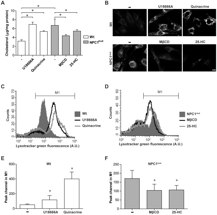
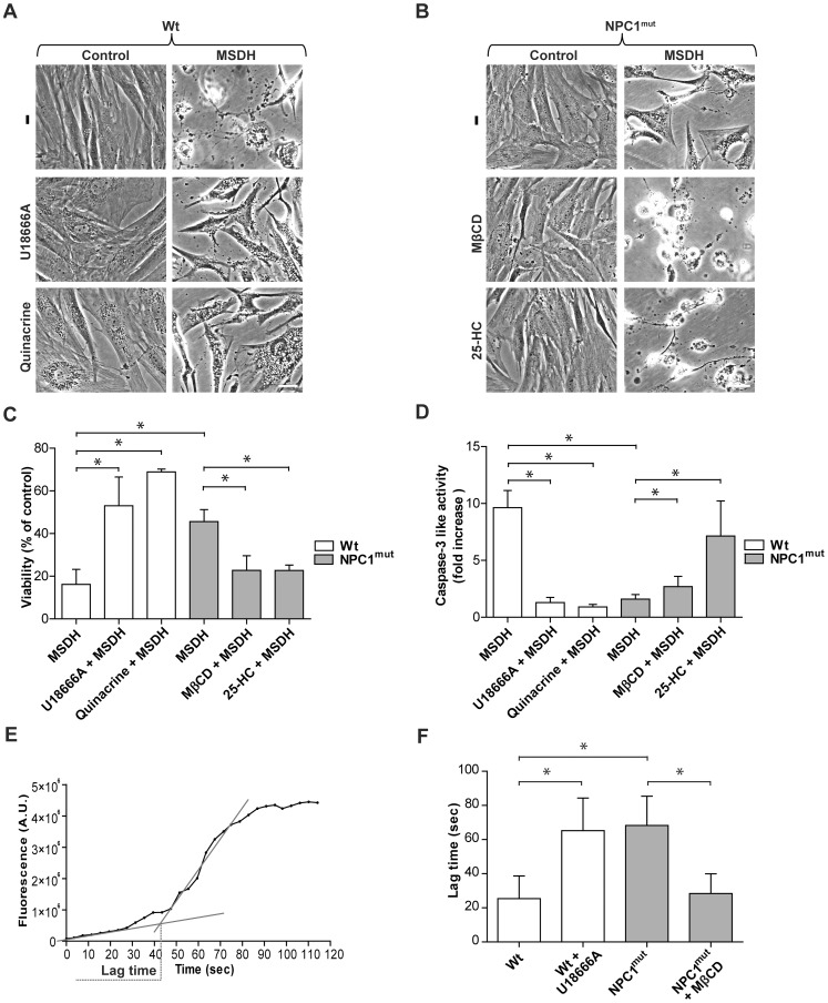
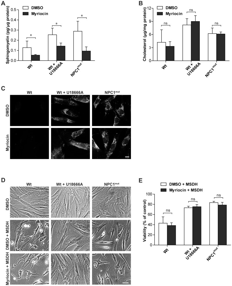
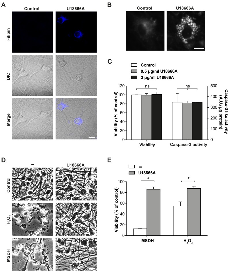
Alterations in lipid homeostasis are implicated in several neurodegenerative diseases, although the mechanisms responsible are poorly understood. We evaluated the impact of cholesterol accumulation, induced by U18666A, quinacrine or mutations in the cholesterol transporting Niemann-Pick disease type C1 (NPC1) protein, on lysosomal stability and sensitivity to lysosome-mediated cell death. We found that neurons with lysosomal cholesterol accumulation were protected from oxidative stress-induced apoptosis. In addition, human fibroblasts with cholesterol-loaded lysosomes showed higher lysosomal membrane stability than controls. Previous studies have shown that cholesterol accumulation is accompanied by the storage of lipids such as sphingomyelin, glycosphingolipids and sphingosine and an up regulation of lysosomal associated membrane protein-2 (LAMP-2), which may also influence lysosomal stability. However, in this study the use of myriocin and LAMP deficient fibroblasts excluded these factors as responsible for the rescuing effect and instead suggested that primarily lysosomal cholesterol content determineD the cellular sensitivity to toxic insults. Further strengthening this concept, depletion of cholesterol using methyl-β-cyclodextrin or 25-hydroxycholesterol decreased the stability of lysosomes and cells became more prone to undergo apoptosis. In conclusion, cholesterol content regulated lysosomal membrane permeabilization and thereby influenced cell death sensitivity. Our data suggests that lysosomal cholesterol modulation might be used as a therapeutic strategy for conditions associated with accelerated or repressed apoptosis.
Overview
The claims and statistics of this paper haven’t been checked yet.
Run the check — free, no account needed. It reads the open-access text and checks the sources it cites.
Integrity signals
Registry
Cites
Statistics
Figures
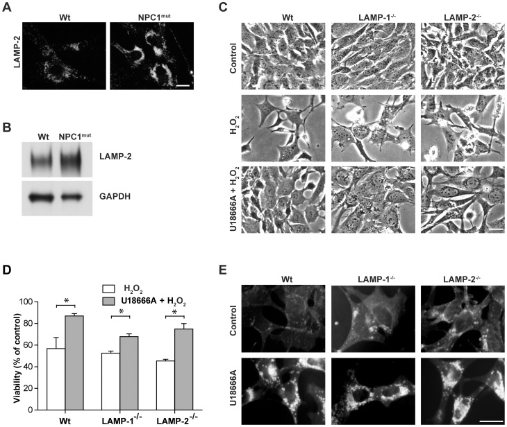
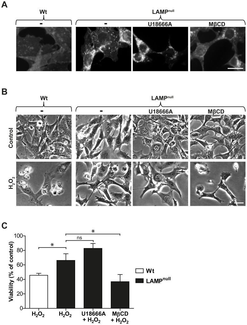
Verification
Claims the paper makes
The claims and statistics of this paper haven’t been checked yet.
Run the check — free, no account needed. It reads the open-access text and checks the sources it cites.
Claims other papers make about this one
Each is checked against this paper’s own text: does it say what the citing paper says it does?
In the citing paper
“Moreover, under physiological conditions, lysosomal membranes can be stabilized by altering lysosomal cholesterol content [788] or by boosting endogenous activity of heat shock protein family A (Hsp70) member 1A (HSPA1A; best known as HSP70) [789, 790].”
From Molecular mechanisms of cell death: recommendations of the nomenclature committee on cell death 2018. (2018) · cites this paper as [788]
Self-check
Percentages, sums and reported differences re-computed from the paper’s own numbers.
The self-check needs the paper’s full text.
Citations
Cites
Lipid rafts as a membrane-organizing principle
Lingwood, D., & Simons, K. (2010) · Science · doi:10.1126/science.1174621 · Cited by 4,417 · cited 1×
“As cholesterol is an important component of all cellular membranes, including specialized lipid raft micro domains [34],”
BackgroundEvidence for the Formation of Symmetric and Asymmetric DLPC-DAPC Lipid Bilayer Domains
Ritter, M., Schmidt, S., Jakab, M., Paulmichl, M., & Henderson, R. M. (2013) · Cellular Physiology and Biochemistry · doi:10.1159/000350122 · Cited by 3,923
Simons, K., & Ikonen, E. (2000) · Science · doi:10.1126/science.290.5497.1721 · Cited by 1,277 · cited 1×
“Normally, cholesterol is released from endocytosed low density lipoprotein (LDL) particles by the action of lysosomal acid lipase and is then transported, via the lysosomal NPC proteins, to the ER where it serves as a sensor for cellular cholesterol homeostasis and may be esterified [6].”
BackgroundLloyd‐Evans, E., Morgan, A. J., He, X., Smith, D. A., Elliot-Smith, E., Sillence, D. J., Churchill, G. C., Schuchman, E. H., Galione, A., & Platt, F. M. (2008) · Nature Medicine · doi:10.1038/nm.1876 · Cited by 832 · cited 3×
“sphingomyelin, glycosphingolipids and sphingosine [9,22], which have been suggested to influence the stability of lysosomes [26,27].”
BackgroundGuicciardi, M. E., Deussing, J. M., Miyoshi, H., Bronk, S. F., Svingen, P. A., Peters, C., Kaufmann, S. H., & Gores, G. J. (2000) · Journal of Clinical Investigation · doi:10.1172/jci9914 · Cited by 710 · cited 1×
“Previous studies have convincingly shown that the presence of lysosomal proteases, cathepsins, in the cytosol mediates apoptosis [2,3,4], implying that the integrity of the lysosomal membrane is of high importance for cell survival.”
BackgroundFoghsgaard, L., Wissing, D., Mauch, D., Lademann, U. A., Bastholm, L., Boes, M. L., Elling, F., Leist, M., & Jäättelä, M. (2001) · The Journal of Cell Biology · doi:10.1083/jcb.153.5.999 · Cited by 629 · cited 1×
“Previous studies have convincingly shown that the presence of lysosomal proteases, cathepsins, in the cytosol mediates apoptosis [2,3,4], implying that the integrity of the lysosomal membrane is of high importance for cell survival.”
BackgroundRegulation of apoptosis-associated lysosomal membrane permeabilization
Johansson, A., Appelqvist, H., Nilsson, C., Kågedal, K., Roberg, K. B., & Öllinger, K. M. (2010) · APOPTOSIS · doi:10.1007/s10495-009-0452-5 · Cited by 479 · cited 1×
“The mechanism underlying LMP is still incompletely understood; however, a number of factors have been described to affect the stability of the lysosomal membrane, including the level of lysosome-associated membrane proteins (LAMP) and cholesterol [5].”
BackgroundSelf-citationDavidson, C. D., Ali, N. F., Micsenyi, M. C., Stephney, G. W., Renault, S., Dobrenis, K., Ory, D. S., Vanier, M. T., & Walkley, S. U. (2009) · PLoS ONE · doi:10.1371/journal.pone.0006951 · Cited by 449 · cited 1×
“b-Cyclodextrin compounds has been shown to correct cholesterol transport in NPC-defective cells [13] and substantially reduce neurodegeneration and increase lifespan in Npc1 mice [14].”
BackgroundLysosomes and lysosomal cathepsins in cell death
Repnik, U., Stoka, V., Türk, V., & Turk, B. (2011) · Biochimica et Biophysica Acta (BBA) - Proteins and Proteomics · doi:10.1016/j.bbapap.2011.08.016 · Cited by 412 · cited 1×
“Lysosomes are acidic organelles involved in several cellular functions, including degradation of macromolecules, repair of the plasma membrane, antigen presentation, recycling of cell surface receptors and apoptosis signaling [1].”
BackgroundSphingosine-induced apoptosis is dependent on lysosomal proteases
Kågedal, K., Ming, Z. H. A. O., Svensson, I., & Brunk, U. T. (2001) · Biochemical Journal · doi:10.1042/0264-6021:3590335 · Cited by 379
Self-citation
Research works printed but not linked: 1
- Hansson E, Rönnbäck L (1989) Primary cultures of astroglia and neurons from different brain regions. In: Shahar A d, Vernadakis A, Haber B, editors. A dissection and tissue culture manual of nervous system. 1st ed. New York: Liss. pp. 92–104.
Cited by
Sorted by citations
Galluzzi, L., Vitale, I., Aaronson, S. A., Abrams, J. M., Adam, D., Agostinis, P., Alnemri, E. S., Altucci, L., Amelio, I., Andrews, D. W., et al. (2018) · Cell Death & Differentiation · doi:10.1038/s41418-017-0012-4 · Cited by 6,680
“siological conditions, lysosomal membranes can be stabilized by altering lysosomal cholesterol content [788] or by boosting endogenous activity of heat shock protein family A (Hsp70) member 1A (HSPA1A; best known as HSP70) [789, 790].”
BackgroundThe role of autophagy in neurodegenerative disease
Nixon, R. A. (2013) · Nature Medicine · doi:10.1038/nm.3232 · Cited by 2,059
The lysosome: from waste bag to potential therapeutic target
Appelqvist, H., Wäster, P., Kågedal, K., & Öllinger, K. (2013) · Journal of Molecular Cell Biology · doi:10.1093/jmcb/mjt022 · Cited by 772
“Multiple cysteine cathepsins (including cathepsin B, D, H, K, L, and S) activate Bid (Cirman et al., 2004; Heinrich et al., 2004; Caruso et al., 2006; Appelqvist et al., 2012a).”
BackgroundInfluentialLysosomal Biology and Function: Modern View of Cellular Debris Bin
Trivedi, P. C., Bartlett, J. J., & Pulinilkunnil, T. (2020) · Cells · doi:10.3390/cells9051131 · Cited by 309
Gangliosides and Gangliosidoses: Principles of Molecular and Metabolic Pathogenesis
Sandhoff, K., & HARZER, K. (2013) · Journal of Neuroscience · doi:10.1523/jneurosci.0822-13.2013 · Cited by 283
“Perimeter membranes are protected by a thick glycocalyx enriched in polylactosamine structures, and by a high cholesterol level (Hay, 1989; Appelqvist et al., 2012), the chaperon HSP70 (Kirkegaard et al., 2010), and presumably also by a high lateral pressure attenuating the interaction with GM2AP,…”
Metadata
- Authors
- Appelqvist, H., Sandin, L., Björnström, K., Saftig, P., Garner, B., Öllinger, K., & Kågedal, K.
- Affiliations
- Linköping University (SE)
- University of Wollongong (AU)
- Corresponding author
- Appelqvist, Hanna
- Venue
- PLoS ONE
- Publisher
- Public Library of Science (PLoS)
- Published
- 2012-11-16
- Volume
- 7
- Issue
- 11
- Pages
- e50262
- ISSN
- 1932-6203 (ISSN-L)
- PMC
- PMC3500374
- OpenAlex
- W2033615443
- Cited by
- 108
- References
- 46
- Citations by year
- 2026: 4 · 2025: 5 · 2024: 8 · 2023: 13 · 2022: 5 · 2021: 13 · 2020: 14 · 2019: 4 · 2018: 4 · 2017: 6 · 2016: 9 · 2015: 7 · 2014: 7 · 2013: 9
- FWCI
- 4.93
- Type
- journal article
- Language
- en
- Open access
- gold
- doi.org
- Locations
- PLoS ONE (publishedVersion, cc-by)
- Europe PMC (PubMed Central) (publishedVersion, cc-by)
- PubMed Central (publishedVersion, cc-by)
- PubMed (publishedVersion)
- Research Online (University of Wollongong) (submittedVersion)
- DOAJ (DOAJ: Directory of Open Access Journals) (submittedVersion, cc-by-sa)
- Figshare (submittedVersion, cc-by)
- Figshare (submittedVersion)
- Funders
- Deutsche Forschungsgemeinschaft
- Vetenskapsrådet (2010-03463)
- Topics
- Health Sciences › Medicine › Genetics (clinical) › Lysosomal Storage Disorders Research
- Life Sciences › Biochemistry, Genetics and Molecular Biology › Cell Biology › Autophagy in Disease and Therapy
- Life Sciences › Biochemistry, Genetics and Molecular Biology › Physiology › Calcium signaling and nucleotide metabolism
- Keywords
- lysosomal cholesterol, lysosome-dependent cell death, apoptosis, lysosomal membrane stability, lysosomal membrane permeabilization, Niemann-Pick disease type C1, NPC1 protein, cholesterol accumulation, lipid homeostasis, neurodegenerative diseases, oxidative stress, myriocin, methyl-β-cyclodextrin, 25-hydroxycholesterol, LAMP2, neurons, fibroblasts
- MeSH terms
- Niemann-Pick C1 Protein, Androstenes, Carrier Proteins, Cholesterol, Fibroblasts, Flow Cytometry, Humans, Hydroxycholesterols, Immunohistochemistry, Lysosomes, Membrane Glycoproteins, Microscopy, Phase-Contrast, Mutation, Neurons, Quinacrine, Blotting, Western, Cell Death, Statistics, Nonparametric, beta-Cyclodextrins, Intracellular Signaling Peptides and Proteins, Lysosomal Membrane Proteins
About these checks
Registry, retraction, Cites and statistics results are computed, not judged, with one exception: a printed reference that no index lists is read once by a model before it counts as a citation error. Claims are extracted by a model and kept only when their quote appears verbatim in the text; each cited source is then checked, with a model’s help, for whether it says what the claim relies on. Those verdicts can be wrong — read the claim’s quote and the cited source before relying on one.
Statistics and arithmetic are recomputed, not judged.
Confidence bands are shown only for fields calibrated on our annotated test set. None is calibrated yet, so no band is shown.
What counts as a claim ›How we measure confidence ›How we check this ›
Are you an author, or do you see a problem with this page? Request a review
