

The DOI matches its Crossref record.
Application of a Novel Alkali-Tolerant Thermostable DyP-Type Peroxidase from Saccharomonospora viridis DSM 43017 in Biobleaching of Eucalyptus Kraft Pulp
Abstract
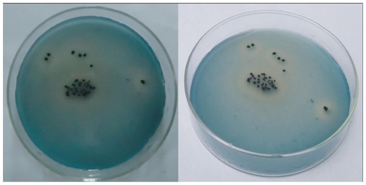
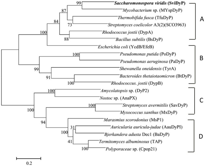
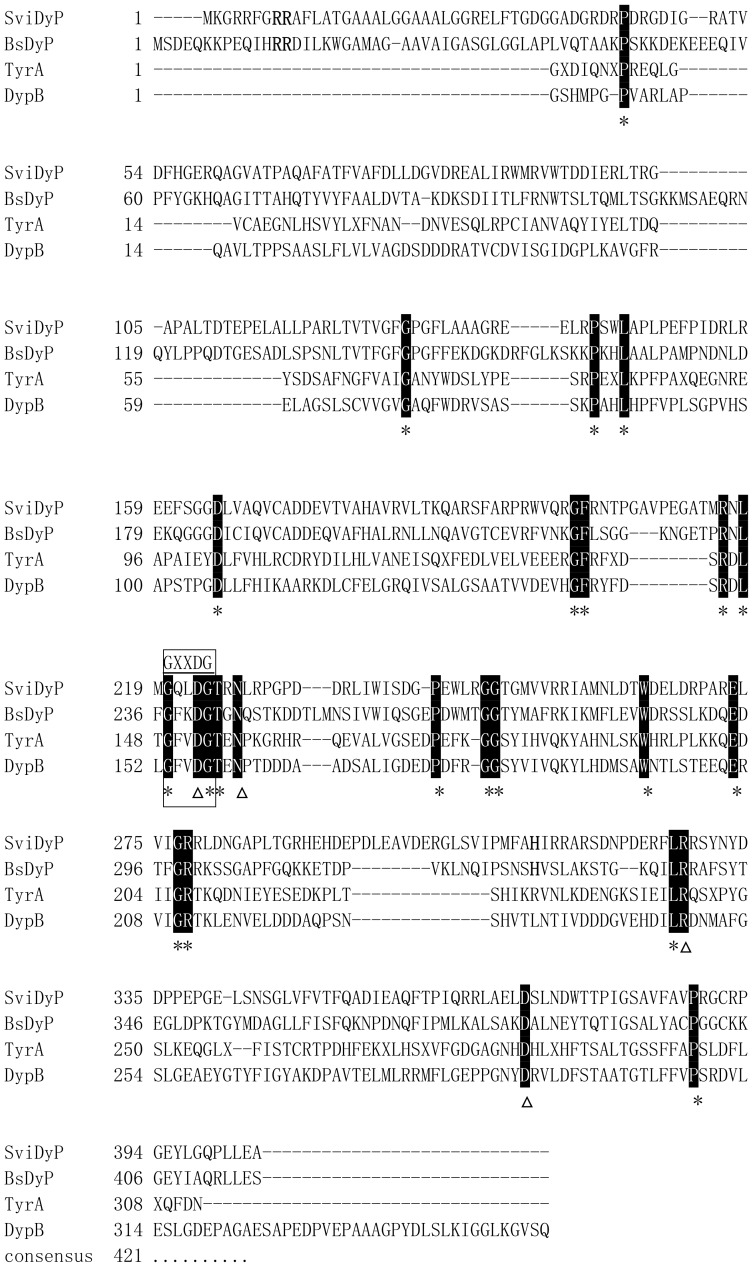
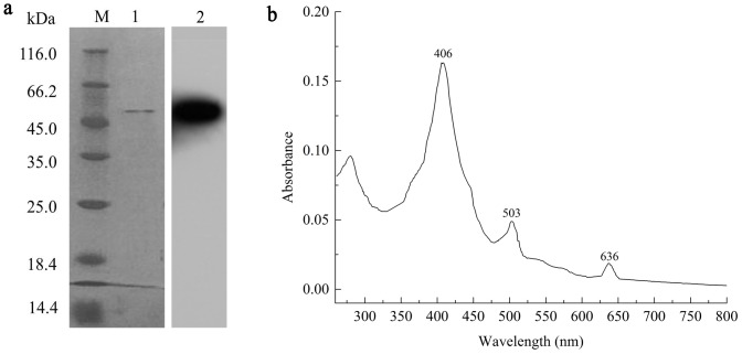
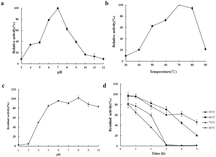
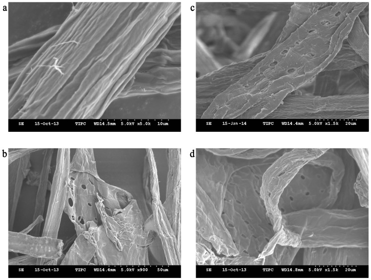
Saccharomonospora viridis is a thermophilic actinomycete that may have biotechnological applications because of its dye decolorizing activity, though the enzymatic oxidative system responsible for this activity remains elusive. Bioinformatic analysis revealed a DyP-type peroxidase gene in the genome of S. viridis DSM 43017 with sequence similarity to peroxidase from dye-decolorizing microbes. This gene, svidyp, consists of 1,215 bp encoding a polypeptide of 404 amino acids. The gene encoding SviDyP was cloned, heterologously expressed in Escherichia coli, and then purified. The recombinant protein could efficiently decolorize several triarylmethane dyes, anthraquinonic and azo dyes under neutral to alkaline conditions. The optimum pH and temperature for SviDyP was pH 7.0 and 70°C, respectively. Compared with other DyP-type peroxidases, SviDyP was more active at high temperatures, retaining>63% of its maximum activity at 50-80°C. It also showed broad pH adaptability (>35% activity at pH 4.0-9.0) and alkali-tolerance (>80% activity after incubation at pH 5-10 for 1 h at 37°C), and was highly thermostable (>60% activity after incubation at 70°C for 2 h at pH 7.0). SviDyP had an accelerated action during the biobleaching of eucalyptus kraft pulp, resulting in a 21.8% reduction in kappa number and an increase of 2.98% (ISO) in brightness. These favorable properties make SviDyP peroxidase a promising enzyme for use in the pulp and paper industries.
Overview
The claims and statistics of this paper haven’t been checked yet.
Run the check — free, no account needed. It reads the open-access text and checks the sources it cites.
Integrity signals
Registry
Cites
Statistics
Figures
Verification
Claims the paper makes
The claims and statistics of this paper haven’t been checked yet.
Run the check — free, no account needed. It reads the open-access text and checks the sources it cites.
Claims other papers make about this one
Each is checked against this paper’s own text: does it say what the citing paper says it does?
The check wasn’t confident either way, so it gives no verdict — never disputed.
In the citing paper
“Although their action mechanism resembles that of other peroxidases, anthraquinone dye degradation is not yet fully mapped 129-134 .”
From Degradation of anthraquinone dyes from effluents: A review focusing on enzymatic dye degradation with industrial potential (2020) · cites this paper as [132]
Experimental — accuracy not yet measured
Self-check
Percentages, sums and reported differences re-computed from the paper’s own numbers.
The self-check needs the paper’s full text.
Citations
Cites
No references could be resolved yet.
Research works printed but not linked: 55
- Dunford HB (1999) Heme peroxidases. Wiley-VCH.
- KG Welinder, 1992, Superfamily of plant, fungal and bacterial peroxidases, Curr Opin Struc Biol, 2, 388, 10.1016/0959-440X(92)90230-5 · DOI
- M Tien, 1988, Lignin peroxidase of Phanerochaete chrysosporium, Method Enzymol, 161, 238, 10.1016/0076-6879(88)61025-1 · DOI · Paper page
- H Wariishi, 1992, Manganese (II) oxidation by manganese peroxidase from the basidiomycete Phanerochaete chrysosporium. Kinetic mechanism and role of chelators, J Biol Chem, 267, 23688, 10.1016/S0021-9258(18)35893-9 · DOI · Paper page
- R Pogni, 2005, Tryptophan-based radical in the catalytic mechanism of versatile peroxidase from Bjerkandera adusta, Biochemistry, 44, 4267, 10.1021/bi047474l · DOI
- DI Colpa, 2014, DyP-type peroxidases: a promising and versatile class of enzymes, J Ind Microbiol Biotechnol, 41, 1, 10.1007/s10295-013-1371-6 · DOI · Paper page
- J Li, 2012, Identification and molecular characterization of a novel DyP-type peroxidase from Pseudomonas aeruginosa PKE117, Appl Biochem Biotech, 166, 774, 10.1007/s12010-011-9466-x · DOI
- D Salvachúa, 2013, Characterization of a novel dye-decolorizing peroxidase (DyP)-Type Enzyme from Irpex lacteus and its application in enzymatic hydrolysis of wheat straw, Appl Environ Microbiol, 79, 4316, 10.1128/AEM.00699-13 · DOI
- T Sato, 2004, A unique dye-decolorizing peroxidase, DyP, from Thanatephorus cucumeris Dec 1: heterologous expression, crystallization and preliminary X-ray analysis, Acta Crystallogr D, 60, 149, 10.1107/S0907444903025472 · DOI
- MF Coughlin, 2003, High performance degradation of azo dye acid orange 7 and sulfanilic acid in laboratory scale reactor after seeding with cultured bacterial strains, Water Res, 37, 2757, 10.1016/S0043-1354(03)00069-1 · DOI
- T Shimokawa, 2009, Purification and characterization of two DyP isozymes from Thanatephorus cucumeris Dec 1 specifically expressed in an air-membrane surface bioreactor, J Biosci Bioeng, 107, 113, 10.1016/j.jbiosc.2008.09.013 · DOI
- Y Sugano, 2009, DyP-type peroxidases comprise a novel heme peroxidase family, Cell Mol Life Sci, 66, 1387, 10.1007/s00018-008-8651-8 · DOI
- C Zubieta, 2007, Identification and structural characterization of heme binding in a novel dye-decolorizing peroxidase, TyrA, Proteins: Structure, Function, and Bioinformatics, 69, 234, 10.1002/prot.21673 · DOI
- C Liers, 2013, Substrate oxidation by dye-decolorizing peroxidases (DyPs) from wood-and litter-degrading agaricomycetes compared to other fungal and plant heme-peroxidases, Appl Microbiol Biotechnol, 97, 5839, 10.1007/s00253-012-4521-2 · DOI
- A Santos, 2014, New dye-decolorizing peroxidases from Bacillus subtilis and Pseudomonas putida MET94: towards biotechnological applications, Appl Microbiol Biotechnol, 98, 2053, 10.1007/s00253-013-5041-4 · DOI · Paper page
- HJO Ogola, 2009, Molecular characterization of a novel peroxidase from the cyanobacterium Anabaena sp. strain PCC 7120, Appl Environ Microbiol, 75, 7509, 10.1128/AEM.01121-09 · DOI · Paper page
- M Ahmad, 2011, Identification of DypB from Rhodococcus jostii RHA1 as a lignin peroxidase, Biochemistry, 50, 5096, 10.1021/bi101892z · DOI · Paper page
- D Fodil, 2011, Purification and characterization of two extracellular peroxidases from Streptomyces sp. strain AM2, a decolorizing actinomycetes responsible for the biodegradation of natural humic acids, Int Biodeter Biodegr, 65, 470, 10.1016/j.ibiod.2011.01.009 · DOI
- D Fodil, 2012, A thermostable humic acid peroxidase from Streptomyces sp. strain AH4: Purification and biochemical characterization, Bioresource Technol, 111, 383, 10.1016/j.biortech.2012.01.153 · DOI
- Roberts JN (2011) Identification and characterization of DyP peroxidases from Rhodococcus jostii RHA1: University of British Columbia. · DOI
- E van Bloois, 2010, A robust and extracellular heme-containing peroxidase from Thermobifida fusca as prototype of a bacterial peroxidase superfamily, Appl Microbiol Biotechnol, 86, 1419, 10.1007/s00253-009-2369-x · DOI · Paper page
- V Antonopoulos, 2001, The use of extracellular enzymes from Streptomyces albus ATCC 3005 for the bleaching of eucalyptus kraft pulp, Appl Microbiol Biotechnol, 57, 92, 10.1007/s002530100740 · DOI
- J Georis, 2000, Purification and properties of three endo-β-1, 4-xylanases produced by Streptomyces sp. strain S38 which differ in their ability to enhance the bleaching of kraft pulps, Enzyme Microb Tech, 26, 178, 10.1016/S0141-0229(99)00141-6 · DOI
- D Wesenberg, 2003, White-rot fungi and their enzymes for the treatment of industrial dye effluents, Biotechnol Adv, 22, 161, 10.1016/j.biotechadv.2003.08.011 · DOI · Paper page
- ID Reid, 1998, Effects of manganese peroxidase on residual lignin of softwood kraft pulp, Appl Environ Microbiol, 64, 2273, 10.1128/AEM.64.6.2273-2274.1998 · DOI
- QK Beg, 2000, Enhanced production of a thermostable xylanase from Streptomyces sp. QG-11-3 and its application in biobleaching of eucalyptus kraft pulp, Enzyme Microb Techn, 27, 459, 10.1016/S0141-0229(00)00231-3 · DOI
- K Harazono, 1996, Bleaching of hardwood kraft pulp with manganese peroxidase from Phanerochaete sordida YK-624 without addition of MnSO (inf4), Appl Environ Microbiol, 62, 913, 10.1128/AEM.62.3.913-917.1996 · DOI
- MD Webb, 2001, Metabolism of pentachlorophenol by Saccharomonospora viridis strains isolated from mushroom compost, Soil Biol Biochem, 33, 1903, 10.1016/S0038-0717(01)00115-8 · DOI
- A Pati, 2009, Complete genome sequence of Saccharomonospora viridis type strain (P101T), Stand Genomic Sci, 1, 141, 10.4056/sigs.20263 · DOI
- C Zubieta, 2007, Crystal structures of two novel dye-decolorizing peroxidases reveal a β-barrel fold with a conserved heme-binding motif, Proteins: Structure, Function, and Bioinformatics, 69, 223, 10.1002/prot.21550 · DOI
- JN Roberts, 2011, Characterization of dye-decolorizing peroxidases from Rhodococcus jostii RHA1, Biochemistry, 50, 5108, 10.1021/bi200427h · DOI
- X Liu, 2011, Crystal structure and biochemical features of EfeB/YcdB from Escherichia coli O157 ASP235 plays divergent roles in different enzyme-catalyzed processes, J Biol Chem, 286, 14922, 10.1074/jbc.M110.197780 · DOI
- JD Thompson, 1994, CLUSTAL W: improving the sensitivity of progressive multiple sequence alignment through sequence weighting, position-specific gap penalties and weight matrix choice, Nucl Acids Res, 22, 4673, 10.1093/nar/22.22.4673 · DOI · Paper page
- S Kumar, 2008, MEGA: a biologist-centric software for evolutionary analysis of DNA and protein sequences, Brief Bioinform, 9, 299, 10.1093/bib/bbn017 · DOI
- K Arnold, 2006, The SWISS-MODEL workspace: a web-based environment for protein structure homology modelling, Bioinformatics, 22, 195, 10.1093/bioinformatics/bti770 · DOI
- F Kiefer, 2009, The SWISS-MODEL repository and associated resources, Nucl Acids Res, 37, D387, 10.1093/nar/gkn750 · DOI
- T Schwede, 2003, SWISS-MODEL: an automated protein homology-modeling server, Nucl Acids Res, 31, 3381, 10.1093/nar/gkg520 · DOI
- MM Bradford, 1976, A rapid and sensitive method for the quantitation of microgram quantities of protein utilizing the principle of protein-dye binding, Anal Biochem, 72, 248, 10.1016/0003-2697(76)90527-3 · DOI · Paper page
- UK Laemmli, 1970, Cleavage of structural proteins during the assembly of the head of bacteriophage T4, Nature, 227, 680, 10.1038/227680a0 · DOI · Paper page
- P Cao, 2005, Production and characterization of a bacterial single-chain antibody fragment specific to B-cell-activating factor of the TNF family, Protein Expr Purif, 43, 157, 10.1016/j.pep.2005.04.022 · DOI
- Y Sugano, 2000, Efficient heterologous expression in Aspergillus oryzae of a unique dye-decolorizing peroxidase, DyP, of Geotrichum candidum Dec 1, Appl Environ Microbiol, 66, 1754, 10.1128/AEM.66.4.1754-1758.2000 · DOI
- H Lineweaver, 1934, The determination of enzyme dissociation constants, J Am Chem Soc, 56, 658, 10.1021/ja01318a036 · DOI · Paper page
- R Kondo, 1994, Bleaching of hardwood kraft pulp with manganese peroxidase secreted from Phanerochaete sordida YK-624, Appl Environ Microbiol, 60, 4359, 10.1128/AEM.60.12.4359-4363.1994 · DOI
- SJ Kim, 1999, Purification and characterization of a novel peroxidase from Geotrichum candidum Dec 1 involved in decolorization of dyes, Appl Environ Microbiol, 65, 1029, 10.1128/AEM.65.3.1029-1035.1999 · DOI · Paper page
- JC Roberts, 1990, Modification of paper properties by the pretreatment of pulp with Saccharomonospora viridis xylanase, Enzyme Microb Tech, 12, 210, 10.1016/0141-0229(90)90040-W · DOI
- Z Wang, 2012, A novel, alkali-tolerant thermostable xylanase from Saccharomonospora viridis: direct gene cloning, expression and enzyme characterization, World J Microbiol Biotechnol, 28, 2741, 10.1007/s11274-012-1085-3 · DOI
- A Sturm, 2006, YcdB from Escherichia coli reveals a novel class of Tat-dependently translocated hemoproteins, J Biol Chem, 281, 13972, 10.1074/jbc.M511891200 · DOI
- S-Y An, 2002, Decolorization of triphenylmethane and azo dyes by Citrobacter sp, Biotechnol Lett, 24, 1037, 10.1023/A:1015610018103 · DOI
- S Ren, 2006, Decolorization of triphenylmethane, azo, and anthraquinone dyes by a newly isolated Aeromonas hydrophila strain, Appl Microbiol Biotechnol, 72, 1316, 10.1007/s00253-006-0418-2 · DOI
- RK Sani, 1999, Decolorization of triphenylmethane dyes and textile and dye-stuff effluent by Kurthia sp, Enzyme and Microbial Technology, 24, 433, 10.1016/S0141-0229(98)00159-8 · DOI
- C Yatome, 1993, Degradation of crystal violet by Nocardia corallina, Appl Microbiol Biotechnol, 38, 565, 10.1007/BF00242956 · DOI
- N Daneshvar, 2007, Biological decolorization of dye solution containing Malachite Green by microalgae Cosmarium sp, Bioresource Technol, 98, 1176, 10.1016/j.biortech.2006.05.025 · DOI · Paper page
- JJ Jones, 2003, Decolorization of malachite green and crystal violet by waterborne pathogenic mycobacteria, Antimicrob Agents Chemother, 47, 2323, 10.1128/AAC.47.7.2323-2326.2003 · DOI
- JX Heck, 2006, Statistical optimization of thermo-tolerant xylanase activity from Amazon isolated Bacillus circulans on solid-state cultivation, Bioresource Technol, 97, 1902, 10.1016/j.biortech.2005.08.013 · DOI
- Y Du, 2013, Characterization of three novel thermophilic xylanases from Humicola insolens Y1 with application potentials in the brewing industry, Bioresource Technol, 130, 161, 10.1016/j.biortech.2012.12.067 · DOI
Cited by
Routoula, E., & Patwardhan, S. V. (2020) · Environmental Science & Technology · doi:10.1021/acs.est.9b03737 · Cited by 579
Explorations and Applications of Enzyme-linked Bioremediation of Synthetic Dyes
Ogola, H. J. O., Ashida, H., Ishikawa, T., & Sawa, Y. (2015) · Advances in Bioremediation of Wastewater and Polluted Soil · doi:10.5772/60753 · Cited by 5
Metadata
- Authors
- Yu, W., Liu, W., Huang, H., Zheng, F., Wang, X., Wu, Y., Li, K., Xie, X., & Jin, Y.
- Venue
- PLoS ONE
- Publisher
- Public Library of Science (PLoS)
- Published
- 2014-10-21
- Volume
- 9
- Issue
- 10
- Pages
- e110319
- ISSN
- 1932-6203
- PMC
- PMC4204856
- Cited by
- 58
- References
- 0
- Language
- en
About these checks
Registry, retraction, Cites and statistics results are computed, not judged, with one exception: a printed reference that no index lists is read once by a model before it counts as a citation error. Claims are extracted by a model and kept only when their quote appears verbatim in the text; each cited source is then checked, with a model’s help, for whether it says what the claim relies on. Those verdicts can be wrong — read the claim’s quote and the cited source before relying on one.
Statistics and arithmetic are recomputed, not judged.
Confidence bands are shown only for fields calibrated on our annotated test set. None is calibrated yet, so no band is shown.
What counts as a claim ›How we measure confidence ›How we check this ›
Are you an author, or do you see a problem with this page? Request a review
Data sources
Bibliographic data: Semantic Scholar, OpenAlex, Crossref.
Contains information from the Semantic Scholar Academic Graph Datasets, made available under the ODC Attribution License (ODC-BY 1.0).
Semantic Scholar’s data is described in: Kinney, R., et al. (2023). The Semantic Scholar Open Data Platform. arXiv:2301.10140.
