

The DOI matches its Crossref record; 56 of the 57 references it cites were found in a registry or an index.
PDGF-AA Promotes Osteogenic Differentiation and Migration of Mesenchymal Stem Cell by Down-Regulating PDGFRα and Derepressing BMP-Smad1/5/8 Signaling
Open accessAbstract
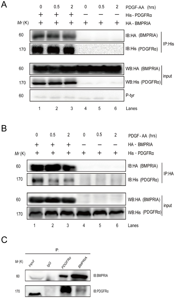
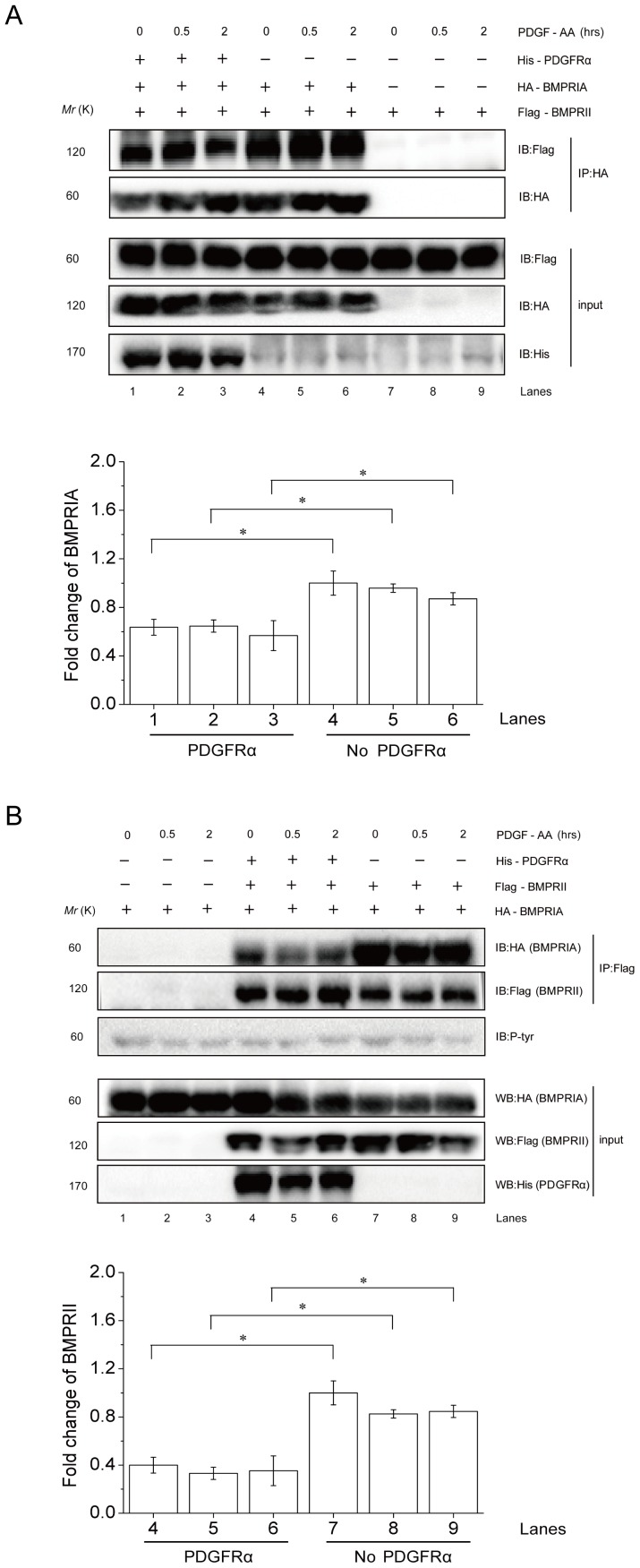
Platelet-derived growth factors (PDGFs) play important roles in skeletal development and bone fracture healing, yet how PDGFs execute their functions remains incompletely understood. Here we show that PDGF-AA, but not -AB or -BB, could activate the BMP-Smad1/5/8 pathway in mesenchymal stem cells (MSCs), which requires BMPRIA as well as PDGFRα. PDGF-AA promotes MSC osteogenic differentiation through the BMP-Smad1/5/8-Runx2/Osx axis and MSC migration via the BMP-Smad1/5/8-Twist1/Atf4 axis. Mechanistic studies show that PDGF-AA activates BMP-Smad1/5/8 signaling by feedback down-regulating PDGFRα, which frees BMPRI and allows for BMPRI-BMPRII complex formation to activate smad1/5/8, using BMP molecules in the microenvironment. This study unravels a physical and functional interaction between PDGFRα and BMPRI, which plays an important role in MSC differentiation and migration, and establishes a link between PDGF-AA and BMPs pathways, two essential regulators of embryonic development and tissue homeostasis.
Overview
The claims and statistics of this paper haven’t been checked yet.
Run the check — free, no account needed. It reads the open-access text and checks the sources it cites.
Integrity signals
Registry
Cites
Statistics
Figures
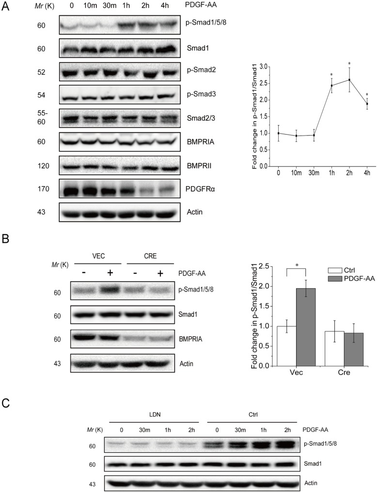
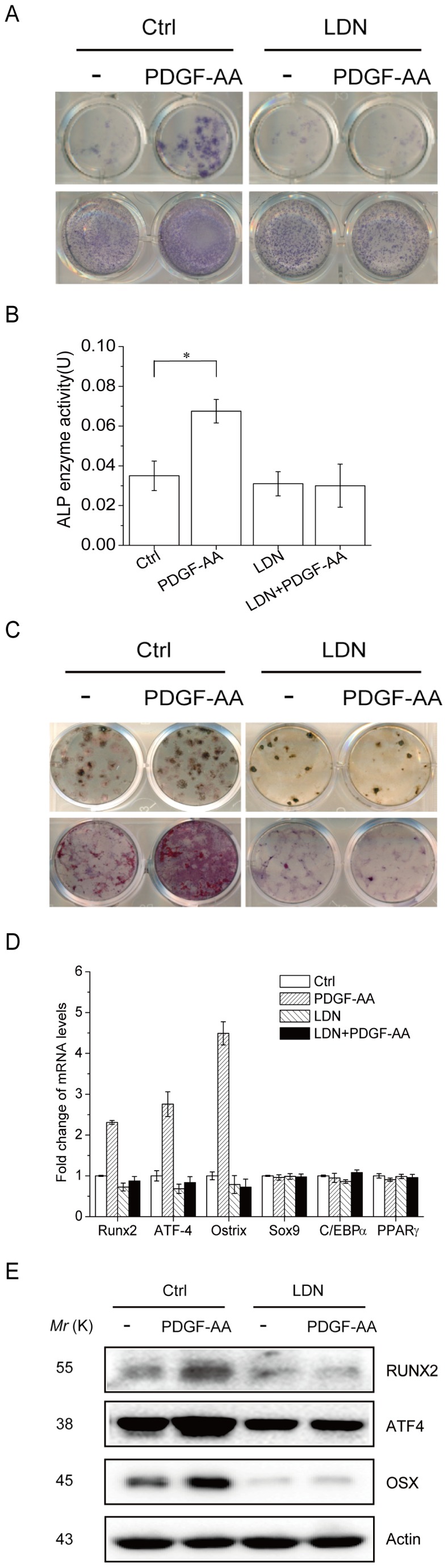
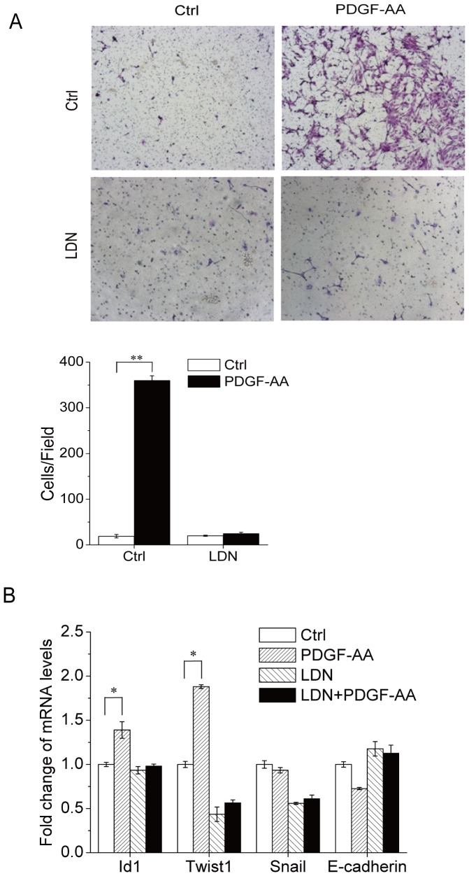
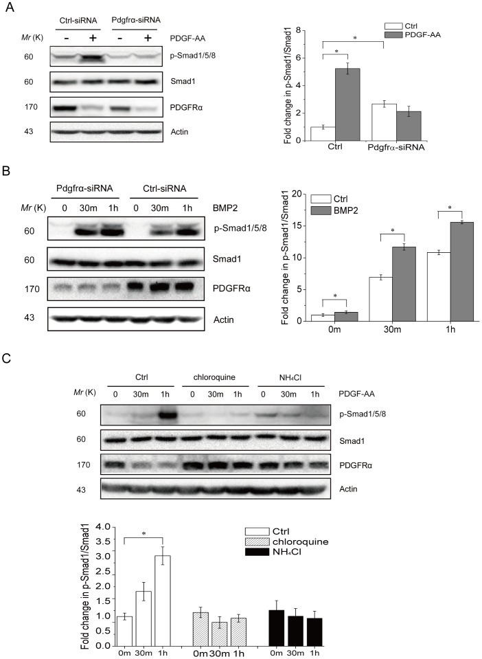
Verification
Claims the paper makes
The claims and statistics of this paper haven’t been checked yet.
Run the check — free, no account needed. It reads the open-access text and checks the sources it cites.
Claims other papers make about this one
Each is checked against this paper’s own text: does it say what the citing paper says it does?
The source backs part of the claim, not all of it — or backs it for a different population, condition or comparison.
In the citing paper
“This process is triggered by the response of MSCs to inflammatory factors released from the bone fracture site, such as PDGFs and BMPs, which activate BMP-Smad1/5/8 signaling, promoting MSC migration [40,77,78].”
From Mesenchymal Stem/Stromal Cells: Immunomodulatory and Bone Regeneration Potential after Tumor Excision in Osteosarcoma Patients (2023) · cites this paper as [78]
Experimental — accuracy not yet measured
Self-check
Percentages, sums and reported differences re-computed from the paper’s own numbers.
The self-check needs the paper’s full text.
Citations
Cites
PERSPECTIVE ARTICLE: Growth factors and cytokines in wound healing
Barrientos, S., Stojadinovic, O., Golinko, M. S., Brem, H., & Tomic‐Canic, M. (2008) · Wound Repair and Regeneration · doi:10.1111/j.1524-475x.2008.00410.x · Cited by 3,583
Massagué, J. (2012) · Nature Reviews Molecular Cell Biology · doi:10.1038/nrm3434 · Cited by 3,239
Role of platelet-derived growth factors in physiology and medicinePreprint
Andræ, J., Gallini, R., & Betsholtz, C. (2008) · Genes & Development · doi:10.1101/gad.1653708 · Cited by 2,534
TGF-β and BMP Signaling in Osteoblast Differentiation and Bone Formation
Chen, G., Deng, C., & Li, Y.-P. (2012) · International Journal of Biological Sciences · doi:10.7150/ijbs.2929 · Cited by 1,756
Mice deficient for PDGF B show renal, cardiovascular, and hematological abnormalities.Preprint
Levéen, P., Pekny, M., Gebré‐Medhin, S., Swolin, B., Larsson, E., & Betsholtz, C. (1994) · Genes & Development · doi:10.1101/gad.8.16.1875 · Cited by 1,130
Dorsomorphin inhibits BMP signals required for embryogenesis and iron metabolism
Yu, P. B., Hong, C. C., Sachidanandan, C., Babitt, J. L., Deng, D. Y., Hoyng, S. A., Lin, H. Y.-F., Bloch, K. D., & Peterson, R. T. (2007) · Nature Chemical Biology · doi:10.1038/nchembio.2007.54 · Cited by 1,059
Soriano, P. M. (1997) · Development · doi:10.1242/dev.124.14.2691 · Cited by 711
Roles of PDGF in animal development
Hoch, R. V., & Soriano, P. M. (2003) · Development · doi:10.1242/dev.00721 · Cited by 593
Integrating Patterning Signals: Wnt/GSK3 Regulates the Duration of the BMP/Smad1 Signal
Fuentealba, L. C., Eivers, E., Ikeda, A., Hurtado, C., Kuroda, H., Pera, E. M., & Robertis, E. M. D. (2007) · Cell · doi:10.1016/j.cell.2007.09.027 · Cited by 555
Recent advances in BMP receptor signaling
Sieber, C., Kopf, J., Hiepen, C., & Knaus, P. I. (2009) · Cytokine & Growth Factor Reviews · doi:10.1016/j.cytogfr.2009.10.007 · Cited by 480
Research works printed but not linked: 2
- J Caverzasio, 2013, Predominant role of PDGF receptor transactivation in Wnt3a-induced osteoblastic cell proliferation, J Bone Miner Res, 28, 260, 10.1002/jbmr.1748 · DOI
- AI Caplan, 2011, PDGF in bone formation and regeneration: new insights into a novel mechanism involving MSCs, J Orthop Res, 29, 1795, 10.1002/jor.21462 · DOI · Paper page
Cited by
Sorted by citations
The roles of signaling pathways in bone repair and regeneration
Majidinia, M., Sadeghpour, A., & Yousefi, B. (2017) · Journal of Cellular Physiology · doi:10.1002/jcp.26042 · Cited by 526
Mesenchymal stem cells: Amazing remedies for bone and cartilage defects
Kangari, P., Talaei-Khozani, T., Razeghian-Jahromi, I., & Razmkhah, M. (2020) · Stem Cell Research & Therapy · doi:10.1186/s13287-020-02001-1 · Cited by 286
Mesenchymal Stem Cell Migration during Bone Formation and Bone Diseases Therapy
Su, P., Tian, Y. E., Yang, C., Ma, X., Wang, X., Pei, J., & Qian, A. (2018) · International Journal of Molecular Sciences · doi:10.3390/ijms19082343 · Cited by 268
Weivoda, M. M., Ruan, M. M., Hachfeld, C. M., Pederson, L. G., Howe, A. K., Davey, R. A., Zajac, J. D., Kobayashi, Y., Williams, B. O., Westendorf, J. J., et al. (2015) · Journal of Bone and Mineral Research · doi:10.1002/jbmr.2599 · Cited by 157
Endogenous cell recruitment strategy for articular cartilage regeneration
Yang, Z., Li, H., Yuan, Z., Fu, L., Jiang, S., Gao, C., Wang, F., Zha, K., Tian, G., Sun, Z., et al. (2020) · Acta Biomaterialia · doi:10.1016/j.actbio.2020.07.008 · Cited by 136
Stem cells, growth factors and scaffolds in craniofacial regenerative medicine
Tollemar, V., Collier, Z. J., Mohammed, M. K., Lee, M. J., Ameer, G. A., & Reid, R. R. (2015) · Genes & Diseases · doi:10.1016/j.gendis.2015.09.004 · Cited by 134
Xu, T., Luo, Y., Wang, J., Zhang, N., Gu, C., Li, L., Qian, D., Cai, W., Fan, J., & Yin, G. (2020) · Journal of Nanobiotechnology · doi:10.1186/s12951-020-00601-w · Cited by 128
Bone Marrow Aspirate Concentrate for Cartilage Defects of the Knee: From Bench to Bedside Evidence
Cotter, E. J., Wang, K. C., Yanke, A. B., & Chubinskaya, S. (2017) · Cartilage · doi:10.1177/1947603517741169 · Cited by 125
Osteogenesis and bone remodeling: A focus on growth factors and bioactive peptides
Toosi, S. H., & Behravan, J. (2019) · BioFactors · doi:10.1002/biof.1598 · Cited by 129
Adkar, S. S., Wu, C., Willard, V. P., Dicks, A. R., Ettyreddy, A. R., Steward, N. S., Bhutani, N., Gersbach, C. A., & Guilak, F. (2018) · Stem Cells · doi:10.1002/stem.2931 · Cited by 96
PM2.5, Fine Particulate Matter: A Novel Player in the Epithelial-Mesenchymal Transition?
Xu, Z., Ding, W., & Deng, X. (2019) · Frontiers in Physiology · doi:10.3389/fphys.2019.01404 · Cited by 97
The basic science of bone marrow aspirate concentrate in chondral injuries
Holton, J., Imam, M. A., Ward, J., & Snow, M. (2016) · Orthopedic Reviews · doi:10.4081/or.2016.6659 · Cited by 87
Human perivascular stem cell-derived extracellular vesicles mediate bone repair
Xu, J., Wang, Y., Hsu, C.-Y., Gao, Y., Meyers, C. A., Chang, L., Zhang, L., Broderick, K. P., Ding, C., Péault, B., et al. (2019) · eLife · doi:10.7554/elife.48191 · Cited by 86
Xu, J., Wang, Y., Zhu, L., Tian, Y., Li, Z., Lu, A. Z., Hsu, C.-Y., Negri, S., Tang, C., Tower, R. J., et al. (2022) · Bone Research · doi:10.1038/s41413-021-00176-8 · Cited by 83
Yan, X., Zhang, Q., Ma, X., Zhong, Y., Tang, H., & Mai, S. (2023) · Japanese Dental Science Review · doi:10.1016/j.jdsr.2023.06.005 · Cited by 81
Xie, Y., Shi, W., Zhou, J., Gao, Y., Li, S.-F., Fang, Q.-Q., Wang, M., Ma, H., Wang, J., Xian, C. J., et al. (2016) · Bone · doi:10.1016/j.bone.2016.09.008 · Cited by 80
Deng, Y., Lei, G., Lin, Z., Yang, Y., Lin, H., & Tuan, R. S. (2018) · Biomaterials · doi:10.1016/j.biomaterials.2018.11.036 · Cited by 80
Li, W., Qiao, W., Liu, X., Bian, D., Shen, D., Zheng, Y., Wu, J., Kwan, K. Y.-H., Wong, T. M., Cheung, K., et al. (2021) · Advanced Science · doi:10.1002/advs.202102035 · Cited by 78
Sending mixed signals: Cilia-dependent signaling during development and disease
Elliott, K. H., & Brugmann, S. A. (2018) · Developmental Biology · doi:10.1016/j.ydbio.2018.03.007 · Cited by 74
Zha, K., Tian, Y., Panayi, A. C., Mi, B., & Liu, G. (2022) · Frontiers in Cell and Developmental Biology · doi:10.3389/fcell.2022.824812 · Cited by 69
Mesenchymal Stem/Progenitor Cells: The Prospect of Human Clinical Translation
Rady, D., Abbass, M. M. S., El‐Rashidy, A. A., Moshy, S. E., Radwan, I. A., Dörfer, C. E., & El‐Sayed, K. M. F. (2020) · Stem Cells International · doi:10.1155/2020/8837654 · Cited by 69
Mesenchymal stem cells and biologic factors leading to bone formation
Bartold, P. M., Gronthos, S., Haynes, D. A., & Ivanovski, S. (2019) · Journal Of Clinical Periodontology · doi:10.1111/jcpe.13053 · Cited by 67
Naaldijk, Y., Johnson, A. A., Ishak, S., Meisel, H. J., Hohaus, C., & Stolzing, A. (2015) · Experimental Cell Research · doi:10.1016/j.yexcr.2015.08.019 · Cited by 62
Crucial Role of Lamin A/C in the Migration and Differentiation of MSCs in Bone
Alcorta-Sevillano, N., Macías, I., Rodrı́guez, C. I., & Infante, A. (2020) · Cells · doi:10.3390/cells9061330 · Cited by 61
The balance of Bmp6 and Wnt10b regulates the telogen-anagen transition of hair follicles
Wu, P., Zhang, Y., Xing, Y., Xu, W., Guo, H., Deng, F., Ma, X., & Li, Y. (2019) · Cell Communication and Signaling · doi:10.1186/s12964-019-0330-x · Cited by 61
External factors influencing mesenchymal stem cell fate in vitro
Sisakhtnezhad, S., Alimoradi, E., & Akrami, H. (2016) · European Journal of Cell Biology · doi:10.1016/j.ejcb.2016.11.003 · Cited by 59
Mechanism of Action of Icariin in Bone Marrow Mesenchymal Stem Cells
Yang, A., Yu, C.-C., Lu, Q., Li, H., Li, Z., & He, C. (2019) · Stem Cells International · doi:10.1155/2019/5747298 · Cited by 57
Wang, Y., Sun, B., Zhao, X., Zhao, N., Sun, R., Zhu, D., Zhang, Y., Li, Y., Gu, Q., Dong, X., et al. (2016) · Oncotarget · doi:10.18632/oncotarget.8328 · Cited by 56
Lu, X., Han, W.-X., & Liu, Y. (2019) · Biomedicine & Pharmacotherapy · doi:10.1016/j.biopha.2019.109378 · Cited by 50
Secretome of Human Fetal Mesenchymal Stem Cell Ameliorates Replicative Senescence
Wang, B., Lee, W., Huang, B., Zhang, J., Wu, T., Jiang, X., Wang, C. C., & Li, G. (2016) · Stem Cells and Development · doi:10.1089/scd.2016.0079 · Cited by 48
Roles of platelet‐derived growth factor in vascular calcification
Ouyang, L., Zhang, K., Chen, J., Wang, J., & Huang, H. (2017) · Journal of Cellular Physiology · doi:10.1002/jcp.25985 · Cited by 44
Yang, Q., Han, Y., Liu, P., Huang, Y., Li, X., Jia, L., Zheng, Y., & Li, W. (2020) · Frontiers in Pharmacology · doi:10.3389/fphar.2020.00701 · Cited by 42
Knuth, C., Sastre, E. A., Fahy, N., Witte‐Bouma, J., Ridwan, Y. R., Strabbing, E., Koudstaal, M. J., van de Peppel, J., Wolvius, E. B., Narcisi, R., et al. (2019) · European Cells and Materials · doi:10.22203/ecm.v038a09 · Cited by 40
Electrical Stimulation in Cartilage Tissue Engineering
Vaičiulevičiūtė, R., Uzielienė, I., Bernotas, P., Novickij, V., Alaburda, A., & Bernotienė, E. (2023) · Bioengineering · doi:10.3390/bioengineering10040454 · Cited by 37
Bartoletti, G., Dong, C., Umar, M., & He, F. (2020) · Developmental Biology · doi:10.1016/j.ydbio.2020.08.004 · Cited by 37
Li, D., Huang, L., Zhang, C.-P., Li, Q., & Wang, J. (2022) · Frontiers in Aging Neuroscience · doi:10.3389/fnagi.2022.890509 · Cited by 38
Xue, D., Chen, E., Zhang, W., Gao, X., Wang, S., Zheng, Q., Pan, Z., Li, H., & Liu, L. (2017) · Oncotarget · doi:10.18632/oncotarget.15473 · Cited by 37
Decellularized Cell Culture ECMs Act as Cell Differentiation Inducers
Parmaksız, M., Elçin, A. E., & Elçin, Y. M. (2020) · Stem Cell Reviews and Reports · doi:10.1007/s12015-020-09963-y · Cited by 35
Cross Talk Between Cells and the Current Bioceramics in Bone Regeneration: A Comprehensive Review
Khayatan, D., Oskouei, A. B., Alam, M. M. R., Mohammadikhah, M., Badkoobeh, A., Golkar, M., Abbasi, K., Karami, S., Soufdoost, R. S., Hakim, L. K., et al. (2024) · Cell Transplantation · doi:10.1177/09636897241236030 · Cited by 33
Park, S.-R., Cho, A., Kim, J.-W., Lee, H., & Hong, I. (2019) · Molecular Therapy · doi:10.1016/j.ymthe.2019.03.010 · Cited by 29
Smad1 promotes colorectal cancer cell migration through Ajuba transactivation
Daming, Y., Hou, T., Li, L., Chu, Y., Zhou, F., Xu, Y., Hou, X., Song, H., Zhu, K., Hou, Z., et al. (2017) · Oncotarget · doi:10.18632/oncotarget.22780 · Cited by 28
Role of Uterine Telocytes During Pregnancy
Soliman, S. A., Abd‐Elhafeez, H. H., Abou-Elhamd, A. S., Kamel, B., Abdellah, N., & Mustafa, F. E. A. (2022) · Microscopy and Microanalysis · doi:10.1093/micmic/ozac001 · Cited by 27
Chabot, V., Dromard, C., Rico, A., Langonné, A., Burlaud‐Gaillard, J., Guilloton, F., Casteilla, L., & Sensebé, L. (2015) · Stem Cell Research & Therapy · doi:10.1186/s13287-015-0163-5 · Cited by 27
The Emerging Role of Mesenchymal Stem Cells in Vascular Calcification
Xie, C., Ouyang, L., Chen, J., Zhang, H., Luo, P., Wang, J., & Huang, H. (2019) · Stem Cells International · doi:10.1155/2019/2875189 · Cited by 26
Zhang, L., Yuan, Y. J., Wu, W., Sun, Z., Lei, L., Fan, J., Gao, B., & Zou, J. (2020) · Frontiers in Cell and Developmental Biology · doi:10.3389/fcell.2020.600639 · Cited by 26
Samuel, S., Ahmad, R. E., Ramasamy, T. S., Karunanithi, P., Naveen, S. V., Murali, M. R., Abbas, A. A., & Kamarul, T. (2016) · PeerJ · doi:10.7717/peerj.2347 · Cited by 24
Mo, G., Baldwin, J. R., Luffer, D. R., Ilaria, R. L., Conti, I., Heathman, M. A., & Cronier, D. M. (2017) · Clinical Pharmacokinetics · doi:10.1007/s40262-017-0562-0 · Cited by 23
Ardebili, M. S. , Hasannia, S., Dabirmanesh, B., & Khavari‐Nejad, R. A. (2024) · Scientific Reports · doi:10.1038/s41598-024-51707-2 · Cited by 22
Friend or Foe? Essential Roles of Osteoclast in Maintaining Skeletal Health
Wang, H., Yang, G., Xiao, Y., Luo, G., Li, G., & Li, Z. (2020) · BioMed Research International · doi:10.1155/2020/4791786 · Cited by 23
Hagmeijer, M. H., Korpershoek, J. V., Crispim, J. F., Chen, L., Jonkheijm, P., Krych, A. J., Saris, D. B. F., & Vonk, L. A. (2021) · Journal of Tissue Engineering and Regenerative Medicine · doi:10.1002/term.3218 · Cited by 21
Hydroxyapatite–collagen augments osteogenic differentiation of dental pulp stem cells
Trivedi, S., Srivastava, K., Saluja, T. S., Shyam, H., Kumar, S., Singh, A., Saxena, S. K., Mehrotra, D., & Singh, S. K. (2019) · Odontology · doi:10.1007/s10266-019-00464-0 · Cited by 21
Kot, M., Musiał-Wysocka, A., Lasota, M., Ulman, A., & Majka, M. (2019) · Acta Biochimica Polonica · doi:10.18388/abp.2019_2895 · Cited by 21
Expression and function of PDGF-C in development and stem cells
Tian, Y., Zhan, Y., Jiang, Q. L., Lu, W., & Li, X. (2021) · Open Biology · doi:10.1098/rsob.210268 · Cited by 21
Baron, M., Drohat, P., Crawford, B., Hornicek, F. J., Best, T. M., & Kouroupis, D. (2023) · Bioengineering · doi:10.3390/bioengineering10101187 · Cited by 19
Ni, L., Kuang, Z., Gong, Z., Xue, D., & Zheng, Q. (2019) · BioMed Research International · doi:10.1155/2019/3456719 · Cited by 18
Xu, L., Willumeit‐Römer, R., & Luthringer-Feyerabend, B. (2019) · Acta Biomaterialia · doi:10.1016/j.actbio.2019.10.018 · Cited by 18
Brown, M. G., Brady, D. J., Healy, K. M., Henry, K. A., Ogunsola, A. S., & Ma, X. (2024) · Cells · doi:10.3390/cells13121045 · Cited by 17
Lu, W., & Li, X. (2017) · Molecular Aspects of Medicine · doi:10.1016/j.mam.2017.10.001 · Cited by 17
Emerging biological functions of Twist1 in cell differentiation
Tu, M., Ge, B., Li, J., Pan, Y., Zhao, B., Han, J., Wu, J., Zhang, K., Liu, G., Hou, M., et al. (2024) · Developmental Dynamics · doi:10.1002/dvdy.736 · Cited by 17
Woodbury, S. M., Swanson, W. B., & Mishina, Y. M. (2023) · Frontiers in Physiology · doi:10.3389/fphys.2023.1220555 · Cited by 16
Self-citationMDA-9/Syntenin in the tumor and microenvironment defines prostate cancer bone metastasis
Maji, S., Pradhan, A. K., Kumar, A., Bhoopathi, P., Mannangatti, P., Guo, C., Windle, J. J., Subler, M. A., Wang, X., Semmes, O. J., et al. (2023) · Proceedings of the National Academy of Sciences · doi:10.1073/pnas.2307094120 · Cited by 15
Primary cilia in hard tissue development and diseases
Li, S., Zhang, H., & Sun, Y. (2021) · Frontiers of Medicine · doi:10.1007/s11684-021-0829-6 · Cited by 15
Shen, C., Quan, Q., Yang, C. H., Wen, Y., & Li, H. (2018) · Stem Cell Research & Therapy · doi:10.1186/s13287-018-0949-3 · Cited by 15
Meghil, M. M., Mandil, O., Nevins, M., Saleh, M. H. A., & Wang, H. (2023) · Medicina · doi:10.3390/medicina59040676 · Cited by 13
Heydarian, M., Oak, P., Zhang, X., Kamgari, N., Kindt, A. S. D., Koschlig, M., Pritzke, T., González, E. G., Förster, K. M., Morty, R. E., et al. (2022) · Thorax · doi:10.1136/thoraxjnl-2021-218083 · Cited by 13
GM1 Ganglioside Promotes Osteogenic Differentiation of Human Tendon Stem Cells
Bergante, S., Creo, P., Piccoli, M., Ghiroldi, A., Menon, A., Cirillo, F., Rota, P., Monasky, M. M., Ciconte, G., Pappone, C., et al. (2018) · Stem Cells International · doi:10.1155/2018/4706943 · Cited by 13
Jakl, V., Popp, T., Haupt, J., Port, M., Roesler, R., Wiese, S., Friemert, B., Rojewski, M., & Schrezenmeier, H. (2023) · Cells · doi:10.3390/cells12162105 · Cited by 12
Zuo, T., Liu, Y., Duan, M., Pu, X., Huang, M., Zhang, D., & Xie, J. (2023) · Biochemistry and Biophysics Reports · doi:10.1016/j.bbrep.2023.101462 · Cited by 12
Jarosz, A., Szyluk, K., Iwanicka, J., Balcerzyk, A., Nowak, T., Iwanicki, T., Negru, M., Kalita, M., Francuz, T., Garczorz, W., et al. (2022) · Journal of Clinical Medicine · doi:10.3390/jcm11123504 · Cited by 11
Ma, C., Tao, C., Zhang, Z., Zhou, H., Fan, C., & Wang, D. (2023) · Materials Today Bio · doi:10.1016/j.mtbio.2023.100893 · Cited by 11
Overcoming barriers confronting application of protein therapeutics in bone fracture healing
Czech, T., & Oyewumi, M. O. (2020) · Drug Delivery and Translational Research · doi:10.1007/s13346-020-00829-x · Cited by 11
Management of osteoarthritis - biological approaches: current concepts
Cotter, E. J., Frank, R. M., & Mandelbaum, B. R. (2019) · Journal of ISAKOS Joint Disorders & Orthopaedic Sports Medicine · doi:10.1136/jisakos-2019-000377 · Cited by 11
The potential role of mesenchymal stem cells in modulating antiageing process
Govindasamy, V., Rajendran, A., LEE, Z., Ooi, G., Then, K.-Y., Then, K., Gayathri, M., Das, A. K., & Cheong, S. (2021) · Cell Biology International · doi:10.1002/cbin.11652 · Cited by 9
Wolfe, P. N., Stoker, A. M., Leary, E. V., Crist, B. D., Bozynski, C. C., & Cook, J. L. (2023) · Cartilage · doi:10.1177/19476035231163032 · Cited by 8
Mechanical stretch promotes antioxidant responses and cardiomyogenic differentiation in P19 cells
Cheng, J., Zou, Q., Xue, Y., Sun, C., & Zhang, D. (2021) · Journal of Tissue Engineering and Regenerative Medicine · doi:10.1002/term.3184 · Cited by 7
RetractedLee, J., Jang, J., Cha, S.-R., Lee, S. B., Hong, S., Bae, H.-S., Lee, Y. J., & Yang, S. (2023) · Immune Network · doi:10.4110/in.2023.23.e48 · Cited by 7
Yao, H., Cui, Y., Li, P., Sun, S., Peng, C., & Wu, D. (2026) · Frontiers in Endocrinology · doi:10.3389/fendo.2026.1783422 · Cited by 5
Regulatory Effects of Senescent Mesenchymal Stem Cells: Endotheliocyte Reaction
Ratushnyy, A. Y., Ездакова, М. И., Константиновна, М. Д., Tyrina, E. A., & Buravkova, L. B. (2024) · Cells · doi:10.3390/cells13161345 · Cited by 5
Unique Regenerative Mechanism to Replace Bone Lost During Dietary Bone Depletion in Weanling Mice
Sheng, M. H. ‐., Lau, K. ‐. W., Lakhan, R., Ahmed, A. S. I., Rundle, C. H., Biswanath, P., & Baylink, D. J. (2017) · Endocrinology · doi:10.1210/en.2016-1379 · Cited by 5
Characterizing the role of Pdgfra in calvarial development
Umar, M., Bartoletti, G., Dong, C., Gahankari, A., Browne, D., Deng, A., Jaramillo, J., Sammarco, M. C., Simkin, J., & He, F. (2023) · Developmental Dynamics · doi:10.1002/dvdy.564 · Cited by 5
Paim, Á., Cardozo, N. S. M., Tessaro, I. C., & Pranke, P. H. L. (2018) · Mathematical Biosciences · doi:10.1016/j.mbs.2018.05.007 · Cited by 5
Kuroda, A., Mineo, A., Shoji, S., Inoue, G., Saito, W., Sekiguchi, H., Takaso, M., & Uchida, K. (2022) · Bio-Medical Materials and Engineering · doi:10.3233/bme-221406 · Cited by 4
Chen, S., Chen, X., Meng, S., Yuan, Q., Wu, H., Hu, W., Zhou, L., & Shan, L. (2025) · Stem Cell Research & Therapy · doi:10.1186/s13287-025-04694-8 · Cited by 3
Zhang, W., Wu, H., Li, Y., Ren, F., & Guo, H. (2017) · Journal of Functional Foods · doi:10.1016/j.jff.2017.07.023 · Cited by 3
Requirement of Pdgfrα+ cells for calvarial bone repair
Xing, X., Li, Z., Xu, J., Chen, A. Z., Archer, M., Wang, Y., Xu, M., Wang, Z., Zhu, M., Qin, Q., et al. (2024) · Stem Cells Translational Medicine · doi:10.1093/stcltm/szae041 · Cited by 3
Effects of a novel biodegredable implant system on a rat tibia fracture model
Yalçınozan, M., Türker, M., & Çırpar, M. (2020) · Acta Orthopaedica et Traumatologica Turcica · doi:10.5152/j.aott.2020.18331 · Cited by 3
Ramos, G. C., Argüelles, D., Vidal, L. O., & Prades, M. (2019) · Repositorio Universidad de Caldas · doi:10.17151/vetzo.2019.13.2.5 · Cited by 2
Antonowicz, B., Borys, J., Roszczyc-Owsiejczuk, K., Łukaszuk, K., Zalewska, A., & Błachnio-Zabielska, A. U. (2025) · International Journal of Molecular Sciences · doi:10.3390/ijms26052020 · Cited by 2
Osteogenic Cells and Microenvironment of Early Bone Development and Clinical Implication
Kim, K., & Lee, C. C. (2023) · IntechOpen eBooks · doi:10.5772/intechopen.1002037 · Cited by 1
Cell–biomaterials interactions: the role of growth factors
Brion, A., & Rammal, H. (2020) · Elsevier eBooks · doi:10.1016/b978-0-08-102967-1.00008-6 · Cited by 1
Korpershoek, J. V. (2022) · doi:10.33540/1207
Wang, S., Dong, X., Zheng, C., Gu, M., Jiang, H., Liang, X., & Zheng, X. (2023) · Research Square · doi:10.21203/rs.3.rs-2410527/v1
Pathobiological analysis of developmental dysplasia of the hip and its progression to osteoarthritis
Wolfe, P. N. (2022) · MOspace Institutional Repository (University of Missouri) · doi:10.32469/10355/93995
Xu, T., Luo, Y., Wang, J., Zhang, N., Gu, C., Li, L., Qian, D., Cai, W., Fan, J., & Yin, G. (2020) · Figshare · doi:10.6084/m9.figshare.11992587
Xu, T., Luo, Y., Wang, J., Zhang, N., Gu, C., Li, L., Qian, D., Cai, W., Fan, J., & Yin, G. (2020) · Figshare · doi:10.6084/m9.figshare.11992563
Xu, T., Luo, Y., Wang, J., Zhang, N., Gu, C., Li, L., Qian, D., Cai, W., Fan, J., & Yin, G. (2020) · Figshare · doi:10.6084/m9.figshare.11992566
Xu, T., Luo, Y., Wang, J., Zhang, N., Gu, C., Li, L., Qian, D., Cai, W., Fan, J., & Yin, G. (2020) · Figshare · doi:10.6084/m9.figshare.11992578
Vafaei, S., Omran, M. M., Yang, Q., Laveaux, S. M., Zota, A. R., Al‐Hendy, A., & Ali, M. K. (2026) · Environmental Toxicology · doi:10.1002/tox.70046
Chen, S., Jiang, Y., Chai, X., & Wu, X. (2026) · Reproductive Sciences · doi:10.1007/s43032-025-02049-5
Kazi, T. (2022) · Institutional Repositories DataBase (IRDB)
Metadata
- Authors
- Li, A., Xia, X., Yeh, J., Kua, H., Liu, H., Mishina, Y., Hao, A., & Li, B.
- Affiliations
- Shandong University (CN)
- Shanghai Jiao Tong University (CN)
- University of Michigan (US)
- Corresponding author
- Hao, Aijun
- Li, Baojie
- Venue
- PLoS ONE
- Publisher
- Public Library of Science (PLoS)
- Published
- 2014-12-03
- Volume
- 9
- Issue
- 12
- Pages
- e113785
- ISSN
- 1932-6203 (ISSN-L)
- PMC
- PMC4254917
- OpenAlex
- W2011964589
- Cited by
- 100
- References
- 57
- Citations by year
- 2026: 4 · 2025: 2 · 2024: 6 · 2023: 14 · 2022: 9 · 2021: 6 · 2020: 16 · 2019: 16 · 2018: 8 · 2017: 9 · 2016: 6 · 2015: 4
- FWCI
- 2.60
- Type
- journal article
- Language
- en
- Open access
- gold
- doi.org
- Locations
- PLoS ONE (publishedVersion, cc-by)
- PubMed Central (publishedVersion, cc-by)
- PubMed (publishedVersion)
- DOAJ (DOAJ: Directory of Open Access Journals) (submittedVersion, cc-by-sa)
- Figshare (submittedVersion, cc-by)
- Funders
- National Natural Science Foundation of China (81121001, 31071229, 81130039)
- Topics
- Life Sciences › Biochemistry, Genetics and Molecular Biology › Molecular Biology › TGF-β signaling in diseases
- Life Sciences › Biochemistry, Genetics and Molecular Biology › Cell Biology › Mesenchymal stem cell research
- Life Sciences › Biochemistry, Genetics and Molecular Biology › Molecular Biology › Bone Metabolism and Diseases
- Keywords
- PDGF-AA, platelet-derived growth factor, mesenchymal stem cells, osteogenic differentiation, cell migration, PDGFRA, BMP signaling, Smad1/5/8, BMPR-IA, BMPR2, TWIST1, ATF4, feedback regulation, skeletal development, fracture healing
- MeSH terms
- Animals, Cell Differentiation, Cell Movement, Cells, Cultured, Mice, Inbred C57BL, Osteogenesis, Platelet-Derived Growth Factor, Protein Binding, Blotting, Western, Signal Transduction, Down-Regulation, Mice, Knockout, Receptor, Platelet-Derived Growth Factor alpha, RNA Interference, Smad Proteins, Smad1 Protein, Smad5 Protein, Smad8 Protein, Bone Morphogenetic Protein Receptors, Type I, Bone Morphogenetic Protein 2, Mesenchymal Stem Cells
About these checks
Registry, retraction, Cites and statistics results are computed, not judged, with one exception: a printed reference that no index lists is read once by a model before it counts as a citation error. Claims are extracted by a model and kept only when their quote appears verbatim in the text; each cited source is then checked, with a model’s help, for whether it says what the claim relies on. Those verdicts can be wrong — read the claim’s quote and the cited source before relying on one.
Statistics and arithmetic are recomputed, not judged.
Confidence bands are shown only for fields calibrated on our annotated test set. None is calibrated yet, so no band is shown.
What counts as a claim ›How we measure confidence ›How we check this ›
Are you an author, or do you see a problem with this page? Request a review
