

The DOI matches its Crossref record; 15 of the 15 references it cites were found in a registry or an index.
CXC Chemokines Function as a Rheostat for Hepatocyte Proliferation and Liver Regeneration
Open accessAbstract
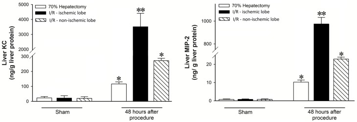
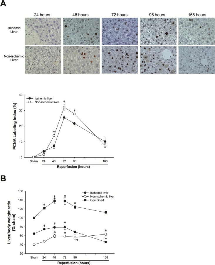
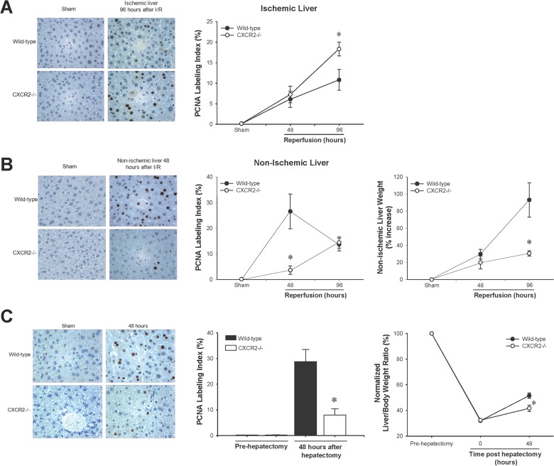
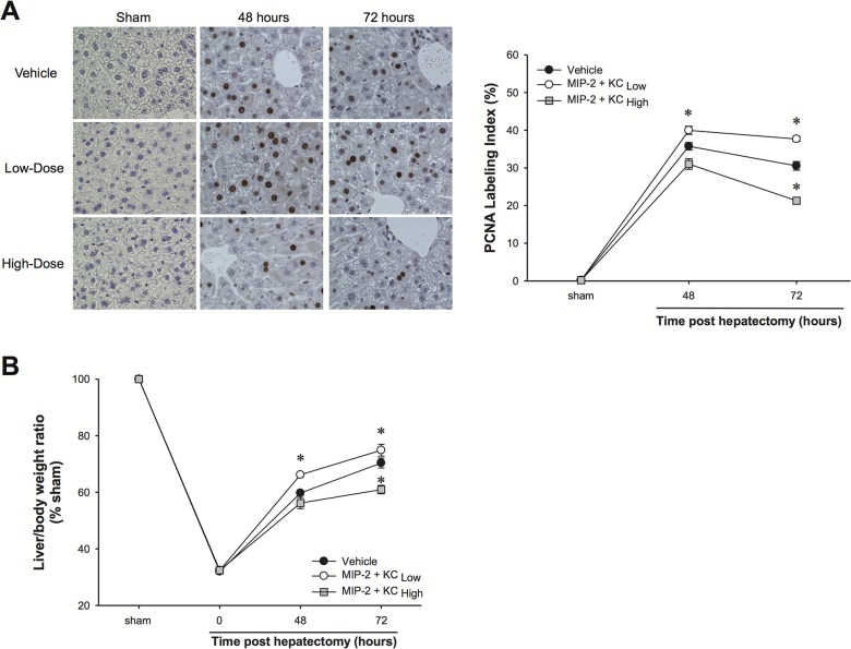
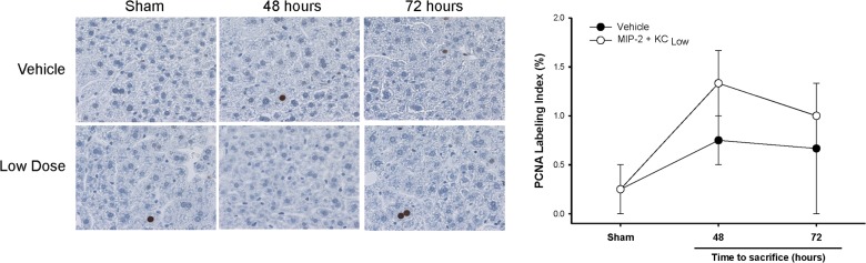
BACKGROUND: Our previous in vitro studies have demonstrated dose-dependent effects of CXCR2 ligands on hepatocyte cell death and proliferation. In the current study, we sought to determine if CXCR2 ligand concentration is responsible for the divergent effects of these mediators on liver regeneration after ischemia/reperfusion injury and partial hepatectomy. METHODS: Murine models of partial ischemia/reperfusion injury and hepatectomy were used to study the effect of CXCR2 ligands on liver regeneration. RESULTS: We found that hepatic expression of the CXCR2 ligands, macrophage inflammatory protein-2 (MIP-2) and keratinocyte-derived chemokine (KC), was significantly increased after both I/R injury and partial hepatectomy. However, expression of these ligands after I/R injury was 30-100-fold greater than after hepatectomy. Interestingly, the same pattern of expression was found in ischemic versus non-ischemic liver lobes following I/R injury with expression significantly greater in the ischemic liver lobes. In both systems, lower ligand expression was associated with increased hepatocyte proliferation and liver regeneration in a CXCR2-dependent fashion. To confirm that these effects were related to ligand concentration, we administered exogenous MIP-2 and KC to mice undergoing partial hepatectomy. Mice received a "high" dose that replicated serum levels found after I/R injury and a "low" dose that was similar to that found after hepatectomy. Mice receiving the "high" dose had reduced levels of hepatocyte proliferation and regeneration whereas the "low" dose promoted hepatocyte proliferation and regeneration. CONCLUSIONS: Together, these data demonstrate that concentrations of CXC chemokines regulate the hepatic proliferative response and subsequent liver regeneration.
Overview
The claims and statistics of this paper haven’t been checked yet.
Run the check — free, no account needed. It reads the open-access text and checks the sources it cites.
Integrity signals
Registry
Cites
Statistics
Figures
Verification
Claims the paper makes
The claims and statistics of this paper haven’t been checked yet.
Run the check — free, no account needed. It reads the open-access text and checks the sources it cites.
Claims other papers make about this one
Each is checked against this paper’s own text: does it say what the citing paper says it does?
The cited source says what the claim attributes to it.
In the citing paper
“CXC chemokines have proliferative effects on hepatocytes, and CXCR2-knockout mice experience delayed liver regeneration after PHx (40, 41).”
From Senescent hepatic stellate cells promote liver regeneration through IL-6 and ligands of CXCR2 (2022) · cites this paper as [41]
Self-check
Percentages, sums and reported differences re-computed from the paper’s own numbers.
The self-check needs the paper’s full text.
Citations
Cites
Addison, C. L., Daniel, T. O., Burdick, M. D., Liu, H., Ehlert, J. E., Xue, Y. Y., Buechi, L., Walz, A., Richmond, A., & Strieter, R. M. (2000) · The Journal of Immunology · doi:10.4049/jimmunol.165.9.5269 · Cited by 575
Block, G. D., Locker, J., Bowen, W. C., Petersen, B. E., Katyal, S., Strom, S. C., Riley, T., Howard, T. A., & Michalopoulos, G. K. (1996) · The Journal of cell biology · doi:10.1083/jcb.132.6.1133 · Cited by 491
Hébert, C. A., Vitangcol, R. V., & Baker, J. B. (1991) · Journal of Biological Chemistry · doi:10.1016/s0021-9258(18)55160-7 · Cited by 369
Lentsch, A. B., Yoshidome, H., Cheadle, W. G., Miller, F. N., & Edwards, M. J. (1998) · Hepatology · doi:10.1002/hep.510270226 · Cited by 336
Self-citationClark‐Lewis, I., Dewald, B., Geiser, T., Moser, B., & Baggiolini, M. (1993) · Proceedings of the National Academy of Sciences · doi:10.1073/pnas.90.8.3574 · Cited by 258
Novel CXCR2‐dependent liver regenerative qualities of ELR‐containing CXC chemokines
Hogaboam, C. M., Bone‐Larson, C. L., Steinhauser, M. L., Lukacs, N. W., Colletti, L. M., Simpson, K. J., Strieter, R. M., & Kunkel, S. L. (1999) · The FASEB Journal · doi:10.1096/fasebj.13.12.1565 · Cited by 107
Kuboki, S., Shin, T., Huber, N., Eismann, T., Galloway, E., Schuster, R. M., Blanchard, J. A., Edwards, M. J., & Lentsch, A. B. (2008) · Hepatology · doi:10.1002/hep.22471 · Cited by 104
Self-citationPROLIFERATIVE EFFECTS OF CXC CHEMOKINES IN RAT HEPATOCYTES IN VITRO AND IN VIVO
Colletti, L. M., Green, M. E., Burdick, M. D., Kunkel, S. L., & Strieter, R. M. (1998) · Shock · doi:10.1097/00024382-199810000-00004 · Cited by 79
Direct Hepatotoxic Effect of KC Chemokine in the Liver Without Infiltration of Neutrophils
Stefanovic, L., Brenner, D. A., & Stefanovic, B. (2005) · Experimental Biology and Medicine · doi:10.1177/153537020523000809 · Cited by 71
CXC chemokine signaling in the liver: Impact on repair and regeneration
Sweringen, H. L. V., Sakai, N., Tevar, A. D., Burns, J. M., Edwards, M. J., & Lentsch, A. B. (2011) · Hepatology · doi:10.1002/hep.24457 · Cited by 68
Self-citation
Cited by
Hepatocyte exosomes mediate liver repair and regeneration via sphingosine-1-phosphate
Nojima, H., Freeman, C. M., Schuster, R. M., Japtok, L., Kleuser, B., Edwards, M. J., Gulbins, E., & Lentsch, A. B. (2016) · Journal of Hepatology · doi:10.1016/j.jhep.2015.07.030 · Cited by 308
Self-citationHepatic Ischemia/Reperfusion: Mechanisms of Tissue Injury, Repair, and Regeneration
Konishi, T., & Lentsch, A. B. (2017) · Gene Expression · doi:10.3727/105221617x15042750874156 · Cited by 237
Self-citationNeutrophils: a cornerstone of liver ischemia and reperfusion injury
de Oliveira, T. H. C., Marques, P. E., Proost, P., & Teixeira, M. M. (2017) · Laboratory Investigation · doi:10.1038/labinvest.2017.90 · Cited by 218
Macrophage inflammatory protein-2 as mediator of inflammation in acute liver injury
Qin, C.-C., Liu, Y.-N., Hu, Y., Yang, Y., & Chen, Z. J. (2017) · World Journal of Gastroenterology · doi:10.3748/wjg.v23.i17.3043 · Cited by 150
Regulation and functional roles of chemokines in liver diseases
Cao, S., Liu, M., Sehrawat, T. S., & Shah, V. (2021) · Nature reviews: Gastroenterology & hepatology · doi:10.1038/s41575-021-00444-2 · Cited by 99
Metadata
- Authors
- Wilson, G. C., Kuboki, S., Freeman, C. M., Nojima, H., Schuster, R. M., Edwards, M. J., & Lentsch, A. B.
- Affiliations
- University of Cincinnati (US)
- Corresponding author
- Lentsch, Alex B.
- Venue
- PLOS ONE
- Publisher
- Public Library of Science (PLoS)
- Published
- 2015-03-10
- Volume
- 10
- Issue
- 3
- Pages
- e0120092
- ISSN
- 1932-6203 (ISSN-L)
- PMC
- PMC4355613
- OpenAlex
- W2067485136
- Cited by
- 29
- References
- 15
- Citations by year
- 2025: 1 · 2024: 2 · 2023: 2 · 2022: 5 · 2021: 3 · 2020: 2 · 2019: 3 · 2018: 4 · 2017: 3 · 2016: 1 · 2015: 3
- FWCI
- 2.17
- Type
- journal article
- Language
- en
- Open access
- gold
- doi.org
- Licence
- Locations
- PLoS ONE (publishedVersion, cc-by)
- Europe PMC (PubMed Central) (publishedVersion, cc-by)
- PubMed Central (publishedVersion, cc-by)
- PubMed (publishedVersion)
- DOAJ (DOAJ: Directory of Open Access Journals) (submittedVersion, cc-by-sa)
- Institutional Repositories DataBase (IRDB) (submittedVersion)
- Funders
- National Institutes of Health
- National Institute on Aging (AG-025881, R01AG025881)
- National Institute of General Medical Sciences (GM08478, T32GM008478)
- National Institute of Diabetes and Digestive and Kidney Diseases (DK 56029, R01DK056029, P30DK078392)
- Topics
- Health Sciences › Medicine › Hepatology › Liver physiology and pathology
- Health Sciences › Medicine › Oncology › Chemokine receptors and signaling
- Health Sciences › Medicine › Surgery › Organ Transplantation Techniques and Outcomes
- Keywords
- CXC chemokines, CXCR2, liver regeneration, hepatocyte proliferation, ischemia-reperfusion injury, partial hepatectomy, CXCL2, keratinocyte-derived chemokine, hepatocyte death
- MeSH terms
- Animals, Liver, Liver Regeneration, Male, Mice, Inbred BALB C, Reperfusion Injury, Mice, Knockout, Hepatocytes, Receptors, Interleukin-8B, Cell Proliferation, Chemokine CXCL2
About these checks
Registry, retraction, Cites and statistics results are computed, not judged, with one exception: a printed reference that no index lists is read once by a model before it counts as a citation error. Claims are extracted by a model and kept only when their quote appears verbatim in the text; each cited source is then checked, with a model’s help, for whether it says what the claim relies on. Those verdicts can be wrong — read the claim’s quote and the cited source before relying on one.
Statistics and arithmetic are recomputed, not judged.
Confidence bands are shown only for fields calibrated on our annotated test set. None is calibrated yet, so no band is shown.
What counts as a claim ›How we measure confidence ›How we check this ›
Are you an author, or do you see a problem with this page? Request a review
How to cite
Cite CoChat for the checks on this page, and the paper for its own findings.
- This page
- CoChat. (2026). Citation check: CXC Chemokines Function as a Rheostat for Hepatocyte Proliferation and Liver Regeneration. https://app.cochat.ai/papers/10.1371/journal.pone.0120092
- The paper
- Wilson, G. C., Kuboki, S., Freeman, C. M., Nojima, H., Schuster, R. M., Edwards, M. J., & Lentsch, A. B. (2015). CXC Chemokines Function as a Rheostat for Hepatocyte Proliferation and Liver Regeneration. PLOS ONE. https://doi.org/10.1371/journal.pone.0120092
Data sources
Bibliographic data: Semantic Scholar, OpenAlex, Crossref.
Contains information from the Semantic Scholar Academic Graph Datasets, made available under the ODC Attribution License (ODC-BY 1.0).
Semantic Scholar’s data is described in: Kinney, R., et al. (2023). The Semantic Scholar Open Data Platform. arXiv:2301.10140.
