

The DOI matches its Crossref record; 71 of the 71 references it cites were found in a registry or an index.
Comparison of insect and human cytochrome b561 proteins: Insights into candidate ferric reductases in insects
Open accessPublished version of a preprint
Abstract
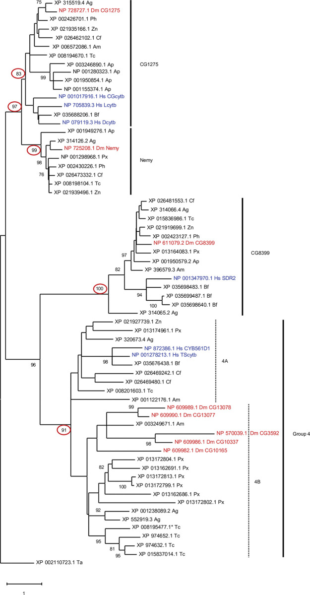
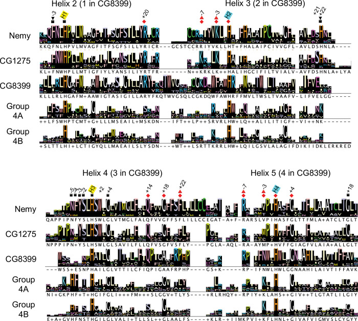
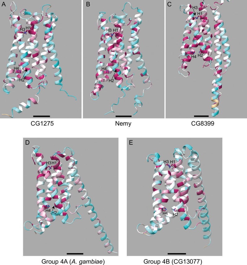
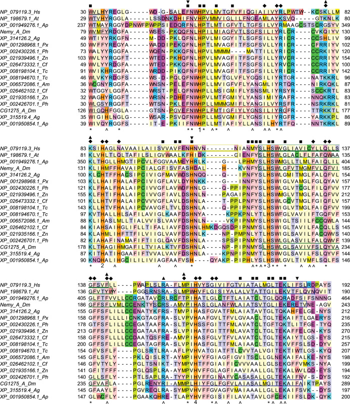
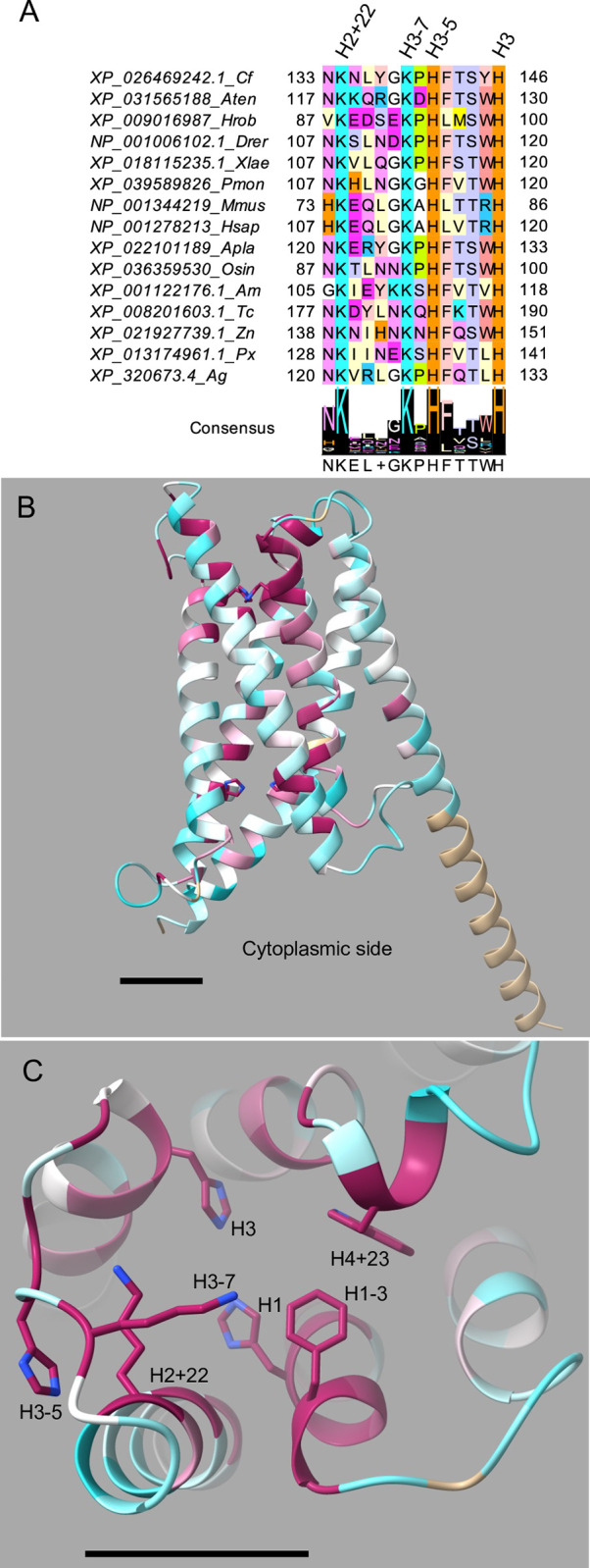
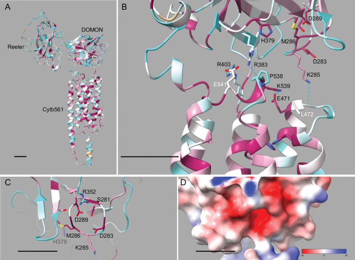
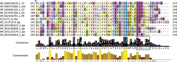
Cytochrome b561 (cytb561) proteins comprise a family of transmembrane oxidoreductases that transfer single electrons across a membrane. Most eukaryotic species, including insects, possess multiple cytb561 homologs. To learn more about this protein family in insects, we carried out a bioinformatics-based investigation of cytb561 family members from nine species representing eight insect orders. We performed a phylogenetic analysis to classify insect cytb561 ortholog groups. We then conducted sequence analyses and analyzed protein models to predict structural elements that may impact the biological functions and localization of these proteins, with a focus on possible ferric reductase activity. Our study revealed three ortholog groups, designated CG1275, Nemy, and CG8399, and a fourth group of less-conserved genes. We found that CG1275 and Nemy proteins are similar to a human ferric reductase, duodenal cytochrome b561 (Dcytb), and have many conserved amino acid residues that function in substrate binding in Dcytb. Notably, CG1275 and Nemy proteins contain a conserved histidine and other residues that play a role in ferric ion reduction by Dcytb. Nemy proteins were distinguished by a novel cysteine-rich cytoplasmic loop sequence. CG8399 orthologs are similar to a putative ferric reductase in humans, stromal cell-derived receptor 2. Like other members of the CYBDOM class of cytb561 proteins, these proteins contain reeler, DOMON, and cytb561 domains. Drosophila melanogaster CG8399 is the only insect cytb561 with known ferric reductase activity. Our investigation of the DOMON domain in CG8399 proteins revealed a probable heme-binding site and a possible site for ferric reduction. The fourth group includes a subgroup of proteins with a conserved “KXXXXKXH” non-cytoplasmic loop motif that may be a substrate binding site and is present in a potential ferric reductase, human tumor suppressor cytochrome b561. This study provides a foundation for future investigations of the biological functions of cytb561 genes in insects.
Overview
The claims and statistics of this paper haven’t been checked yet.
Run the check — free, no account needed. It reads the open-access text and checks the sources it cites.
Integrity signals
Registry
Cites
Statistics
Figures
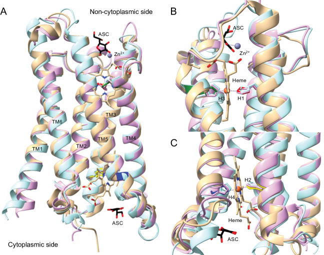
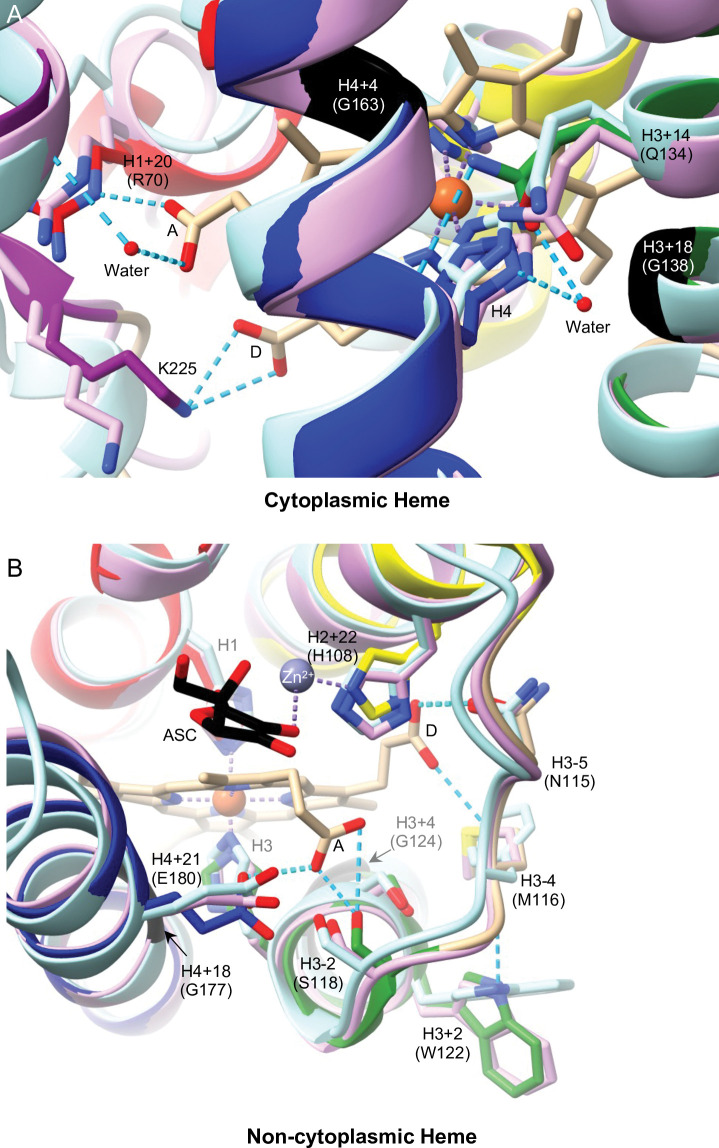
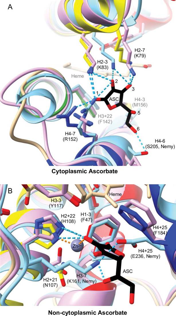
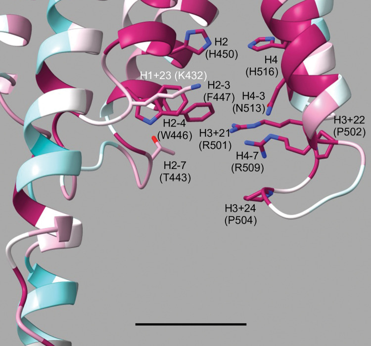
Verification
Claims the paper makes
The claims and statistics of this paper haven’t been checked yet.
Run the check — free, no account needed. It reads the open-access text and checks the sources it cites.
Self-check
Percentages, sums and reported differences re-computed from the paper’s own numbers.
The self-check needs the paper’s full text.
Citations
Cites
Highly accurate protein structure prediction with AlphaFold
Jumper, J., Evans, R., Pritzel, A., Green, T., Figurnov, M., Ronneberger, O., Tunyasuvunakool, K., Bates, R., Žídek, A., Potapenko, A., et al. (2021) · Nature · doi:10.1038/s41586-021-03819-2 · Cited by 43,764
MEGA X: Molecular Evolutionary Genetics Analysis across Computing Platforms
Kumar, S., Stecher, G., Li, M., Knyaz, C., & Tamura, K. (2018) · Molecular Biology and Evolution · doi:10.1093/molbev/msy096 · Cited by 37,529
Fast, scalable generation of high‐quality protein multiple sequence alignments using Clustal Omega
Sievers, F., Wilm, A., Dineen, D., Gibson, T. J., Karplus, K., Li, W., Lopez, R., McWilliam, H., Remmert, M., Söding, J., et al. (2011) · Molecular Systems Biology · doi:10.1038/msb.2011.75 · Cited by 15,355
Database resources of the National Center for Biotechnology Information: update
Wheeler, D. L. (2004) · Nucleic Acids Research · doi:10.1093/nar/gkh073 · Cited by 10,641
Jalview Version 2—a multiple sequence alignment editor and analysis workbench
Waterhouse, A. M., Procter, J. B., Martin, D. M. A., Clamp, M., & Barton, G. J. (2009) · Bioinformatics · doi:10.1093/bioinformatics/btp033 · Cited by 10,235
UCSF ChimeraX: Structure visualization for researchers, educators, and developers
Pettersen, E. F., Goddard, T. D., Huang, C. C., Meng, E. C., Couch, G. S., Croll, T. I., Morris, J. H., & Ferrin, T. E. (2021) · Protein Science · doi:10.1002/pro.3943 · Cited by 9,514
Varadi, M., Anyango, S., Deshpande, M., Nair, S., Natassia, C., Yordanova, G., Yuan, D., Stroe, O., Wood, G., Laydon, A., et al. (2022) · Nucleic Acids Research · doi:10.1093/nar/gkab1061 · Cited by 7,850
UCSF ChimeraX: Meeting modern challenges in visualization and analysis
Goddard, T. D., Huang, C. C., Meng, E. C., Pettersen, E. F., Couch, G. S., Morris, J. H., & Ferrin, T. E. (2018) · Protein Science · doi:10.1002/pro.3235 · Cited by 5,267
SignalP 5.0 improves signal peptide predictions using deep neural networks
Armenteros, J. J. A., Tsirigos, K. D., Sønderby, C. K., Petersen, T. N., Winther, O., Brunak, S., von HEIJNE, G., & Nielsen, H. (2019) · Nature Biotechnology · doi:10.1038/s41587-019-0036-z · Cited by 4,354
SignalP 6.0 predicts all five types of signal peptides using protein language models
Teufel, F., Armenteros, J. J. A., Johansen, A. R., Gíslason, M. H., Pihl, S. I., Tsirigos, K. D., Winther, O., Brunak, S., von HEIJNE, G., & Nielsen, H. (2022) · Nature Biotechnology · doi:10.1038/s41587-021-01156-3 · Cited by 3,194
Metadata
- Authors
- Holst, J. D., Murphy, L. G., Gorman, M. J., & Ragan, E. J.
- Affiliations
- Metropolitan State University of Denver (US)
- Kansas State University (US)
- Corresponding author
- Ragan, Emily J.
- Venue
- PLOS ONE
- Publisher
- Public Library of Science (PLoS)
- Published
- 2023-12-01
- Volume
- 18
- Issue
- 12
- Pages
- e0291564
- ISSN
- 1932-6203 (ISSN-L)
- PMC
- PMC10691727
- OpenAlex
- W4389234010
- Cited by
- 6
- References
- 71
- Citations by year
- 2025: 4 · 2024: 2
- FWCI
- 1.23
- Type
- journal article
- Language
- en
- Open access
- gold
- doi.org
- Licence
- Locations
- PLoS ONE (publishedVersion, cc-by)
- bioRxiv (Cold Spring Harbor Laboratory) (acceptedVersion, cc-by)
- PubMed Central (publishedVersion, cc-by)
- PubMed (publishedVersion)
- DOAJ (DOAJ: Directory of Open Access Journals) (submittedVersion, cc-by-sa)
- Funders
- Division of Integrative Organismal Systems (NSF IOS-1656388, NSF IOS-1656407)
- Foundation for the National Institutes of Health (NIH R35GM141859, R01-GM129325, 5U41HG000739)
- Topics
- Health Sciences › Medicine › Hematology › Iron Metabolism and Disorders
- Life Sciences › Biochemistry, Genetics and Molecular Biology › Molecular Biology › Protein Structure and Dynamics
- Life Sciences › Biochemistry, Genetics and Molecular Biology › Molecular Biology › Insect Resistance and Genetics
- Keywords
- cytochrome b561, Dcytb, heme binding site, substrate binding site, Drosophila melanogaster
- MeSH terms
- Animals, Cytochrome b Group, Drosophila melanogaster, Humans, Iron, Oxidoreductases, Phylogeny, FMN Reductase
About these checks
Registry, retraction, Cites and statistics results are computed, not judged, with one exception: a printed reference that no index lists is read once by a model before it counts as a citation error. Claims are extracted by a model and kept only when their quote appears verbatim in the text; each cited source is then checked, with a model’s help, for whether it says what the claim relies on. Those verdicts can be wrong — read the claim’s quote and the cited source before relying on one.
Statistics and arithmetic are recomputed, not judged.
Confidence bands are shown only for fields calibrated on our annotated test set. None is calibrated yet, so no band is shown.
What counts as a claim ›How we measure confidence ›How we check this ›
Are you an author, or do you see a problem with this page? Request a review
How to cite
Cite CoChat for the checks on this page, and the paper for its own findings.
- This page
- CoChat. (2026). Citation check: Comparison of insect and human cytochrome b561 proteins: Insights into candidate ferric reductases in insects. https://app.cochat.ai/papers/10.1371/journal.pone.0291564
- The paper
- Holst, J. D., Murphy, L. G., Gorman, M. J., & Ragan, E. J. (2023). Comparison of insect and human cytochrome b561 proteins: Insights into candidate ferric reductases in insects. PLOS ONE. https://doi.org/10.1371/journal.pone.0291564
Data sources
Bibliographic data: Semantic Scholar, OpenAlex, Crossref.
Contains information from the Semantic Scholar Academic Graph Datasets, made available under the ODC Attribution License (ODC-BY 1.0).
Semantic Scholar’s data is described in: Kinney, R., et al. (2023). The Semantic Scholar Open Data Platform. arXiv:2301.10140.
