

Evaluating deep learning-based melanoma classification using immunohistochemistry and routine histology: A three center study
Open accessThe DOI matches its Crossref record; the references it cites are not checked yet.
Abstract
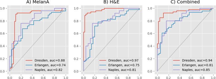
Pathologists routinely use immunohistochemical (IHC)-stained tissue slides against MelanA in addition to hematoxylin and eosin (H&E)-stained slides to improve their accuracy in diagnosing melanomas. The use of diagnostic Deep Learning (DL)-based support systems for automated examination of tissue morphology and cellular composition has been well studied in standard H&E-stained tissue slides. In contrast, there are few studies that analyze IHC slides using DL. Therefore, we investigated the separate and joint performance of ResNets trained on MelanA and corresponding H&E-stained slides. The MelanA classifier achieved an area under receiver operating characteristics curve (AUROC) of 0.82 and 0.74 on out of distribution (OOD)-datasets, similar to the H&E-based benchmark classification of 0.81 and 0.75, respectively. A combined classifier using MelanA and H&E achieved AUROCs of 0.85 and 0.81 on the OOD datasets. DL MelanA-based assistance systems show the same performance as the benchmark H&E classification and may be improved by multi stain classification to assist pathologists in their clinical routine.
Overview
The claims and statistics of this paper haven’t been checked yet.
Free: it reads the open-access text and checks the first 5 claims that cite a source we can check. Create a free account to check every claim.
Integrity signals
Registry
Cites
Statistics
Figures
Verification
Claims the paper makes
The claims and statistics of this paper haven’t been checked yet.
Free: it reads the open-access text and checks the first 5 claims that cite a source we can check. Create a free account to check every claim.
Claims other papers make about this one
Each is checked against this paper’s own text: does it say what the citing paper says it does?
The source backs part of the claim, not all of it — or backs it for a different population, condition or comparison.
In the citing paper
“combining Melan A and H&E images further improved performance (AUROC of 0.85 and 0.81) [38].”
From Artificial Intelligence in Dermatopathology: An Update and Review of the Current Literature (2026) · cites this paper as [38]
Experimental — accuracy not yet measuredThe source backs part of the claim, not all of it — or backs it for a different population, condition or comparison.
In the citing paper
“In dermatopathology, applications include assessing proliferation markers (e.g., Ki-67) in melanocytic lesions [37], quantifying lineage markers (Melan-A, SOX10) [38] for melanocytic lesion evaluation, and analyzing immune-related markers (CD3, CD8, PD-L1) [39,40] in both cutaneous tumors and inflammatory dermatoses.”
From Artificial Intelligence in Dermatopathology: An Update and Review of the Current Literature (2026) · cites this paper as [38]
Experimental — accuracy not yet measuredThe source backs part of the claim, not all of it — or backs it for a different population, condition or comparison.
In the citing paper
“A multicenter study evaluated a ResNet classifier trained on Melan A IHC to detect melanomas, achieving area under the receiver operating characteristic curves (AUROCs) of 0.82 and 0.74 on external datasets; combining Melan A and H&E images further improved performance (AUROC of 0.85 and 0.81) [38].”
From Artificial Intelligence in Dermatopathology: An Update and Review of the Current Literature (2026) · cites this paper as [38]
Experimental — accuracy not yet measured
Self-check
Percentages, sums and reported differences re-computed from the paper’s own numbers.
The self-check needs the paper’s full text.
Citations
The citation lists are queued
They are fetched later and appear here then.
References 37 · Cited by 16
Research works printed but not linked: 1
- R Wightman, 2023, rwightman/pytorch-image-models: v0.8.10dev0 Release, Zenodo
Metadata
- Authors
- Wies, C., Schneider, L., Haggenmüller, S., Bucher, T.-C., Hobelsberger, S., Heppt, M. V., Ferrara, G., Krieghoff-Henning, E. I., & Brinker, T. J.
- Affiliations
- German Cancer Research Center (DE)
- University Hospital Carl Gustav Carus (DE)
- Friedrich-Alexander-Universität Erlangen-Nürnberg (DE)
- Istituto Nazionale Tumori IRCCS "Fondazione G. Pascale" (IT)
- Corresponding author
- Brinker, Titus J.
- ORCID
- Venue
- PLOS ONE
- Publisher
- Public Library of Science (PLoS)
- Published
- 2024-01-19
- Volume
- 19
- Issue
- 1
- Pages
- e0297146
- ISSN
- 1932-6203 (ISSN-L)
- PMC
- PMC10798511
- OpenAlex
- W4391045194
- Semantic Scholar
- 0e0f56ca2336d0226eba02332cdd0df1f5e93090
- Cited by
- 16
- References
- 37
- Citations by year
- 2026: 3 · 2025: 8 · 2024: 5
- FWCI
- 2.35
- Type
- journal article
- Language
- en
- Open access
- gold
- doi.org
- Licence
- Locations
- PLoS ONE (publishedVersion, cc-by)
- PubMed Central (publishedVersion, cc-by)
- PubMed (publishedVersion)
- arXiv (Cornell University) (submittedVersion, public-domain)
- DOAJ (DOAJ: Directory of Open Access Journals) (submittedVersion, cc-by-sa)
- arXiv (Cornell University) (public-domain)
- Funders
- Federal Ministry of Health, Berlin, Germany (TPI, SCP2)
- Ministry of Social Affairs, Health and Integration of the Federal State Baden-Württemberg, Germany (KTI)
- Topics
- Physical Sciences › Computer Science › Artificial Intelligence › AI in cancer detection
- Health Sciences › Medicine › Oncology › Cutaneous Melanoma Detection and Management
- Health Sciences › Medicine › Radiology, Nuclear Medicine and Imaging › Radiomics and Machine Learning in Medical Imaging
- Keywords
- melanoma classification, H&E staining, deep learning, ResNet, out-of-distribution generalization, computer-aided diagnosis, histopathology
- MeSH terms
- Deep Learning, Humans, Immunohistochemistry, Melanoma, ROC Curve, MART-1 Antigen
About these checks
Registry, retraction, Cites and statistics results are computed, not judged, with one exception: a printed reference that no index lists is read once by a model before it counts as a citation error. Claims are extracted by a model and kept only when their quote appears verbatim in the text; each cited source is then checked, with a model’s help, for whether it says what the claim relies on. Those verdicts can be wrong — read the claim’s quote and the cited source before relying on one.
Statistics and arithmetic are recomputed, not judged.
Confidence bands are shown only for fields calibrated on our annotated test set. None is calibrated yet, so no band is shown.
What counts as a claim ›How we measure confidence ›How we check this ›
Are you an author, or do you see a problem with this page? Request a review
How to cite
Cite CoChat for the checks on this page, and the paper for its own findings.
- This page
- CoChat. (2026). Citation check: Evaluating deep learning-based melanoma classification using immunohistochemistry and routine histology: A three center study. https://app.cochat.ai/papers/10.1371/journal.pone.0297146
- The paper
- Wies, C., Schneider, L., Haggenmüller, S., Bucher, T.-C., Hobelsberger, S., Heppt, M. V., Ferrara, G., Krieghoff-Henning, E. I., & Brinker, T. J. (2024). Evaluating deep learning-based melanoma classification using immunohistochemistry and routine histology: A three center study. PLOS ONE. https://doi.org/10.1371/journal.pone.0297146
Data sources
Bibliographic data: Semantic Scholar, OpenAlex, Crossref.
Contains information from the Semantic Scholar Academic Graph Datasets, made available under the ODC Attribution License (ODC-BY 1.0).
Semantic Scholar’s data is described in: Kinney, R., et al. (2023). The Semantic Scholar Open Data Platform. arXiv:2301.10140.
