

The DOI matches its Crossref record; 22 of the 22 references it cites were found in a registry or an index.
Tracing the function expansion for a primordial protein fold in the era of fold-based function prediction: β-trefoil
Open accessPublished version of a preprint
Abstract
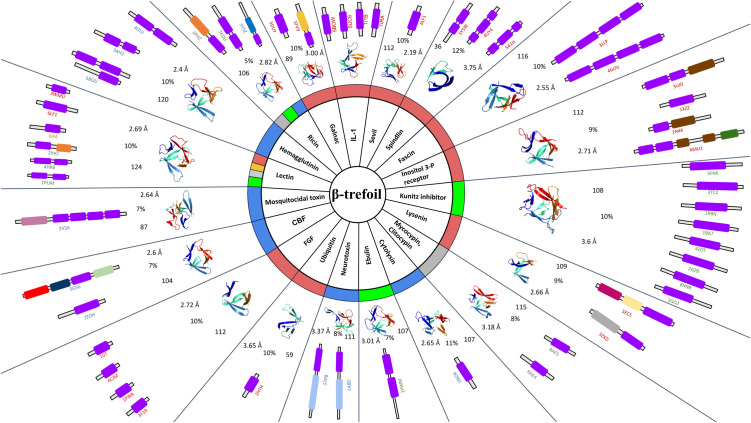
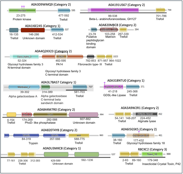
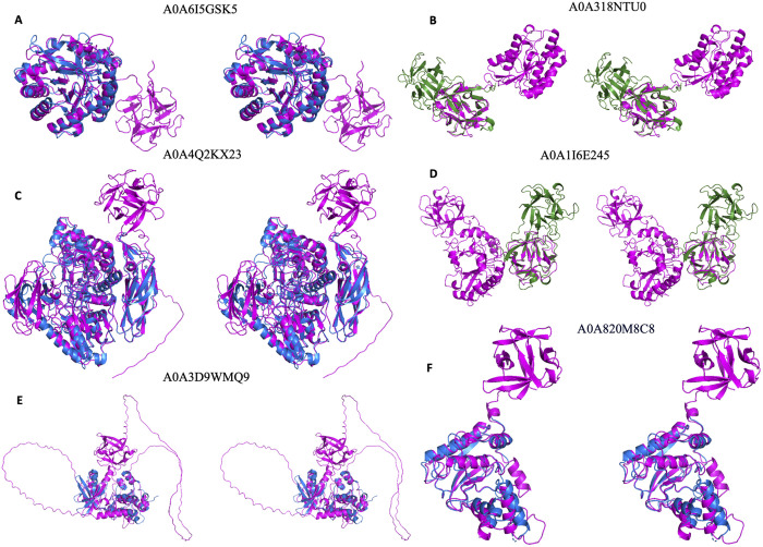
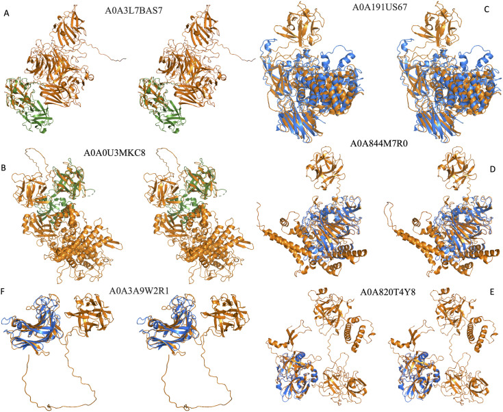
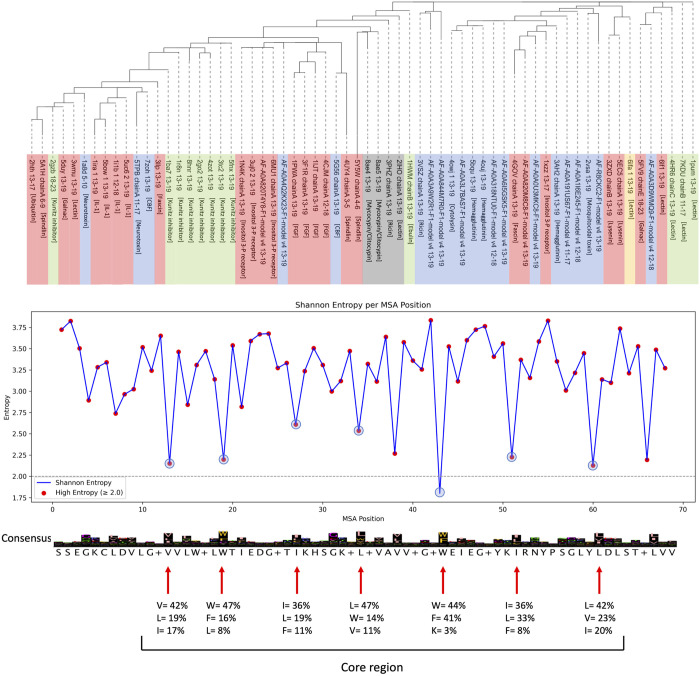
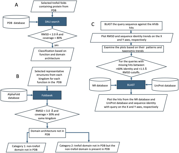
The incredibly narrow protein fold bottleneck, which separates the billions of unique proteins on one side to deliver diverse biological functions on the other, arises from folds that tolerate mutations during evolution. One such fold, called the β-trefoil, is present in functionally diverse proteins including cytokines involved in the immune system such as interleukin-1. The unrecognizable sequence-level diversity, even among paralogs of interleukin-1 within the same chromosomal locus, suggests the resilience of this fold to mutational onslaught. Furthermore, β-trefoil domain containing-proteins are known to coexist with other domains to achieve functional diversity. In this study, we challenge the reach and limitations of function prediction using fold-fold comparison using β-trefoil fold as an example. We identified proteins containing β-trefoil fold belonging to thirty-two distinct functional classes based on diverse domain architecture and/or functional annotation by mining both the PDB and AlphaFold databases using fold-fold comparison. Among the proteins with novel domain architecture we find β-trefoil along with chitinase, lipase, β-glucosidase, protein kinase, peptidoglycan-binding + peptidase matrixin, glycosyl hydrolases family 3 + PA14 + fibronectin type- III, alpha galactosidase A, PhoD-like phosphatase, insecticidal crystal toxin, trypsin, alginate lyase and two novel structurally uncharacterized domains. We demonstrate that fold-fold comparison can extend function prediction beyond the reach of sequence-based approach and provides an opportunity to discover novel domain architecture associated with known folds. However, since extending fold similarity to functional similarity may be challenged by convergent fold evolution, we explore if β-trefoil may be a convergent evolution and share our hypothesis.
Overview
The claims and statistics of this paper haven’t been checked yet.
Run the check — free, no account needed. It reads the open-access text and checks the sources it cites.
Integrity signals
Registry
Cites
Statistics
Figures
Verification
Claims the paper makes
The claims and statistics of this paper haven’t been checked yet.
Run the check — free, no account needed. It reads the open-access text and checks the sources it cites.
Self-check
Percentages, sums and reported differences re-computed from the paper’s own numbers.
The self-check needs the paper’s full text.
Citations
Cites
Basic local alignment search tool
Altschul, S. F., Gish, W., Miller, W., Myers, E. W., & Lipman, D. J. (1990) · Journal of Molecular Biology · doi:10.1006/jmbi.1990.9999 · Cited by 98,669
Highly accurate protein structure prediction with AlphaFold
Jumper, J., Evans, R., Pritzel, A., Green, T., Figurnov, M., Ronneberger, O., Tunyasuvunakool, K., Bates, R., Žídek, A., Potapenko, A., et al. (2021) · Nature · doi:10.1038/s41586-021-03819-2 · Cited by 43,764
Atlas of Protein Sequence and Structure, 1966.
Hersh, R. T., Eck, R. V., & Dayhoff, M. O. (1967) · Systematic Zoology · doi:10.2307/2412074 · Cited by 1,798
Dali: a network tool for protein structure comparison
Holm, L., & Sander, C. (1995) · Trends in Biochemical Sciences · doi:10.1016/s0968-0004(00)89105-7 · Cited by 1,462
Eddy, S. R. (1996) · Current Opinion in Structural Biology · doi:10.1016/s0959-440x(96)80056-x · Cited by 1,430
One thousand families for the molecular biologist
Chothia, C. (1992) · Nature · doi:10.1038/357543a0 · Cited by 1,018
Kamisetty, H., Ovchinnikov, S., & Baker, D. (2013) · Proceedings of the National Academy of Sciences · doi:10.1073/pnas.1314045110 · Cited by 672
SCOP: a Structural Classification of Proteins database
Conte, L. L. (2000) · Nucleic Acids Research · doi:10.1093/nar/28.1.257 · Cited by 581
Critical assessment of methods of protein structure prediction (CASP)—Round XIV
Kryshtafovych, A., Schwede, T., Topf, M., Fidelis, K., & Moult, J. (2021) · Proteins: Structure, Function, and Bioinformatics · doi:10.1002/prot.26237 · Cited by 503
Clustering predicted structures at the scale of the known protein universe
Barrio-Hernandez, I., Yeo, J., Jänes, J., Mirdita, M., Gilchrist, C. L. M., Wein, T., Varadi, M., Velankar, S., Beltrao, P., & Steinegger, M. (2023) · Nature · doi:10.1038/s41586-023-06510-w · Cited by 362
Metadata
- Authors
- Goswami, M., & Srinivasan, S.
- Affiliations
- Institute of Bioinformatics and Applied Biotechnology (IN)
- Corresponding author
- Srinivasan, Subhashini
- Venue
- PLOS One
- Publisher
- Public Library of Science (PLoS)
- Published
- 2025-07-03
- Volume
- 20
- Issue
- 7
- Pages
- e0320177
- ISSN
- 1932-6203 (ISSN-L)
- PMC
- PMC12225799
- OpenAlex
- W4411995712
- Cited by
- 0
- References
- 22
- FWCI
- 0.00
- Type
- journal article
- Language
- en
- Open access
- gold
- doi.org
- Licence
- Locations
- PLoS ONE (publishedVersion, cc-by)
- bioRxiv (Cold Spring Harbor Laboratory) (acceptedVersion, cc-by)
- PubMed Central (publishedVersion, cc-by)
- PubMed (publishedVersion)
- DOAJ (DOAJ: Directory of Open Access Journals) (submittedVersion, cc-by-sa)
- DOAJ (DOAJ: Directory of Open Access Journals) (submittedVersion)
- Funders
- Department of Biotechnology, Ministry of Science and Technology, India (BT/PR40212/BTIS/137/40/2022)
- Topics
- Life Sciences › Biochemistry, Genetics and Molecular Biology › Molecular Biology › Protein Structure and Dynamics
- Life Sciences › Biochemistry, Genetics and Molecular Biology › Structural Biology › Enzyme Structure and Function
- Life Sciences › Biochemistry, Genetics and Molecular Biology › Molecular Biology › Genomics and Phylogenetic Studies
- Keywords
- β-trefoil fold, protein function prediction, domain architecture, interleukin-1, cytokines, convergent evolution, protein fold evolution, protein domains, structural genomics
- MeSH terms
- Protein Domains, Models, Molecular, Proteins, Protein Folding, Databases, Protein
About these checks
Registry, retraction, Cites and statistics results are computed, not judged, with one exception: a printed reference that no index lists is read once by a model before it counts as a citation error. Claims are extracted by a model and kept only when their quote appears verbatim in the text; each cited source is then checked, with a model’s help, for whether it says what the claim relies on. Those verdicts can be wrong — read the claim’s quote and the cited source before relying on one.
Statistics and arithmetic are recomputed, not judged.
Confidence bands are shown only for fields calibrated on our annotated test set. None is calibrated yet, so no band is shown.
What counts as a claim ›How we measure confidence ›How we check this ›
Are you an author, or do you see a problem with this page? Request a review
How to cite
Cite CoChat for the checks on this page, and the paper for its own findings.
- This page
- CoChat. (2026). Citation check: Tracing the function expansion for a primordial protein fold in the era of fold-based function prediction: β-trefoil. https://app.cochat.ai/papers/10.1371/journal.pone.0320177
- The paper
- Goswami, M., & Srinivasan, S. (2025). Tracing the function expansion for a primordial protein fold in the era of fold-based function prediction: β-trefoil. PLOS One. https://doi.org/10.1371/journal.pone.0320177
Data sources
Bibliographic data: Semantic Scholar, OpenAlex, Crossref.
Contains information from the Semantic Scholar Academic Graph Datasets, made available under the ODC Attribution License (ODC-BY 1.0).
Semantic Scholar’s data is described in: Kinney, R., et al. (2023). The Semantic Scholar Open Data Platform. arXiv:2301.10140.
