Cited by 166

The DOI matches its Crossref record.

HDP—A Novel Heme Detoxification Protein from the Malaria Parasite

Jani, Dewal, Nagarkatti, Rana, Beatty, Wandy, Angel, Ross, Slebodnick, Carla, Andersen, John, Kumar, Sanjai, Rathore, Dharmendar·2008·PLoS Pathogens

Overview

The claims and statistics of this paper haven’t been checked yet.

Run the check — free, no account needed. It reads the open-access text and checks the sources it cites.

A few free checks a day · papers already verified open instantly · results are public

Integrity signals

Registry

Crossref · 10.1371/journal.ppat.1000053

Cites

Statistics

Figures

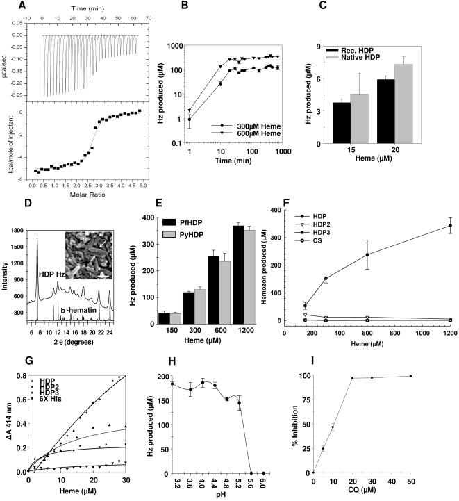
Figure 1 HDP rapidly detoxifies and sequesters heme as Hz. A, Heme (100 µM) solution was titrated into protein (5 µM) and the heat evolved was measured by Isothermal titration calorimetry. Binding isotherm was generated by integrating the data from the top panel. B, HDP-mediated Hz formation was concentration dependent and occurred rapidly with up to 75% of the total Hz being produced within the first 20 minutes. C, Native HDP (grey) can produce Hz at levels comparable to the recombinant protein (black). D, Powder X-ray diffraction pattern of HDP-produced Hz matches with that of β-hematin. Inset–Scanning electron microscopy image of HDP-produced Hz crystallites that are 0.1–0.2 µm in length (arrows) with a width of approx.0.05 µm (Bar, 0.1 µm) E, Hz formation activity of 0.5 µM P. yoelii HDP (grey bars) is indistinguishable from its P. falciparum ortholog, utilized at identical concentration (black bars). F, Full length HDP (closed circle) is necessary for Hz formation as HDP2 (open circle) and HDP3 (closed triangle) alone could not produce Hz. 0.5 µM of each of the three proteins was utilized for analysis. G, Heme in 2 µM increments was added to a 10 µM solution of either HDP, HDP2, HDP3 or polyhistidine peptide in 100 mM Sodium acetate buffer pH 5.2. Change in absorbance at 414 nm was plotted against the concentration of heme. The curve depicts the non-linear fit of the data. H, HDP-mediated Hz production is restricted to a pH range found inside the FV. I, CQ inhibits HDP-mediated Hz production. Values are mean±s. d
Figure 1 HDP rapidly detoxifies and sequesters heme as Hz. A, Heme (100 µM) solution was titrated into protein (5 µM) and the heat evolved was measured by Isothermal titration calorimetry. Binding isotherm was generated by integrating the data from the top panel. B, HDP-mediated Hz formation was concentration dependent and occurred rapidly with up to 75% of the total Hz being produced within the first 20 minutes. C, Native HDP (grey) can produce Hz at levels comparable to the recombinant protein (black). D, Powder X-ray diffraction pattern of HDP-produced Hz matches with that of β-hematin. Inset–Scanning electron microscopy image of HDP-produced Hz crystallites that are 0.1–0.2 µm in length (arrows) with a width of approx.0.05 µm (Bar, 0.1 µm) E, Hz formation activity of 0.5 µM P. yoelii HDP (grey bars) is indistinguishable from its P. falciparum ortholog, utilized at identical concentration (black bars). F, Full length HDP (closed circle) is necessary for Hz formation as HDP2 (open circle) and HDP3 (closed triangle) alone could not produce Hz. 0.5 µM of each of the three proteins was utilized for analysis. G, Heme in 2 µM increments was added to a 10 µM solution of either HDP, HDP2, HDP3 or polyhistidine peptide in 100 mM Sodium acetate buffer pH 5.2. Change in absorbance at 414 nm was plotted against the concentration of heme. The curve depicts the non-linear fit of the data. H, HDP-mediated Hz production is restricted to a pH range found inside the FV. I, CQ inhibits HDP-mediated Hz production. Values are mean±s. dCC0 · PubMed Central
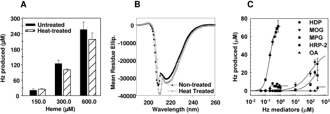
Figure 2 Structural and biochemical analysis of HDP-mediated Hz formation. A, HDP (Black bars) retained its Hz production activity after heat treatment (Hashed Bars) at 94°C for 10 minutes. B, CD spectra of heat-treated HDP is qualitatively similar to non-treated HDP, which was incubated at room temperature. Mean residue ellipticity is expressed as degree cm−1 dmol−1. C, When compared to MPG (square), MOG (inverted triangle), HRP 2 (diamond) and OA (triangle), HDP (circle) was most effective in producing Hz.
Figure 2 Structural and biochemical analysis of HDP-mediated Hz formation. A, HDP (Black bars) retained its Hz production activity after heat treatment (Hashed Bars) at 94°C for 10 minutes. B, CD spectra of heat-treated HDP is qualitatively similar to non-treated HDP, which was incubated at room temperature. Mean residue ellipticity is expressed as degree cm−1 dmol−1. C, When compared to MPG (square), MOG (inverted triangle), HRP 2 (diamond) and OA (triangle), HDP (circle) was most effective in producing Hz.CC0 · PubMed Central
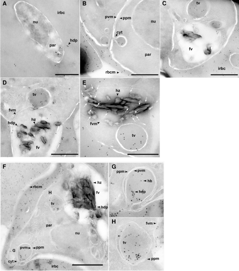
Figure 3 Circuitous transport and delivery of HDP to the FV. A, HDP is secreted into the cytosol of infected erythrocytes (arrowhead) in early ring stages before any Hz could be detected inside the parasite. B, After secreting it into the host cell cytosol, parasite intakes HDP through the cytostome. C, HDP could be found in transport vesicles destined to the FV. D, Transport vesicles deliver HDP to the FV. E, HDP (arrows) was found in close proximity of Hz crystals. F, HDP (18 nm particles) is transported to FV along with hemoglobin (12 nm particles). Inset G, HDP is being internalized along with hemoglobin. Inset H, Transport vesicle ready to deliver both, HDP and hemoglobin, to the FV. cyt, cytostome; fv, food vacuole; fvm, fv membrane; hz, hemozoin; hdp, heme detoxification protein; hb, hemoglobin; nu, nucleus; par, parasite; ppm, parasite plasma membrane; pvm, parasitophorous vacuole membrane; irbc, infected red blood cell; rbcm, RBC membrane; tv, transport vesicle. Bar, 0.5 µm.
Figure 3 Circuitous transport and delivery of HDP to the FV. A, HDP is secreted into the cytosol of infected erythrocytes (arrowhead) in early ring stages before any Hz could be detected inside the parasite. B, After secreting it into the host cell cytosol, parasite intakes HDP through the cytostome. C, HDP could be found in transport vesicles destined to the FV. D, Transport vesicles deliver HDP to the FV. E, HDP (arrows) was found in close proximity of Hz crystals. F, HDP (18 nm particles) is transported to FV along with hemoglobin (12 nm particles). Inset G, HDP is being internalized along with hemoglobin. Inset H, Transport vesicle ready to deliver both, HDP and hemoglobin, to the FV. cyt, cytostome; fv, food vacuole; fvm, fv membrane; hz, hemozoin; hdp, heme detoxification protein; hb, hemoglobin; nu, nucleus; par, parasite; ppm, parasite plasma membrane; pvm, parasitophorous vacuole membrane; irbc, infected red blood cell; rbcm, RBC membrane; tv, transport vesicle. Bar, 0.5 µm.CC0 · PubMed Central
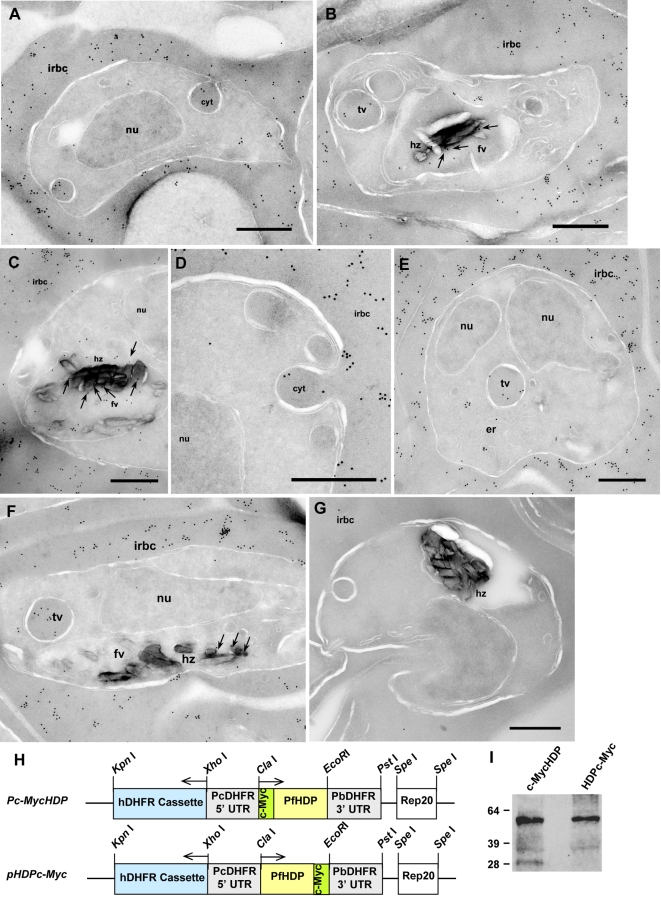
Figure 4 c-Myc tagged HDP has an identical trafficking pathway to HDP. Panels A–C depicts intracellular location of c-MycHDP fusion protein in parasite-infected RBC. Cytostomal uptake of c-MycHDP (A) followed by its transport (B) and its presence within the FV (C) in close proximity of Hz (shown by arrows). Panels D–F show intracellular location of HDPc-Myc, where, like HDP, chimera was present in the infected RBC cytosol (D), detected in the process of being internalized (D), transported (E), and found in the FV (F), where it presence has been marked with arrows. G, c-Myc antibodies specifically recognized the two chimeras as they did not recognize any protein in the non-transfected 3D7 P. falciparum parasites. H, Schematic representation of the two c-Myc based fusion constructs utilized for expression. I, c-myc tag was still attached to HDP when the protein was secreted out into the infected RBC cytosol. HDP was primarily present as a dimer, with little monomeric form also detected in c-MycHDP preparation. cyt, cytostome; fv, food vacuole; hz, hemozoin; nu, nucleus; irbc, infected red blood cell; tv, transport vesicle. Bar, 0.5 µm
Figure 4 c-Myc tagged HDP has an identical trafficking pathway to HDP. Panels A–C depicts intracellular location of c-MycHDP fusion protein in parasite-infected RBC. Cytostomal uptake of c-MycHDP (A) followed by its transport (B) and its presence within the FV (C) in close proximity of Hz (shown by arrows). Panels D–F show intracellular location of HDPc-Myc, where, like HDP, chimera was present in the infected RBC cytosol (D), detected in the process of being internalized (D), transported (E), and found in the FV (F), where it presence has been marked with arrows. G, c-Myc antibodies specifically recognized the two chimeras as they did not recognize any protein in the non-transfected 3D7 P. falciparum parasites. H, Schematic representation of the two c-Myc based fusion constructs utilized for expression. I, c-myc tag was still attached to HDP when the protein was secreted out into the infected RBC cytosol. HDP was primarily present as a dimer, with little monomeric form also detected in c-MycHDP preparation. cyt, cytostome; fv, food vacuole; hz, hemozoin; nu, nucleus; irbc, infected red blood cell; tv, transport vesicle. Bar, 0.5 µmCC0 · PubMed Central
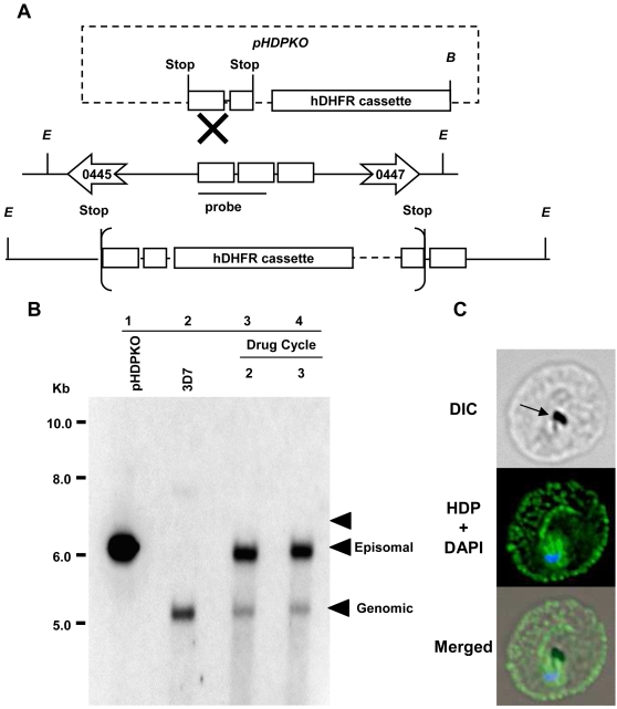
Figure 5 Genetic manipulation of HDP locus. Schematic representation of the strategy used for targeting HDP locus through single cross over recombination. A, Schematic representation of the plasmid and the genomic locus showing the enzyme sites and the genes flanking HDP. The anticipated cross-over event at the HDP locus and restriction enzyme sites for Eco RV, E and Bam HI, B are shown. Eco RV site is unique to the parasite DNA while Bam HI site is unique to the plasmid. B, Lanes 1 and 2, depict Bam HI-linearized pHDPKO (6.3 kb) and Eco RV and Bam HI digested DNA from wild type P. falciparum parasites containing the HDP locus (5.3 kb), respectively. Lanes 3 and 4 depict the transfected parasites after 2nd and 3rd drug cycle, respectively. Parasites surviving after three selection cycles (lanes 3, 4) had an intact HDP locus and an episomal copy of the pHDPKO plasmid expressing hDHFR. The expected size on integration is indicated by the arrow (6657 bp). C, Immuno-fluorescence staining using anti-HDP antibodies showed that the transfected parasites continually expressed HDP (green) even after the third drug cycle thus indicating that the locus was not disrupted. Arrows on the DIC image indicate hemozoin. Parasite nucleus (blue) is stained with DAPI.
Figure 5 Genetic manipulation of HDP locus. Schematic representation of the strategy used for targeting HDP locus through single cross over recombination. A, Schematic representation of the plasmid and the genomic locus showing the enzyme sites and the genes flanking HDP. The anticipated cross-over event at the HDP locus and restriction enzyme sites for Eco RV, E and Bam HI, B are shown. Eco RV site is unique to the parasite DNA while Bam HI site is unique to the plasmid. B, Lanes 1 and 2, depict Bam HI-linearized pHDPKO (6.3 kb) and Eco RV and Bam HI digested DNA from wild type P. falciparum parasites containing the HDP locus (5.3 kb), respectively. Lanes 3 and 4 depict the transfected parasites after 2nd and 3rd drug cycle, respectively. Parasites surviving after three selection cycles (lanes 3, 4) had an intact HDP locus and an episomal copy of the pHDPKO plasmid expressing hDHFR. The expected size on integration is indicated by the arrow (6657 bp). C, Immuno-fluorescence staining using anti-HDP antibodies showed that the transfected parasites continually expressed HDP (green) even after the third drug cycle thus indicating that the locus was not disrupted. Arrows on the DIC image indicate hemozoin. Parasite nucleus (blue) is stained with DAPI.CC0 · PubMed Central
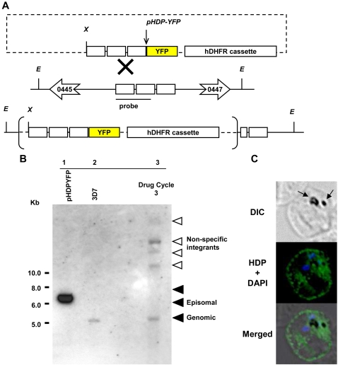
Figure 6 Non-specific recombination of HDP-YFP chimera. A, Schematic representation of the gene replacement strategy employed for obtaining HDP knocked-in parasites. The anticipated cross-over event at the HDP locus and the restriction enzyme sites for Eco RV, E and Xho I, X are shown. B, Lanes 1 and 2 depict Xho I linearized pHDP-YFP (6.32 kb) and Xho I and Eco RV digested DNA from wild type P. falciparum parasites containing the HDP locus (5.3kb), respectively. Lane 3 depicts the double digested DNA from transfected parasites after the 3rd drug cycle. Integration at non specific sites was observed as seen by higher size bands in comparison to the episomal plasmid, represented by the open arrows. As the genomic DNA was digested with restriction enzymes unique to the flanking regions of the HDP locus (Eco RV) and the plasmid (Xho I) these bands do not appear to be site specific integrations of concatameric forms of pHDP-YFP. No integration was observed at the expected size as indicated by the arrow at 7588 bp. Parasites surviving after three selection cycles (lane 3) had the non-recombined HDP locus and no YFP expression, as determined by fluorescence microscopy. C, Immuno-fluorescence staining of these parasites with anti HDP antibodies also indicated the expression of the native HDP (green) after the third drug cycle. Arrows on the DIC image indicate hemozoin. Parasite nucleus (blue) is stained with DAPI.
Figure 6 Non-specific recombination of HDP-YFP chimera. A, Schematic representation of the gene replacement strategy employed for obtaining HDP knocked-in parasites. The anticipated cross-over event at the HDP locus and the restriction enzyme sites for Eco RV, E and Xho I, X are shown. B, Lanes 1 and 2 depict Xho I linearized pHDP-YFP (6.32 kb) and Xho I and Eco RV digested DNA from wild type P. falciparum parasites containing the HDP locus (5.3kb), respectively. Lane 3 depicts the double digested DNA from transfected parasites after the 3rd drug cycle. Integration at non specific sites was observed as seen by higher size bands in comparison to the episomal plasmid, represented by the open arrows. As the genomic DNA was digested with restriction enzymes unique to the flanking regions of the HDP locus (Eco RV) and the plasmid (Xho I) these bands do not appear to be site specific integrations of concatameric forms of pHDP-YFP. No integration was observed at the expected size as indicated by the arrow at 7588 bp. Parasites surviving after three selection cycles (lane 3) had the non-recombined HDP locus and no YFP expression, as determined by fluorescence microscopy. C, Immuno-fluorescence staining of these parasites with anti HDP antibodies also indicated the expression of the native HDP (green) after the third drug cycle. Arrows on the DIC image indicate hemozoin. Parasite nucleus (blue) is stained with DAPI.CC0 · PubMed Central
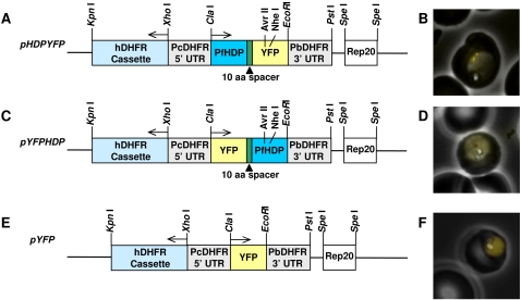
Figure 7 Episomal expression of the YFP-based chimera. P. falciparum 3D7 parasites were independently transfected with plasmids pHDP-YFP (A), pYFP-HDP (C) and pYFP (E) containing the hDHFR cassette as selection marker. Culture was maintained in the presence of WR92210 and the expression of yellow fluorescent protein was investigated by live cell imaging. On expression, transport of both the fusion proteins was found to be disrupted (B and D) with protein exclusively localized within the parasite cytosol. pYFP, when expressed alone (F) showed YFP, like GFP [50], to be localized within the parasite.
Figure 7 Episomal expression of the YFP-based chimera. P. falciparum 3D7 parasites were independently transfected with plasmids pHDP-YFP (A), pYFP-HDP (C) and pYFP (E) containing the hDHFR cassette as selection marker. Culture was maintained in the presence of WR92210 and the expression of yellow fluorescent protein was investigated by live cell imaging. On expression, transport of both the fusion proteins was found to be disrupted (B and D) with protein exclusively localized within the parasite cytosol. pYFP, when expressed alone (F) showed YFP, like GFP [50], to be localized within the parasite.CC0 · PubMed Central

Verification

Claims the paper makes

The claims and statistics of this paper haven’t been checked yet.

Run the check — free, no account needed. It reads the open-access text and checks the sources it cites.

A few free checks a day · papers already verified open instantly · results are public

Claims other papers make about this one

Each is checked against this paper’s own text: does it say what the citing paper says it does?

Self-check

Percentages, sums and reported differences re-computed from the paper’s own numbers.

The self-check needs the paper’s full text.

Citations

Cites

No references could be resolved yet.

Research works printed but not linked: 47

  1. SE Francis, 1997, Hemoglobin metabolism in the malaria parasite Plasmodium falciparum., Annu Rev Microbiol, 51, 97, 10.1146/annurev.micro.51.1.97 · DOI
  2. DE Goldberg, 1990, Hemoglobin degradation in the malaria parasite Plasmodium falciparum: an ordered process in a unique organelle., Proc Natl Acad Sci U S A, 87, 2931, 10.1073/pnas.87.8.2931 · DOI
  3. S Pagola, 2000, The structure of malaria pigment beta-haematin., Nature, 404, 307, 10.1038/35005132 · DOI
  4. TJ Egan, 2002, Fate of haem iron in the malaria parasite Plasmodium falciparum., Biochem J, 365, 343, 10.1042/bj20020793 · DOI
  5. G Kikuchi, 2005, Heme oxygenase and heme degradation., Biochem Biophys Res Commun, 338, 558, 10.1016/j.bbrc.2005.08.020 · DOI
  6. D Rathore, 2005, Antimalarial drugs: current status and new developments., Expert Opin Investig Drugs, 14, 871, 10.1517/13543784.14.7.871 · DOI
  7. A Leed, 2002, Solution structures of antimalarial drug-heme complexes., Biochemistry, 41, 10245, 10.1021/bi020195i · DOI
  8. DJ Sullivan Jr, 1996, On the molecular mechanism of chloroquine's antimalarial action., Proc Natl Acad Sci U S A, 93, 11865, 10.1073/pnas.93.21.11865 · DOI
  9. R Kannan, 2002, Heme-artemisinin adducts are crucial mediators of the ability of artemisinin to inhibit heme polymerization., Chem Biol, 9, 321, 10.1016/S1074-5521(02)00117-5 · DOI
  10. DJ Sullivan Jr, 1996, Plasmodium hemozoin formation mediated by histidine-rich proteins., Science, 271, 219, 10.1126/science.271.5246.219 · DOI
  11. K Bendrat, 1995, Haem polymerization in malaria., Nature, 378, 138, 10.1038/378138a0 · DOI
  12. CD Fitch, 1999, Involvement of lipids in ferriprotoporphyrin IX polymerization in malaria., Biochim Biophys Acta, 1454, 31, 10.1016/S0925-4439(99)00017-4 · DOI
  13. A Dorn, 1995, Malarial haemozoin/beta-haematin supports haem polymerization in the absence of protein., Nature, 374, 269, 10.1038/374269a0 · DOI
  14. DJ Sullivan, 2002, Theories on malarial pigment formation and quinoline action., Int J Parasitol, 32, 1645, 10.1016/S0020-7519(02)00193-5 · DOI
  15. BL Tekwani, 2005, Targeting the hemozoin synthesis pathway for new antimalarial drug discovery: technologies for in vitro beta-hematin formation assay., Comb Chem High Throughput Screen, 8, 63, 10.2174/1386207053328101 · DOI
  16. JM Pisciotta, 2007, The role of neutral lipid nanospheres in Plasmodium falciparum haem crystallization., Biochem J, 402, 197, 10.1042/BJ20060986 · DOI
  17. TJ Egan, 2006, Haemozoin (beta-haematin) biomineralization occurs by self-assembly near the lipid/water interface., FEBS Lett, 580, 5105, 10.1016/j.febslet.2006.08.043 · DOI
  18. MJ Gardner, 2002, Genome sequence of the human malaria parasite Plasmodium falciparum., Nature, 419, 498, 10.1038/nature01097 · DOI · Paper page
  19. JM Carlton, 2002, Genome sequence and comparative analysis of the model rodent malaria parasite Plasmodium yoelii yoelii., Nature, 419, 512, 10.1038/nature01099 · DOI
  20. A Pain, 2005, Genome of the host-cell transforming parasite Theileria annulata compared with T. parva., Science, 309, 131, 10.1126/science.1110418 · DOI
  21. JC Kissinger, 2003, ToxoDB: accessing the Toxoplasma gondii genome., Nucleic Acids Res, 31, 234, 10.1093/nar/gkg072 · DOI
  22. JC Ulstrup, 1995, Relationship of secretion pattern and MPB70 homology with osteoblast-specific factor 2 to osteitis following Mycobacterium bovis BCG vaccination., Infect Immun, 63, 672, 10.1128/IAI.63.2.672-675.1995 · DOI
  23. JE Kim, 2000, Identification of motifs for cell adhesion within the repeated domains of transforming growth factor-beta-induced gene, betaig-h3., J Biol Chem, 275, 30907, 10.1074/jbc.M002752200 · DOI
  24. A Yayon, 1984, Identification of the acidic compartment of Plasmodium falciparum-infected human erythrocytes as the target of the antimalarial drug chloroquine., Embo J, 3, 2695, 10.1002/j.1460-2075.1984.tb02195.x · DOI
  25. M Marti, 2004, Targeting malaria virulence and remodeling proteins to the host erythrocyte., Science, 306, 1930, 10.1126/science.1102452 · DOI
  26. NL Hiller, 2004, A host-targeting signal in virulence proteins reveals a secretome in malarial infection., Science, 306, 1934, 10.1126/science.1102737 · DOI
  27. A Khattab, 2006, Maurer's clefts-restricted localization, orientation and export of a Plasmodium falciparum RIFIN., Traffic, 7, 1654, 10.1111/j.1600-0854.2006.00494.x · DOI
  28. JD Bendtsen, 2004, Improved prediction of signal peptides: SignalP 3.0., J Mol Biol, 340, 783, 10.1016/j.jmb.2004.05.028 · DOI · Paper page
  29. JD Bendtsen, 2004, Feature-based prediction of non-classical and leaderless protein secretion., Protein Eng Des Sel, 17, 349, 10.1093/protein/gzh037 · DOI
  30. A Krogh, 2001, Predicting transmembrane protein topology with a hidden Markov model: application to complete genomes., J Mol Biol, 305, 567, 10.1006/jmbi.2000.4315 · DOI · Paper page
  31. J Schultz, 1998, SMART, a simple modular architecture research tool: identification of signaling domains., Proc Natl Acad Sci U S A, 95, 5857, 10.1073/pnas.95.11.5857 · DOI
  32. A Marchler-Bauer, 2003, CDD: a curated Entrez database of conserved domain alignments., Nucleic Acids Res, 31, 383, 10.1093/nar/gkg087 · DOI
  33. EL Schneider, 2005, Heme binding to the histidine-rich protein II from Plasmodium falciparum., Biochemistry, 44, 979, 10.1021/bi048570p · DOI
  34. S Subramanian, 2007, Falcipain cysteine proteases require bipartite motifs for trafficking to the Plasmodium falciparum food vacuole., J Biol Chem, 282, 24961, 10.1074/jbc.M703316200 · DOI
  35. M Klemba, 2004, Trafficking of plasmepsin II to the food vacuole of the malaria parasite Plasmodium falciparum., J Cell Biol, 164, 47, 10.1083/jcb200307147 · DOI
  36. J Liu, 2005, The role of Plasmodium falciparum food vacuole plasmepsins., J Biol Chem, 280, 1432, 10.1074/jbc.M409740200 · DOI
  37. J Liu, 2006, Plasmodium falciparum ensures its amino acid supply with multiple acquisition pathways and redundant proteolytic enzyme systems., Proc Natl Acad Sci U S A, 103, 8840, 10.1073/pnas.0601876103 · DOI
  38. AF Slater, 1992, Inhibition by chloroquine of a novel haem polymerase enzyme activity in malaria trophozoites., Nature, 355, 167, 10.1038/355167a0 · DOI
  39. TE Wellems, 1991, Genetic mapping of the chloroquine-resistance locus on Plasmodium falciparum chromosome 7., Proc Natl Acad Sci U S A, 88, 3382, 10.1073/pnas.88.8.3382 · DOI
  40. RJ Howard, 1986, Secretion of a malarial histidine-rich protein (Pf HRP II) from Plasmodium falciparum-infected erythrocytes., J Cell Biol, 103, 1269, 10.1083/jcb.103.4.1269 · DOI
  41. TJ Egan, 2007, Recent advances in understanding the mechanism of hemozoin (malaria pigment) formation., J Inorg Biochem
  42. T Akompong, 2002, Trans expression of a Plasmodium falciparum histidine-rich protein II (HRPII) reveals sorting of soluble proteins in the periphery of the host erythrocyte and disrupts transport to the malarial food vacuole., J Biol Chem, 277, 28923, 10.1074/jbc.M201968200 · DOI
  43. L Florens, 2002, A proteomic view of the Plasmodium falciparum life cycle., Nature, 419, 520, 10.1038/nature01107 · DOI
  44. D Rathore, 2003, Molecular mechanism of host specificity in Plasmodium falciparum infection: role of circumsporozoite protein., J Biol Chem, 278, 40905, 10.1074/jbc.M306250200 · DOI
  45. DA Fidock, 1997, Transformation with human dihydrofolate reductase renders malaria parasites insensitive to WR99210 but does not affect the intrinsic activity of proguanil., Proc Natl Acad Sci U S A, 94, 10931, 10.1073/pnas.94.20.10931 · DOI
  46. RA O'Donnell, 2002, A genetic screen for improved plasmid segregation reveals a role for Rep20 in the interaction of Plasmodium falciparum chromosomes., Embo J, 21, 1231, 10.1093/emboj/21.5.1231 · DOI
  47. M Kadekoppala, 2000, Stable expression of a new chimeric fluorescent reporter in the human malaria parasite Plasmodium falciparum., Infect Immun, 68, 2328, 10.1128/IAI.68.4.2328-2332.2000 · DOI

Cited by

Sorted by citations

See all 166

Metadata

Authors
Jani, D., Nagarkatti, R., Beatty, W., Angel, R., Slebodnick, C., Andersen, J., Kumar, S., & Rathore, D.
Venue
PLoS Pathogens
Published
2008-04-25
Volume
4
Issue
4
Pages
e1000053
ISSN
1553-7374
Cited by
166
References
0
Language
en

About these checks

Registry, retraction, Cites and statistics results are computed, not judged, with one exception: a printed reference that no index lists is read once by a model before it counts as a citation error. Claims are extracted by a model and kept only when their quote appears verbatim in the text; each cited source is then checked, with a model’s help, for whether it says what the claim relies on. Those verdicts can be wrong — read the claim’s quote and the cited source before relying on one.

Statistics and arithmetic are recomputed, not judged.

Confidence bands are shown only for fields calibrated on our annotated test set. None is calibrated yet, so no band is shown.

What counts as a claim ›How we measure confidence ›How we check this ›

Are you an author, or do you see a problem with this page? Request a review

How to cite

Cite CoChat for the checks on this page, and the paper for its own findings.

This page
CoChat. (2026). Citation check: HDP—A Novel Heme Detoxification Protein from the Malaria Parasite. https://app.cochat.ai/papers/10.1371/journal.ppat.1000053
The paper
Jani, D., Nagarkatti, R., Beatty, W., Angel, R., Slebodnick, C., Andersen, J., Kumar, S., & Rathore, D. (2008). HDP—A Novel Heme Detoxification Protein from the Malaria Parasite. PLoS Pathogens. https://doi.org/10.1371/journal.ppat.1000053