Cited by 261Legionella and Acanthamoeba researchinterferon and immune responsesImmune responses and vaccinations

The DOI matches its Crossref record; 71 of the 71 references it cites were found in a registry or an index.

IFNs Modify the Proteome of Legionella-Containing Vacuoles and Restrict Infection Via IRG1-Derived Itaconic Acid

Open access

Abstract

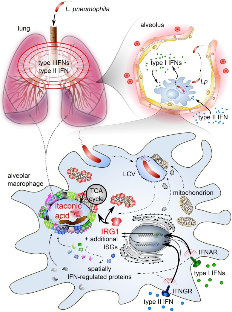
Macrophages can be niches for bacterial pathogens or antibacterial effector cells depending on the pathogen and signals from the immune system. Here we show that type I and II IFNs are master regulators of gene expression during Legionella pneumophila infection, and activators of an alveolar macrophage-intrinsic immune response that restricts bacterial growth during pneumonia. Quantitative mass spectrometry revealed that both IFNs substantially modify Legionella-containing vacuoles, and comparative analyses reveal distinct subsets of transcriptionally and spatially IFN-regulated proteins. Immune-responsive gene (IRG)1 is induced by IFNs in mitochondria that closely associate with Legionella-containing vacuoles, and mediates production of itaconic acid. This metabolite is bactericidal against intravacuolar L. pneumophila as well as extracellular multidrug-resistant Gram-positive and -negative bacteria. Our study explores the overall role IFNs play in inducing substantial remodeling of bacterial vacuoles and in stimulating production of IRG1-derived itaconic acid which targets intravacuolar pathogens. IRG1 or its product itaconic acid might be therapeutically targetable to fight intracellular and drug-resistant bacteria.

Overview

The claims and statistics of this paper haven’t been checked yet.

Run the check — free, no account needed. It reads the open-access text and checks the sources it cites.

A few free checks a day · papers already verified open instantly · results are public

Integrity signals

Registry

Crossref · 10.1371/journal.ppat.1005408

Cites

71 of 71 references verified0 citation errors · 0 not checkable

Statistics

Figures

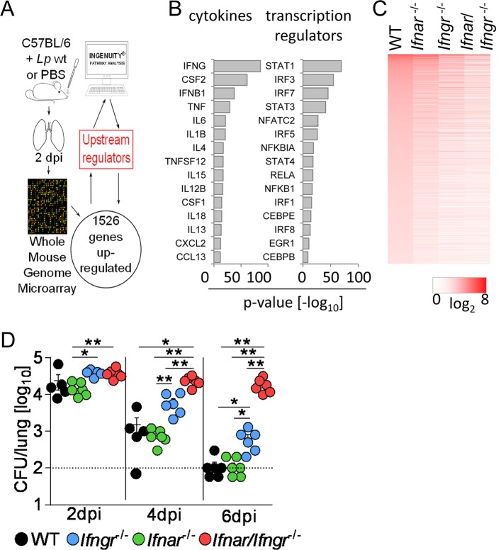
Fig 1 Type I and II IFNs are main regulators of gene expression during L. pneumophila infection. (A) Workflow for upstream regulator analysis. (B, C) WT (B) or WT, Ifnar -/-, Ifngr -/- and Ifnar/Ifngr -/- mice (C) were infected with L. pneumophila or treated with PBS. Mice were sacrificed 2 d p.i., and microarray analysis performed from lung RNA. Up-regulated genes in L. pneumophila-infected WT mice compared to controls were analyzed for their predicted upstream regulators (B). Heat map of microarray analysis of infected versus PBS-treated mice for wild-type and respective knock-out strains (C). 5 mice per group, pooled for RNA extraction and subsequent analysis. (D) Bacterial loads in the lungs of L. pneumophila infected WT, Ifnar -/-, Ifngr -/- and Ifnar/Ifngr -/- mice are depicted. Data represent mean + s.e.m. of 5–6 mice per group. Dotted line indicates lower detection limit, * p<0.05, ** p<0.01 (Kruskal-Wallis analysis of variance followed by Mann-Whitney U test with Bonferroni correction for multiple comparisons).
Fig 1 Type I and II IFNs are main regulators of gene expression during L. pneumophila infection. (A) Workflow for upstream regulator analysis. (B, C) WT (B) or WT, Ifnar -/-, Ifngr -/- and Ifnar/Ifngr -/- mice (C) were infected with L. pneumophila or treated with PBS. Mice were sacrificed 2 d p.i., and microarray analysis performed from lung RNA. Up-regulated genes in L. pneumophila-infected WT mice compared to controls were analyzed for their predicted upstream regulators (B). Heat map of microarray analysis of infected versus PBS-treated mice for wild-type and respective knock-out strains (C). 5 mice per group, pooled for RNA extraction and subsequent analysis. (D) Bacterial loads in the lungs of L. pneumophila infected WT, Ifnar -/-, Ifngr -/- and Ifnar/Ifngr -/- mice are depicted. Data represent mean + s.e.m. of 5–6 mice per group. Dotted line indicates lower detection limit, * p<0.05, ** p<0.01 (Kruskal-Wallis analysis of variance followed by Mann-Whitney U test with Bonferroni correction for multiple comparisons).CC BY · PubMed Central
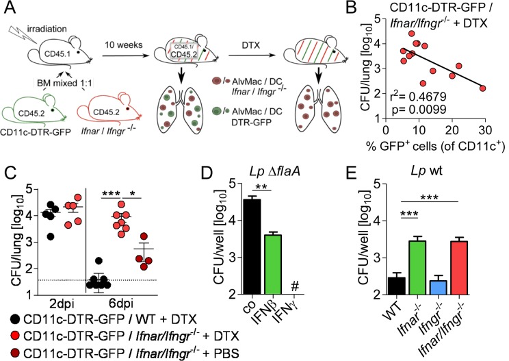
Fig 2 Type I and II IFNs restrict L. pneumophila infection through an alveolar macrophage-intrinsic mechanism. (A) Overview of generation of CD11c-DTR-GFP / Ifnar/Ifngr -/- mixed bone marrow chimeric mice followed by DTX mediated depletion of CD11c-DTR-GFP+ cells. (B) Frequency of remaining CD11c+ GFP+ wild-type cells was correlated to bacterial load in the lungs of CD11c-DTR-GFP / Ifnar/Ifngr -/- + DTX chimeras including all DTX-treated mice (13 mice) at 6 d p.i.. (C) Only mice with <10% GFP+ (of all CD11c+ cells) were considered for analysis and bacterial loads in the lungs of L. pneumophila infected mixed bone marrow-chimeric mice at indicated time points are shown. (D, E) Alveolar macrophages of WT, Ifnar -/-, Ifngr -/- and Ifnar/Ifngr -/- mice were left untreated (E) or were treated with 50 U/ml IFNβ or IFNγ (D) 16–18 h prior to and during infection with L. pneumophila ΔflaA (D) or wt (E). Bacterial growth was determined by CFU counting after 72 h. Data represent 3 independent experiments done in triplicates. * p<0.05, ** p<0.01, *** p<0.001 (Pearson correlation (B), Kruskal-Wallis analysis of variance, Dunn’s multiple comparison (C) or Mann-Whitney U test (D, E)). # No bacteria were detected.
Fig 2 Type I and II IFNs restrict L. pneumophila infection through an alveolar macrophage-intrinsic mechanism. (A) Overview of generation of CD11c-DTR-GFP / Ifnar/Ifngr -/- mixed bone marrow chimeric mice followed by DTX mediated depletion of CD11c-DTR-GFP+ cells. (B) Frequency of remaining CD11c+ GFP+ wild-type cells was correlated to bacterial load in the lungs of CD11c-DTR-GFP / Ifnar/Ifngr -/- + DTX chimeras including all DTX-treated mice (13 mice) at 6 d p.i.. (C) Only mice with <10% GFP+ (of all CD11c+ cells) were considered for analysis and bacterial loads in the lungs of L. pneumophila infected mixed bone marrow-chimeric mice at indicated time points are shown. (D, E) Alveolar macrophages of WT, Ifnar -/-, Ifngr -/- and Ifnar/Ifngr -/- mice were left untreated (E) or were treated with 50 U/ml IFNβ or IFNγ (D) 16–18 h prior to and during infection with L. pneumophila ΔflaA (D) or wt (E). Bacterial growth was determined by CFU counting after 72 h. Data represent 3 independent experiments done in triplicates. * p<0.05, ** p<0.01, *** p<0.001 (Pearson correlation (B), Kruskal-Wallis analysis of variance, Dunn’s multiple comparison (C) or Mann-Whitney U test (D, E)). # No bacteria were detected.CC BY · PubMed Central
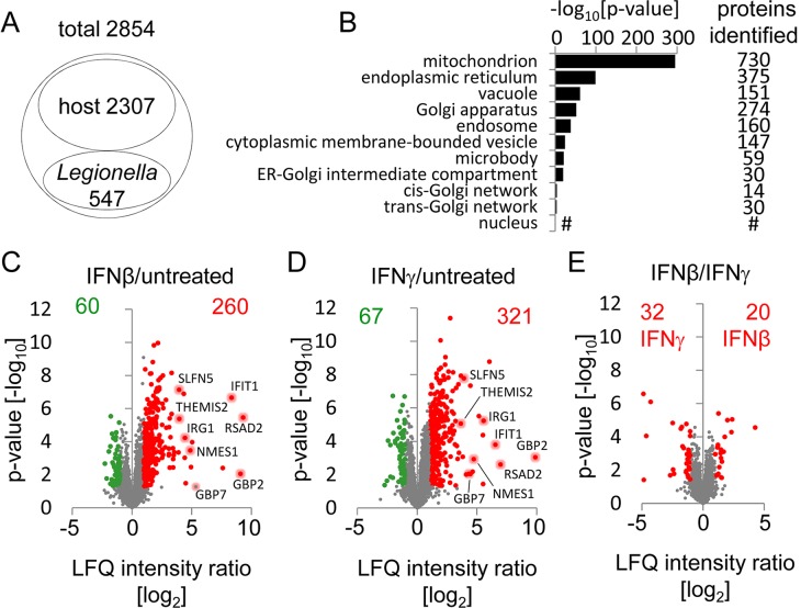
Fig 3 Type I and II IFNs alter the protein composition of LCVs. (A) Proteomic analysis of LCVs isolated 2 h p.i. from untreated BMMs infected with L. pneumophila ΔflaA detected 2854 proteins in all six replicates of which 2307 were identified as host- and 547 as L. pneumophila-derived. (B) GO enrichment analysis for overrepresented cellular components of the host proteins was done and overrepresented child terms of GO:0043231 ‘intracellular membrane-bounded organelle’ were extracted. Depicted are p-values for the indicated GO terms as well as the number of identified proteins annotated with each term; # no p-value for GO term ‘nucleus’ was computed. (C-E) Quantitative proteomic analysis of LCVs isolated 2 h p.i. with L. pneumophila ΔflaA from BMMs left untreated or treated with 50 U/ml IFNβ or IFNγ 16–18 h prior to and during infection. Volcano plots show unchanged proteins (grey) and proteins with a significant higher (red) or lower (green) abundance at LCVs from IFNβ- (C) or IFNγ- (D) treated BMMs compared to untreated cells, and direct comparison of IFNβ- versus IFNγ-treated samples (E). See also S6 Fig for detailed list of top 20 proteins for each condition. Proteomic analysis was done from 6 (untreated), 5 (IFNγ) and 4 (IFNβ) individual LCV isolations.
Fig 3 Type I and II IFNs alter the protein composition of LCVs. (A) Proteomic analysis of LCVs isolated 2 h p.i. from untreated BMMs infected with L. pneumophila ΔflaA detected 2854 proteins in all six replicates of which 2307 were identified as host- and 547 as L. pneumophila-derived. (B) GO enrichment analysis for overrepresented cellular components of the host proteins was done and overrepresented child terms of GO:0043231 ‘intracellular membrane-bounded organelle’ were extracted. Depicted are p-values for the indicated GO terms as well as the number of identified proteins annotated with each term; # no p-value for GO term ‘nucleus’ was computed. (C-E) Quantitative proteomic analysis of LCVs isolated 2 h p.i. with L. pneumophila ΔflaA from BMMs left untreated or treated with 50 U/ml IFNβ or IFNγ 16–18 h prior to and during infection. Volcano plots show unchanged proteins (grey) and proteins with a significant higher (red) or lower (green) abundance at LCVs from IFNβ- (C) or IFNγ- (D) treated BMMs compared to untreated cells, and direct comparison of IFNβ- versus IFNγ-treated samples (E). See also S6 Fig for detailed list of top 20 proteins for each condition. Proteomic analysis was done from 6 (untreated), 5 (IFNγ) and 4 (IFNβ) individual LCV isolations.CC BY · PubMed Central
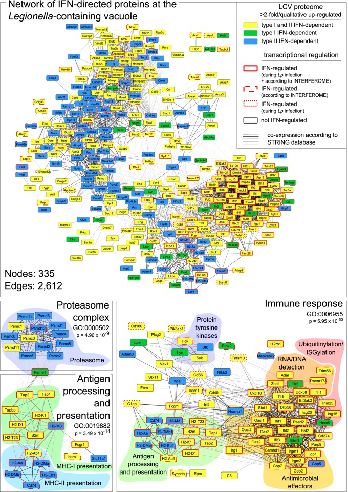
Fig 4 Integrated network analysis of IFN-regulated proteins of the LCVs. Proteins with higher abundance at LCVs from IFNβ- and/or IFNγ-treated compared to untreated cells were analyzed with the STRING database. The proteome data were further compared with the whole genome microarray data (S1 Dataset; genes > 2-fold higher expressed and p < 0.05 in infected WT vs. Ifnar/Ifngr -/- mice) and the INTERFEROME database to indicate molecules which are also transcriptionally regulated by IFNs. A GO enrichment analysis was performed for extracting significant subnetworks of a complex network composed of 335 nodes and 2,612 edges. Shown are subnetworks positively affected by IFNβ and/or IFNγ activation such as ‘immune response’, ‘antigen processing and presentation’ and the ‘proteasome complex’.
Fig 4 Integrated network analysis of IFN-regulated proteins of the LCVs. Proteins with higher abundance at LCVs from IFNβ- and/or IFNγ-treated compared to untreated cells were analyzed with the STRING database. The proteome data were further compared with the whole genome microarray data (S1 Dataset; genes > 2-fold higher expressed and p < 0.05 in infected WT vs. Ifnar/Ifngr -/- mice) and the INTERFEROME database to indicate molecules which are also transcriptionally regulated by IFNs. A GO enrichment analysis was performed for extracting significant subnetworks of a complex network composed of 335 nodes and 2,612 edges. Shown are subnetworks positively affected by IFNβ and/or IFNγ activation such as ‘immune response’, ‘antigen processing and presentation’ and the ‘proteasome complex’.CC BY · PubMed Central
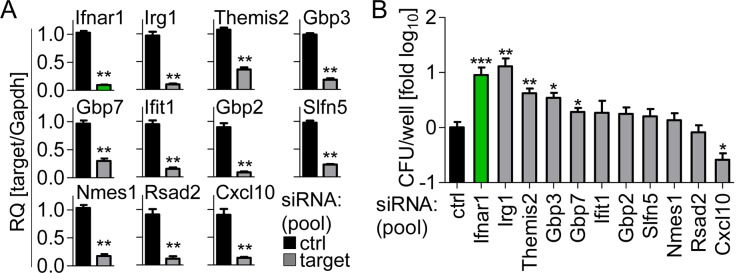
Fig 5 RNAi screen identifies IRG1 as crucial L. pneumophila restricting factor. (A, B) BMMs were transfected with control siRNA or a pool of two siRNAs per gene 24 h prior to infection and infected with L. pneumophila. Expression of targeted genes was assessed 24 h p.i. by qRT-PCR (A), and CFUs were counted 72 h p.i. (B). Data are mean + s.e.m. of 2 (A) or 4 (B) independent experiments done in triplicates. * p<0.05, ** p<0.01, *** p<0.001, no indication if not significant (Mann-Whitney U test).
Fig 5 RNAi screen identifies IRG1 as crucial L. pneumophila restricting factor. (A, B) BMMs were transfected with control siRNA or a pool of two siRNAs per gene 24 h prior to infection and infected with L. pneumophila. Expression of targeted genes was assessed 24 h p.i. by qRT-PCR (A), and CFUs were counted 72 h p.i. (B). Data are mean + s.e.m. of 2 (A) or 4 (B) independent experiments done in triplicates. * p<0.05, ** p<0.01, *** p<0.001, no indication if not significant (Mann-Whitney U test).CC BY · PubMed Central
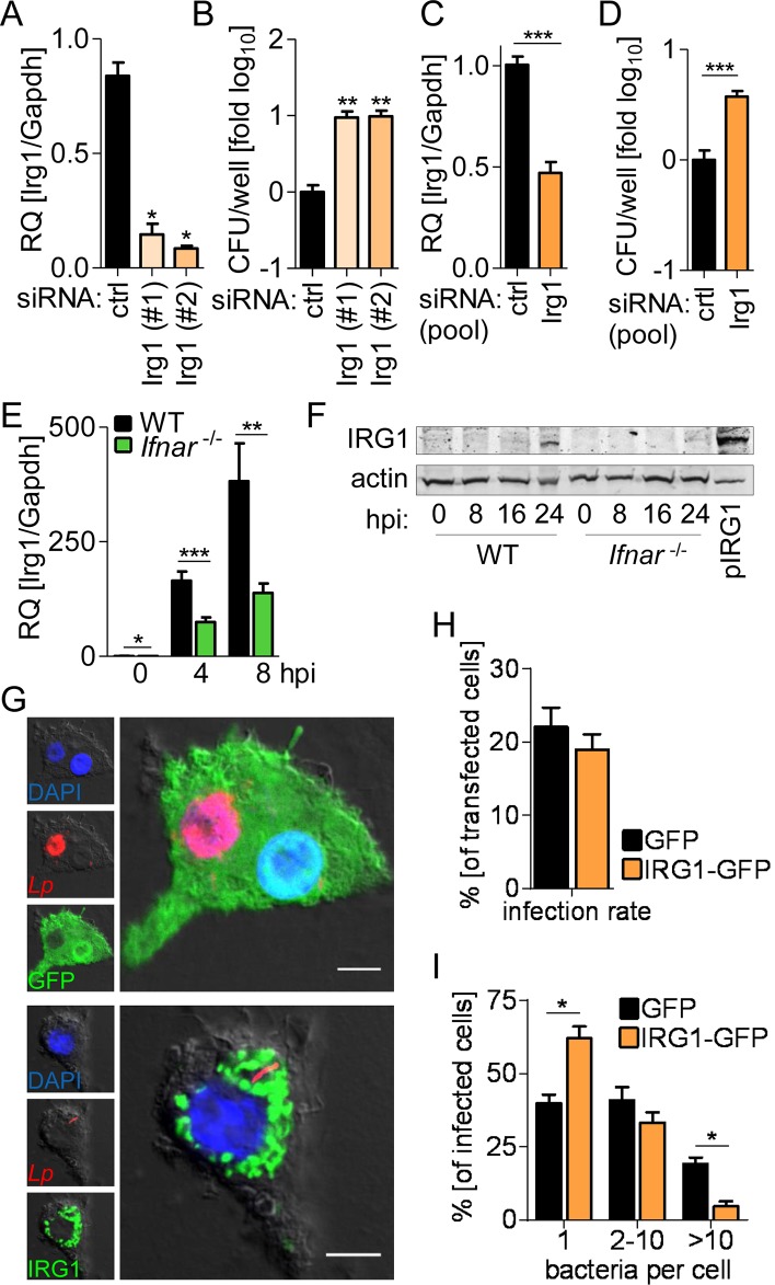
Fig 6 IRG1 is regulated by IFNs and restricts L. pneumophila within their vacuoles. (A-D) BMMs (A, B) or alveolar macrophages (C, D) were transfected with control or two IRG1 siRNAs separately (A, B) or pooled (C, D), infected with L. pneumophila, Irg1 expression was assessed 24 h p.i. (A, C), and CFUs were counted 72 h p.i. (B, D). (E, F) Irg1 gene and IRG1 protein expression in WT and Ifnar -/- BMMs upon infection with L. pneumophila was determined at indicated time points by qRT-PCR (E) and western blot (F). Cells overexpressing IRG1-Myc-DDK (pIRG1) were loaded as a positive control (F). (G-I) Ifnar -/- BMMs overexpressing IRG1-GFP or GFP only were infected with DsRed-expressing L. pneumophila (red). Cells were fixed 24 h p.i., representative images taken (G), and overall infection rate (H) as well as number of intracellular bacteria per cell determined by manual counting (I). (G) Scale bars indicate 5 μm. Data are mean + s.e.m. of 2 (A, B) or 4 (E) independent experiments done in triplicates or 3 independent experiments done in quadruplicates (C, D). * p<0.05, ** p<0.01, *** p<0.001, no indication if not significant (Mann-Whitney U test). (F) Representative blot of 3 independent experiments. (H, I) > 100 GFP- / IRG1-GFP-expressing cells were counted manually for intracellular bacterial numbers, data represent mean + s.e.m. of 2 independent experiments done in duplicates * p<0.05, no indication if not significant (Mann-Whitney U test).
Fig 6 IRG1 is regulated by IFNs and restricts L. pneumophila within their vacuoles. (A-D) BMMs (A, B) or alveolar macrophages (C, D) were transfected with control or two IRG1 siRNAs separately (A, B) or pooled (C, D), infected with L. pneumophila, Irg1 expression was assessed 24 h p.i. (A, C), and CFUs were counted 72 h p.i. (B, D). (E, F) Irg1 gene and IRG1 protein expression in WT and Ifnar -/- BMMs upon infection with L. pneumophila was determined at indicated time points by qRT-PCR (E) and western blot (F). Cells overexpressing IRG1-Myc-DDK (pIRG1) were loaded as a positive control (F). (G-I) Ifnar -/- BMMs overexpressing IRG1-GFP or GFP only were infected with DsRed-expressing L. pneumophila (red). Cells were fixed 24 h p.i., representative images taken (G), and overall infection rate (H) as well as number of intracellular bacteria per cell determined by manual counting (I). (G) Scale bars indicate 5 μm. Data are mean + s.e.m. of 2 (A, B) or 4 (E) independent experiments done in triplicates or 3 independent experiments done in quadruplicates (C, D). * p<0.05, ** p<0.01, *** p<0.001, no indication if not significant (Mann-Whitney U test). (F) Representative blot of 3 independent experiments. (H, I) > 100 GFP- / IRG1-GFP-expressing cells were counted manually for intracellular bacterial numbers, data represent mean + s.e.m. of 2 independent experiments done in duplicates * p<0.05, no indication if not significant (Mann-Whitney U test).CC BY · PubMed Central
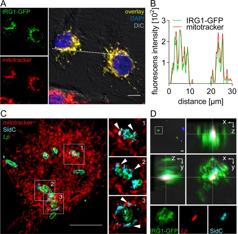
Fig 7 Mitochondrial IRG1 closely associates with LCVs. (A, B) Ifnar -/- BMMs overexpressing IRG1-GFP (green) were stained with mitotracker (red), fixed and nuclei were visualized by DAPI (blue) staining. Images represent maximum intensity projections of z-stacks of 10 individual 1.4 μm thick sections. (B) Fluorescence intensity profiles of IRG1-GFP and mitotracker along the dotted line in (A) are depicted. (C) Super-resolution fluorescent image generated by structured illumination microscopy of BMMs stained with mitotracker (red) and subsequently infected with L. pneumophila for 2 h. Bacteria and LCVs were visualized in fixed cells by L. pneumophila (green) and SidC (LCV-located L. pneumophila-protein; cyan) staining, respectively. Large overview image represents maximum intensity projection of a z-stack of 30 individual 0.126 μm thick sections. Enlarged sections in the left panel depict one single confocal plane for each selected LCV according to numbers in the overview image. White arrowheads point towards close associations of mitochondria with the LCV membrane. (D) Ifnar -/- BMMs overexpressing IRG1-GFP were infected with DsRed-expressing L. pneumophila wt (red). 2 h p.i. cells were homogenized and LCVs and nuclei were visualized by SidC (LCV-located L. pneumophila-protein; cyan) and DAPI (blue) staining. Framed area from the upper left overview image is shown as orthogonal view of a z-stack of 8 individual 0.68 μm thick sections and as single channels for depicted section. (A, C, D) Scale bars indicate 5 μm.
Fig 7 Mitochondrial IRG1 closely associates with LCVs. (A, B) Ifnar -/- BMMs overexpressing IRG1-GFP (green) were stained with mitotracker (red), fixed and nuclei were visualized by DAPI (blue) staining. Images represent maximum intensity projections of z-stacks of 10 individual 1.4 μm thick sections. (B) Fluorescence intensity profiles of IRG1-GFP and mitotracker along the dotted line in (A) are depicted. (C) Super-resolution fluorescent image generated by structured illumination microscopy of BMMs stained with mitotracker (red) and subsequently infected with L. pneumophila for 2 h. Bacteria and LCVs were visualized in fixed cells by L. pneumophila (green) and SidC (LCV-located L. pneumophila-protein; cyan) staining, respectively. Large overview image represents maximum intensity projection of a z-stack of 30 individual 0.126 μm thick sections. Enlarged sections in the left panel depict one single confocal plane for each selected LCV according to numbers in the overview image. White arrowheads point towards close associations of mitochondria with the LCV membrane. (D) Ifnar -/- BMMs overexpressing IRG1-GFP were infected with DsRed-expressing L. pneumophila wt (red). 2 h p.i. cells were homogenized and LCVs and nuclei were visualized by SidC (LCV-located L. pneumophila-protein; cyan) and DAPI (blue) staining. Framed area from the upper left overview image is shown as orthogonal view of a z-stack of 8 individual 0.68 μm thick sections and as single channels for depicted section. (A, C, D) Scale bars indicate 5 μm.CC BY · PubMed Central
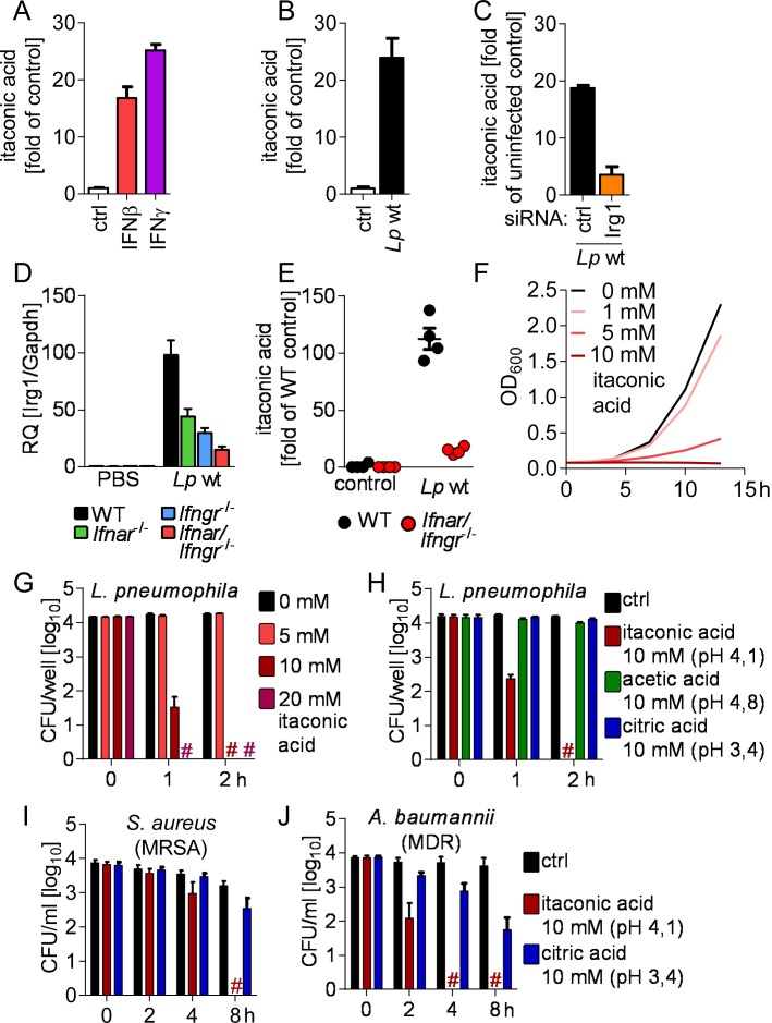
Fig 8 IRG1 mediates production of itaconic acid, which is bactericidal against L. pneumophila as well as multidrug-resistant Gram-positive and -negative bacteria. (A-C) Intracellular levels of itaconic acid were measured by GC-MS in BMMs after stimulation with 50 U/ml IFNβ or IFNγ for 16–18 h (A), infection with L. pneumophila for 24 h (B), and in BMMs transfected with siRNAs 24 h prior infection and infected with L. pneumophila for 24 h (C). (D, E) Irg1 expression (D) and itaconic acid levels (E) in lungs of L. pneumophila-infected or control mice measured by qRT-PCR or GC-MS, respectively, 2 d p.i.. (F) Itaconic acid was added to L. pneumophila in liquid culture and bacterial growth was assessed. (G-J) L. pneumophila, S. aureus (MRSA) and A. baumannii (MDR) were incubated in PBS containing itaconic acid or related acids, and numbers of viable bacteria were determined by CFU counting. (A-C) Data represent mean + s.e.m. of 3 (A) or 4 (B, C) experiments, done in sextuplicates pooled for GC-MS analysis. (D, E) Data represent mean + s.e.m. of 4 (E) or 5 (D) mice per group. (F) Representative graph of 3 independent experiments. (E, F) Data represent mean + s.e.m. of 2 (G) or 3 (H-J) experiments done in triplicates. # No viable bacteria were detected.
Fig 8 IRG1 mediates production of itaconic acid, which is bactericidal against L. pneumophila as well as multidrug-resistant Gram-positive and -negative bacteria. (A-C) Intracellular levels of itaconic acid were measured by GC-MS in BMMs after stimulation with 50 U/ml IFNβ or IFNγ for 16–18 h (A), infection with L. pneumophila for 24 h (B), and in BMMs transfected with siRNAs 24 h prior infection and infected with L. pneumophila for 24 h (C). (D, E) Irg1 expression (D) and itaconic acid levels (E) in lungs of L. pneumophila-infected or control mice measured by qRT-PCR or GC-MS, respectively, 2 d p.i.. (F) Itaconic acid was added to L. pneumophila in liquid culture and bacterial growth was assessed. (G-J) L. pneumophila, S. aureus (MRSA) and A. baumannii (MDR) were incubated in PBS containing itaconic acid or related acids, and numbers of viable bacteria were determined by CFU counting. (A-C) Data represent mean + s.e.m. of 3 (A) or 4 (B, C) experiments, done in sextuplicates pooled for GC-MS analysis. (D, E) Data represent mean + s.e.m. of 4 (E) or 5 (D) mice per group. (F) Representative graph of 3 independent experiments. (E, F) Data represent mean + s.e.m. of 2 (G) or 3 (H-J) experiments done in triplicates. # No viable bacteria were detected.CC BY · PubMed Central
Fig 9 Overview of the type I and II IFN-driven, alveolar macrophage-intrinsic defense pathway that restricts L. pneumophila during lung infection (as discussed in the text).
Fig 9 Overview of the type I and II IFN-driven, alveolar macrophage-intrinsic defense pathway that restricts L. pneumophila during lung infection (as discussed in the text).CC BY · PubMed Central

Verification

Claims the paper makes

The claims and statistics of this paper haven’t been checked yet.

Run the check — free, no account needed. It reads the open-access text and checks the sources it cites.

A few free checks a day · papers already verified open instantly · results are public

Claims other papers make about this one

Each is checked against this paper’s own text: does it say what the citing paper says it does?

Self-check

Percentages, sums and reported differences re-computed from the paper’s own numbers.

The self-check needs the paper’s full text.

Citations

Cites

Show all 71 references

Research works printed but not linked: 1

  1. C Hoffmann, 2013, Functional analysis of novel Rab GTPases identified in the proteome of purified Legionella-containing vacuoles from macrophages, Cell Microbiol, 16, 1034

Cited by

Sorted by citations

See all 261

Metadata

Authors
Naujoks, J., Tabeling, C., Dill, B. D., Hoffmann, C., Brown, A. S., Kunze, M., Kempa, S., Peter, A., Mollenkopf, H.-J., Dorhoi, A., Kershaw, O., Gruber, A. D., Sander, L. E., Witzenrath, M., Herold, S., Nerlich, A., Hocke, A. C., van Driel, I., Suttorp, N., Bedoui, S., Hilbi, H., Trost, M., & Opitz, B.
Affiliations
  • Charité – Universitätsmedizin Berlin (DE)
  • University of Dundee (GB)
  • Max-von-Pettenkofer-Institut für Hygiene und Medizinische Mikrobiologie (DE)
  • The University of Melbourne (AU)
  • Max Delbrück Center (DE)
  • Max Planck Institute for Infection Biology (DE)
  • Freie Universität Berlin (DE)
  • Justus-Liebig-Universität Gießen (DE)
  • University of Zurich (CH)
Corresponding author
Opitz, Bastian
Venue
PLOS Pathogens
Published
2016-02-01
Volume
12
Issue
2
Pages
e1005408
ISSN
  • 1553-7366 (ISSN-L)
  • 1553-7374
OpenAlex
W2270624586
Cited by
261
References
71
Citations by year
2026: 17 · 2025: 30 · 2024: 24 · 2023: 19 · 2022: 34 · 2021: 33 · 2020: 25 · 2019: 27 · 2018: 22 · 2017: 20 · 2016: 10
FWCI
25.45
Type
journal article
Language
en
Open access
Locations
  • PLoS Pathogens (publishedVersion, cc-by)
  • PubMed Central (publishedVersion, cc-by)
  • PubMed (publishedVersion)
  • Discovery Research Portal (University of Dundee) (publishedVersion, cc-by)
  • MDC Repository (Max-Delbrueck-Center for Molecular Medicine) (acceptedVersion)
  • Zurich Open Repository and Archive (University of Zurich) (acceptedVersion, cc-by)
  • MPG.PuRe (Max Planck Society) (submittedVersion, other-oa)
  • DOAJ (DOAJ: Directory of Open Access Journals) (submittedVersion, cc-by-sa)
  • Open access LMU (Ludwid Maxmilian's Universitat Munchen) (submittedVersion)
  • Max Planck Digital Library (submittedVersion)
  • Figshare (submittedVersion, cc-by)
  • Minerva Access (University of Melbourne) (submittedVersion, cc-by)
  • Refubium (Universitätsbibliothek der Freien Universität Berlin) (submittedVersion, cc-by)
  • Zurich Open Repository and Archive (University of Zurich) (submittedVersion, cc-by)
  • Universitätsbibliothek der FU Berlin Hochschulschriftenstelle u. Dokumentenserver (cc-by)
  • Zurich Open Repository and Archive (University of Zurich)
Funders
  • International Max Planck Research School for Environmental, Cellular and Molecular Microbiology
  • Deutsche Forschungsgemeinschaft (SPP1580/OP86/10-1, SPP 1580, SFB‐TR84)
  • International Max Planck Research School for Advanced Methods in Process and Systems Engineering
Topics
  • Life Sciences › Biochemistry, Genetics and Molecular Biology › Endocrinology › Legionella and Acanthamoeba research
  • Life Sciences › Immunology and Microbiology › Immunology › interferon and immune responses
  • Life Sciences › Immunology and Microbiology › Immunology › Immune responses and vaccinations
Keywords
type I interferon, interferon-gamma, Legionella pneumophila, Legionnaires' disease, Legionella-containing vacuole, IRG1, itaconic acid, alveolar macrophages, intracellular bacterial infection, antimicrobial metabolites, multidrug-resistant bacteria, host-pathogen interaction, innate immunity
MeSH terms
Animals, Disease Models, Animal, Female, Gene Expression Regulation, Hydro-Lyases, Immunity, Innate, Interferons, Legionnaires' Disease, Mice, Inbred C57BL, Mice, Transgenic, Mitochondria, Succinates, Vacuoles, Macrophages, Alveolar, Legionella pneumophila, Reactive Oxygen Species, Models, Immunological, Proteome, Mice, Gene Ontology

About these checks

Registry, retraction, Cites and statistics results are computed, not judged, with one exception: a printed reference that no index lists is read once by a model before it counts as a citation error. Claims are extracted by a model and kept only when their quote appears verbatim in the text; each cited source is then checked, with a model’s help, for whether it says what the claim relies on. Those verdicts can be wrong — read the claim’s quote and the cited source before relying on one.

Statistics and arithmetic are recomputed, not judged.

Confidence bands are shown only for fields calibrated on our annotated test set. None is calibrated yet, so no band is shown.

What counts as a claim ›How we measure confidence ›How we check this ›

Are you an author, or do you see a problem with this page? Request a review