

The DOI matches its Crossref record; 52 of the 52 references it cites were found in a registry or an index.
The astrovirus N-terminal nonstructural protein anchors replication complexes to the perinuclear ER membranes
Open accessPublished version of a preprint
Abstract
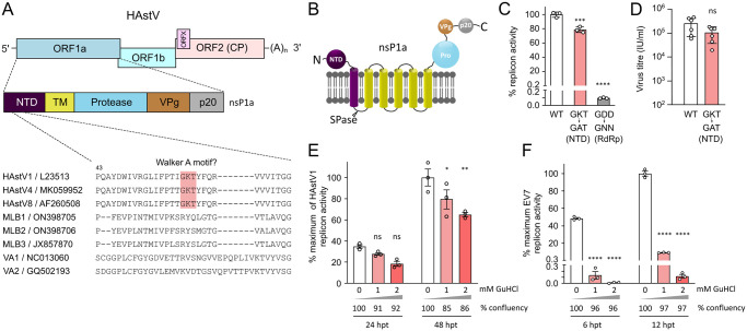
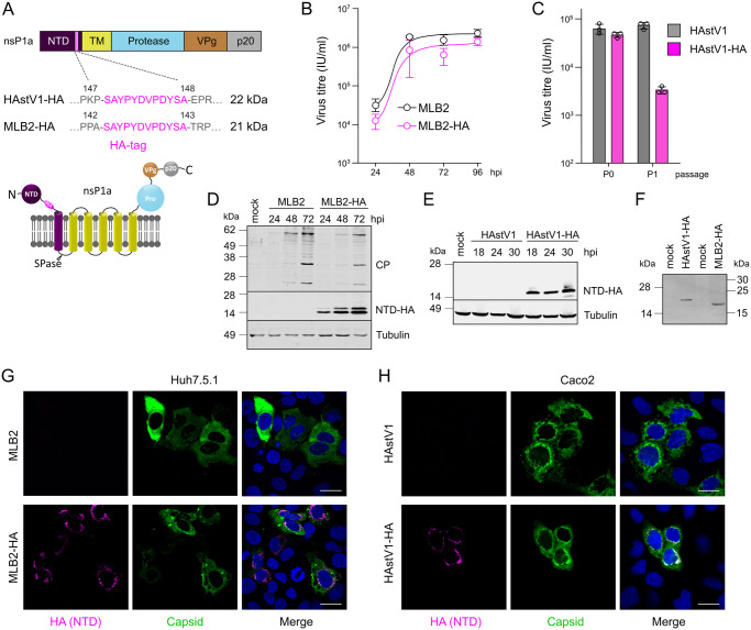
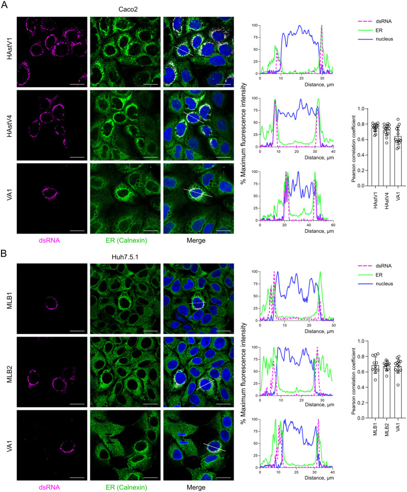
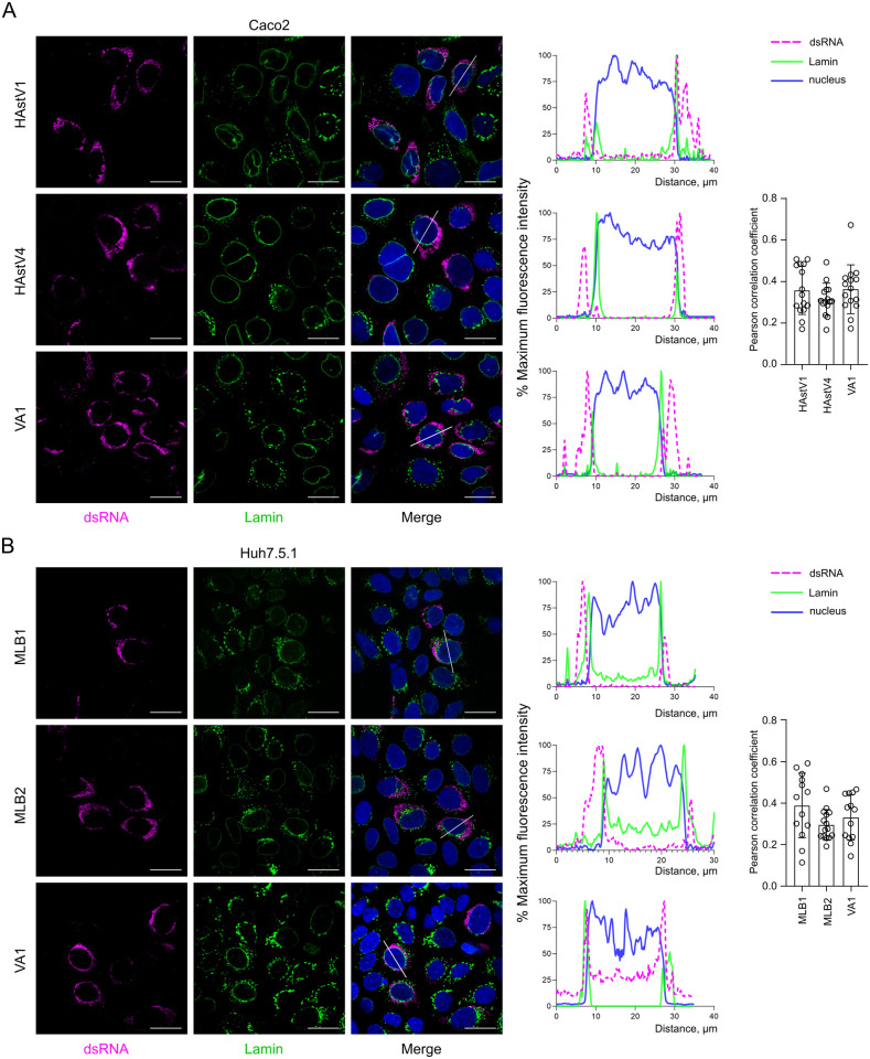
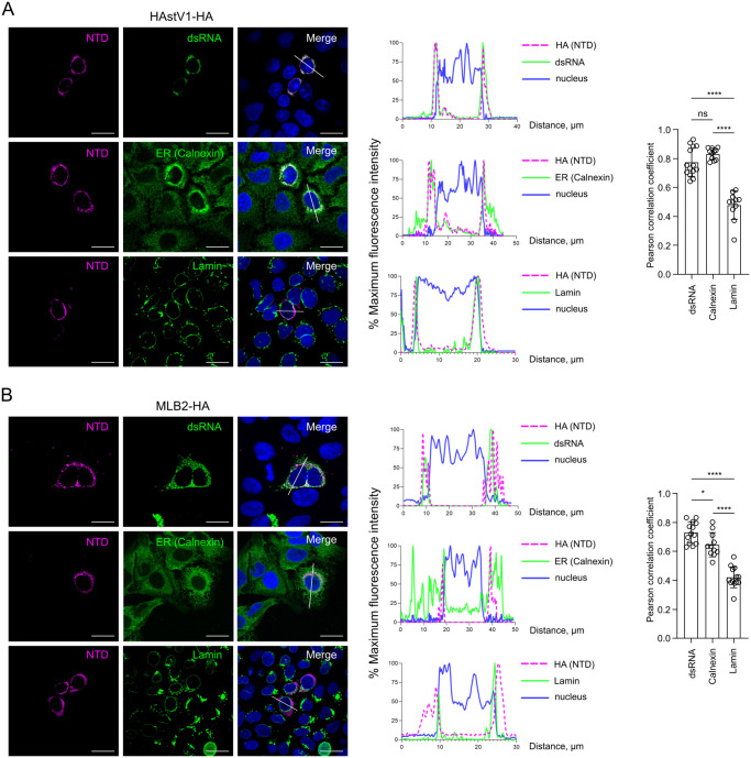
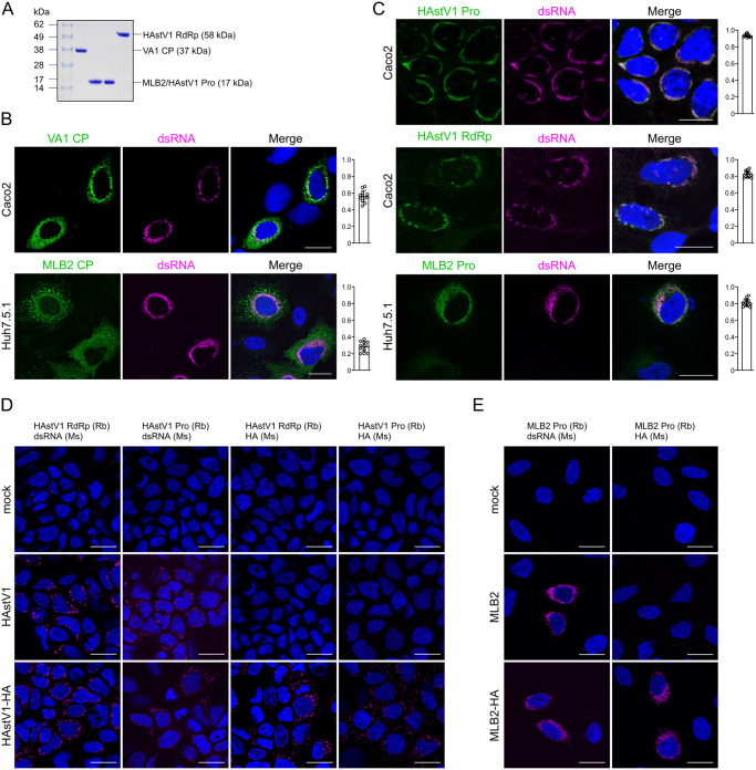
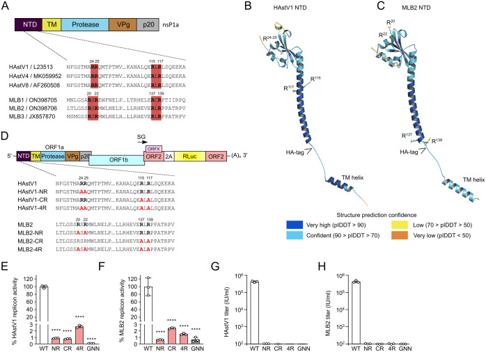
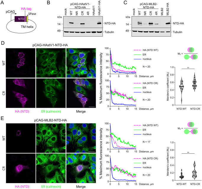
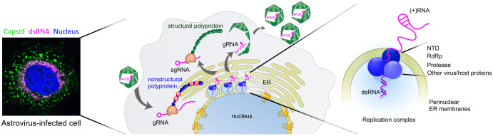
An essential aspect of positive-sense RNA virus replication is anchoring the replication complex (RC) to cellular membranes. Positive-sense RNA viruses employ diverse strategies, including co-translational membrane targeting through signal peptides and co-opting cellular membrane trafficking components. Often, N-terminal nonstructural proteins play a crucial role in linking the RC to membranes, facilitating the early association of the replication machinery. Astroviruses utilize a polyprotein strategy to synthesize nonstructural proteins, relying on subsequent processing to form replication-competent complexes. This study provides evidence for the perinuclear ER membrane association of RCs in five distinct human astrovirus strains. Using tagged recombinant classical human astrovirus 1 and neurotropic MLB2 strains, we establish that the N-terminal domain guides the ER membrane association. We identified di-arginine motifs responsible for the perinuclear ER retention and formation of functional RCs through mutational analysis of the N-terminal domain in replicon and reverse genetics systems. In addition, we demonstrate the association of key components of the astrovirus replication complex: double-stranded RNA, RNA-dependent RNA polymerase, protease, and N-terminal protein. Our findings highlight the intricate virus-ER interaction mechanism employed by astroviruses, potentially leading to the development of novel antiviral intervention strategies.
Overview
The claims and statistics of this paper haven’t been checked yet.
Run the check — free, no account needed. It reads the open-access text and checks the sources it cites.
Integrity signals
Registry
Cites
Statistics
Figures
Verification
Claims the paper makes
The claims and statistics of this paper haven’t been checked yet.
Run the check — free, no account needed. It reads the open-access text and checks the sources it cites.
Self-check
Percentages, sums and reported differences re-computed from the paper’s own numbers.
The self-check needs the paper’s full text.
Citations
Cites
Highly accurate protein structure prediction with AlphaFold
Jumper, J., Evans, R., Pritzel, A., Green, T., Figurnov, M., Ronneberger, O., Tunyasuvunakool, K., Bates, R., Žídek, A., Potapenko, A., et al. (2021) · Nature · doi:10.1038/s41586-021-03819-2 · Cited by 43,764
Accurate structure prediction of biomolecular interactions with AlphaFold 3
Abramson, J., Adler, J., Dunger, J., Evans, R., Green, T., Pritzel, A., Ronneberger, O., Willmore, L., Ballard, A. J., Bambrick, J., et al. (2024) · Nature · doi:10.1038/s41586-024-07487-w · Cited by 15,861
Varadi, M., Anyango, S., Deshpande, M., Nair, S., Natassia, C., Yordanova, G., Yuan, D., Stroe, O., Wood, G., Laydon, A., et al. (2022) · Nucleic Acids Research · doi:10.1093/nar/gkab1061 · Cited by 7,850
Walker, J. E., Saraste, M., Runswick, M. J., & Gay, N. J. (1982) · The EMBO Journal · doi:10.1002/j.1460-2075.1982.tb01276.x · Cited by 5,312
ELM server: a new resource for investigating short functional sites in modular eukaryotic proteins
Puntervoll, P. (2003) · Nucleic Acids Research · doi:10.1093/nar/gkg545 · Cited by 602
Sionov, E., Lee, H., Chang, Y. C., & Kwon-Chung, K. J. (2010) · PLoS Pathogens · doi:10.1371/journal.ppat.1000848 · Cited by 479
The dependence of viral RNA replication on co-opted host factors
Nagy, P. D., & Pogany, J. (2012) · Nature Reviews Microbiology · doi:10.1038/nrmicro2692 · Cited by 449
Bosch, A., Pintó, R. M., & Guix, S. (2014) · Clinical Microbiology Reviews · doi:10.1128/cmr.00013-14 · Cited by 371
Cytoplasmic Viral Replication Complexes
den Boon, J. A., Diaz, A., & Ahlquist, P. (2010) · Cell Host & Microbe · doi:10.1016/j.chom.2010.06.010 · Cited by 341
Double-Membrane Vesicles as Platforms for Viral Replication
Wolff, G., Melia, C. E., Snijder, E. J., & Bárcena, M. (2020) · Trends in Microbiology · doi:10.1016/j.tim.2020.05.009 · Cited by 321
Schutze, M. P., Peterson, P. A., & Jackson, M. R. (1994) · The EMBO Journal · doi:10.1002/j.1460-2075.1994.tb06434.x · Cited by 313
Emara, M. M., & Brinton, M. A. (2007) · Proceedings of the National Academy of Sciences · doi:10.1073/pnas.0703348104 · Cited by 301
Jiang, B., Monroe, S. S., Koonin, E. V., Stine, S. E., & Glass, R. I. (1993) · Proceedings of the National Academy of Sciences · doi:10.1073/pnas.90.22.10539 · Cited by 282
The mechanisms of integral membrane protein biogenesis
Hegde, R. S., & Keenan, R. J. (2021) · Nature Reviews Molecular Cell Biology · doi:10.1038/s41580-021-00413-2 · Cited by 262
Michelsen, K., Yuan, H., & Schwappach, B. (2005) · The EMBO Reports · doi:10.1038/sj.embor.7400480 · Cited by 235
Viral and cellular RNA helicases as antiviral targets
Kwong, A. D., Rao, B. G., & Jeang, K. (2005) · Nature Reviews Drug Discovery · doi:10.1038/nrd1853 · Cited by 221
Epidemiology of Classic and Novel Human Astrovirus: Gastroenteritis and Beyond
Vu, D., Bosch, A., Pintó, R. M., & Guix, S. (2017) · Viruses · doi:10.3390/v9020033 · Cited by 205
Human stool contains a previously unrecognized diversity of novel astroviruses
Finkbeiner, S. R., Holtz, L. R., Jiang, Y., Rajendran, P., Franz, C., Zhao, G., Kang, G., & Wang, D. (2009) · Virology Journal · doi:10.1186/1743-422x-6-161 · Cited by 195
Monroe, S. S., Jiang, B., Stine, S. E., Koopmans, M., & Glass, R. I. (1993) · Journal of Virology · doi:10.1128/jvi.67.6.3611-3614.1993 · Cited by 190
Pfister, T. D., & Wimmer, E. (1999) · Journal of Biological Chemistry · doi:10.1074/jbc.274.11.6992 · Cited by 180
Teo, C. S. H., & Chu, J. J. H. (2013) · Journal of Virology · doi:10.1128/jvi.01249-13 · Cited by 169
Cis-acting RNA elements in human and animal plus-strand RNA viruses
Liu, Y., Wimmer, E., & Paul, A. V. (2009) · Biochimica et Biophysica Acta (BBA) - Gene Regulatory Mechanisms · doi:10.1016/j.bbagrm.2009.09.007 · Cited by 169
An upstream protein-coding region in enteroviruses modulates virus infection in gut epithelial cells
Lulla, V., Dinan, A. M., Hosmillo, M. D. T., Chaudhry, Y., Sherry, L., Irigoyen, N., Nayak, K., Stonehouse, N. J., Zilbauer, M., Goodfellow, I., et al. (2018) · Nature Microbiology · doi:10.1038/s41564-018-0297-1 · Cited by 151
Self-citationDiaz, A., Wang, X., & Ahlquist, P. (2010) · Proceedings of the National Academy of Sciences · doi:10.1073/pnas.1011105107 · Cited by 142
Johnson, C. N., Hargest, V., Cortez, V. A., Meliopoulos, V., & Schultz‐Cherry, S. (2017) · Viruses · doi:10.3390/v9010022 · Cited by 119
Opportunistic intruders: how viruses orchestrate ER functions to infect cells
Ravindran, M. S., Bagchi, P., Cunningham, C. N., & Tsai, B. (2016) · Nature Reviews Microbiology · doi:10.1038/nrmicro.2016.60 · Cited by 117
Marczinke, B., Bloys, A. J., Brown, T. D. K., Willcocks, M. M., Carter, M. J., & Brierley, I. (1994) · Journal of Virology · doi:10.1128/jvi.68.9.5588-5595.1994 · Cited by 116
The Host Protein Reticulon 3.1A Is Utilized by Flaviviruses to Facilitate Membrane Remodelling
Aktepe, T. E., Liebscher, S., Prier, J. E., Simmons, C. P., & Mackenzie, J. M. (2017) · Cell Reports · doi:10.1016/j.celrep.2017.10.055 · Cited by 112
Xia, H., Wang, P., Wang, G., Yang, J., Sun, X. G., Wu, W., Qiu, Y., Shu, T., Zhao, X., Yin, L., et al. (2015) · PLoS Pathogens · doi:10.1371/journal.ppat.1005067 · Cited by 105
Seeking Membranes: Positive-Strand RNA Virus Replication Complexes
Denison, M. R. (2008) · PLoS Biology · doi:10.1371/journal.pbio.0060270 · Cited by 100
Gu, B., Liu, C., Lin-Goerke, J., Maley, D. R., Gutshall, L. L., Feltenberger, C. A., & Vecchio, A. M. D. (2000) · Journal of Virology · doi:10.1128/jvi.74.4.1794-1800.2000 · Cited by 93
Identification of Human Astrovirus Genome-Linked Protein (VPg) Essential for Virus Infectivity
Fuentes, C., Bosch, A., Pintó, R. M., & Guix, S. (2012) · Journal of Virology · doi:10.1128/jvi.00797-12 · Cited by 74
Geigenmüller, U., Ginzton, N. H., & Matsui, S. M. (1997) · Journal of Virology · doi:10.1128/jvi.71.2.1713-1717.1997 · Cited by 71
Ranji, A., & Boris-Lawrie, K. (2010) · RNA Biology · doi:10.4161/rna.7.6.14249 · Cited by 70
A hidden gene in astroviruses encodes a viroporin
Lulla, V., & Firth, A. E. (2020) · Nature Communications · doi:10.1038/s41467-020-17906-x · Cited by 64
Self-citationXu, K., & Nagy, P. D. (2014) · Current Opinion in Virology · doi:10.1016/j.coviro.2014.09.015 · Cited by 61
Guix, S., Caballero, S., Bosch, A., & Pintó, R. M. (2004) · Journal of Virology · doi:10.1128/jvi.78.24.13627-13636.2004 · Cited by 58
RNA Virus Replication Complexes
Tao, Y. J., & Ye, Q. (2010) · PLoS Pathogens · doi:10.1371/journal.ppat.1000943 · Cited by 57
Identification of Host Cell Factors Associated with Astrovirus Replication in Caco-2 Cells
Murillo, A., Vera‐Estrella, R., Barkla, B. J., Méndez, E., & Arias, C. F. (2015) · Journal of Virology · doi:10.1128/jvi.01225-15 · Cited by 55
Positive-strand RNA virus genome replication organelles: structure, assembly, control
den Boon, J. A., Nishikiori, M., Zhan, H., & Ahlquist, P. (2024) · Trends in Genetics · doi:10.1016/j.tig.2024.04.003 · Cited by 52
Genome Prediction of Putative Genome-Linked Viral Protein (VPg) of Astroviruses
Al-Mutairy, B., Walter, J. E., Pothen, A., & Mitchell, D. K. (2005) · Virus Genes · doi:10.1007/s11262-004-2196-1 · Cited by 51
Large-scale phage-based screening reveals extensive pan-viral mimicry of host short linear motifs
Mihalič, F., Simonetti, L., Giudice, G., Sander, M. R., Lindqvist, R., Peters, M. B. A., Benz, C., Kassa, E., Badgujar, D. C., Inturi, R., et al. (2023) · Nature Communications · doi:10.1038/s41467-023-38015-5 · Cited by 51
Boulaflous, A., Saint‐Jore‐Dupas, C., Herranz, M. C., Pagny, S., Plasson, C., Garidou, F., Kiefer‐Meyer, M., Ritzenthaler, C., Faye, L. J.-Y., & Gomord, V. (2009) · BMC Plant Biology · doi:10.1186/1471-2229-9-144 · Cited by 45
Hidden Viral Sequences in Public Sequencing Data and Warning for Future Emerging Diseases
Kawasaki, J., Kojima, S., Tomonaga, K., & Horie, M. (2021) · mBio · doi:10.1128/mbio.01638-21 · Cited by 37
Beyond the Gastrointestinal Tract: The Emerging and Diverse Tissue Tropisms of Astroviruses
Janowski, A. B. (2021) · Viruses · doi:10.3390/v13050732 · Cited by 36
Speroni, S., Rohayem, J., Nenci, S., Bonivento, D., Robel, I., Barthel, J., Luzhkov, V. B., Coutard, B., Canard, B., & Mattevi, A. (2009) · Journal of Molecular Biology · doi:10.1016/j.jmb.2009.02.044 · Cited by 34
Li, J., Fuchs, S., Zhang, J., Wellford, S. A., Schuldiner, M., & Wang, X. (2016) · Journal of Cell Science · doi:10.1242/jcs.190082 · Cited by 33
Hachmann, J. P., & Amshey, J. W. (2005) · Analytical Biochemistry · doi:10.1016/j.ab.2005.04.015 · Cited by 30
Zeyen, L., Döring, T., Stieler, J., & Prange, R. (2020) · Cellular Microbiology · doi:10.1111/cmi.13181 · Cited by 28
ER functions are exploited by viruses to support distinct stages of their life cycle
Chen, Y.-J., Bagchi, P., & Tsai, B. (2020) · Biochemical Society Transactions · doi:10.1042/bst20200395 · Cited by 22
Bub, T., Hargest, V., Tan, S., Smith, M. A., Vazquez-Pagan, A. G., Flerlage, T., Brigleb, P. H., Meliopoulos, V., Lindenbach, B. D., Ramanathan, H. N., et al. (2023) · Journal of Virology · doi:10.1128/jvi.01025-23 · Cited by 18
Attenuation hotspots in neurotropic human astroviruses
Ali, H., Lulla, A., Nicholson, A. S., Hankinson, J., Wignall-Fleming, E. B., O’Connor, R. L., Vu, D., Graham, S. C., Deane, J. E., Guix, S., et al. (2023) · PLoS Biology · doi:10.1371/journal.pbio.3001815 · Cited by 12
Self-citation
Metadata
- Authors
- Ali, H., Noyvert, D., Hankinson, J., Lindsey, G., Lulla, A., & Lulla, V.
- Affiliations
- University of Cambridge (GB)
- Corresponding author
- Lulla, Valeria
- Venue
- PLOS Pathogens
- Publisher
- Public Library of Science (PLoS)
- Published
- 2024-07-15
- Volume
- 20
- Issue
- 7
- Pages
- e1011959
- ISSN
- 1553-7366 (ISSN-L)
- 1553-7374
- PMC
- PMC11271882
- OpenAlex
- W4400647539
- Cited by
- 11
- References
- 52
- Citations by year
- 2026: 4 · 2025: 7
- FWCI
- 3.80
- Type
- journal article
- Language
- en
- Update notices
- new_version (2024-07-25)
- Open access
- gold
- doi.org
- Licence
- Locations
- PLoS Pathogens (publishedVersion, cc-by)
- bioRxiv (Cold Spring Harbor Laboratory) (acceptedVersion, cc-by)
- PubMed Central (publishedVersion, cc-by)
- PubMed (publishedVersion)
- UCL Discovery (University College London) (submittedVersion, cc-by)
- Apollo (University of Cambridge) (submittedVersion, cc-by)
- DOAJ (DOAJ: Directory of Open Access Journals) (submittedVersion)
- Funders
- Wellcome Trust (220620/Z/20/Z)
- Isaac Newton Trust
- Medical Research Foundation (MR/T000376/1)
- Studentship from Elisabeth Mann Fund
- Topics
- Health Sciences › Medicine › Infectious Diseases › Viral gastroenteritis research and epidemiology
- Life Sciences › Agricultural and Biological Sciences › Animal Science and Zoology › Animal Virus Infections Studies
- Health Sciences › Medicine › Epidemiology › Virology and Viral Diseases
- Keywords
- astrovirus, viral replication complex, ER retention signal, polyprotein processing, RNA-dependent RNA polymerase, double-stranded RNA, antiviral drug targets
- MeSH terms
- Mamastrovirus, Endoplasmic Reticulum, Humans, Intracellular Membranes, Virus Replication, Viral Nonstructural Proteins, Astroviridae Infections
About these checks
Registry, retraction, Cites and statistics results are computed, not judged, with one exception: a printed reference that no index lists is read once by a model before it counts as a citation error. Claims are extracted by a model and kept only when their quote appears verbatim in the text; each cited source is then checked, with a model’s help, for whether it says what the claim relies on. Those verdicts can be wrong — read the claim’s quote and the cited source before relying on one.
Statistics and arithmetic are recomputed, not judged.
Confidence bands are shown only for fields calibrated on our annotated test set. None is calibrated yet, so no band is shown.
What counts as a claim ›How we measure confidence ›How we check this ›
Are you an author, or do you see a problem with this page? Request a review
Data sources
Bibliographic data: Semantic Scholar, OpenAlex, Crossref.
Contains information from the Semantic Scholar Academic Graph Datasets, made available under the ODC Attribution License (ODC-BY 1.0).
Semantic Scholar’s data is described in: Kinney, R., et al. (2023). The Semantic Scholar Open Data Platform. arXiv:2301.10140.
