Student or educator? Sign up with your school email and get 6 months of Pro free.
Cited by 0interferon and immune responsesVirology and Viral DiseasesImmune Response and Inflammation

The DOI matches its Crossref record; 66 of the 66 references it cites were found in a registry or an index.

Viral piracy of host RNA phosphatase DUSP11 by avipoxviruses

Open access

Published version of a preprint

Abstract

Proper recognition of viral pathogens is an essential part of the innate immune response. A common viral replicative intermediate and chemical signal that cells use to identify pathogens is the presence of a triphosphorylated 5’ end (5’ppp) RNA, which activates the cytosolic RNA sensor RIG-I and initiates downstream antiviral signaling. While 5’pppRNA generated by viral RNA-dependent RNA polymerases (RdRps) can be a potent activator of the immune response, endogenous RNA polymerase III (RNAPIII) transcripts can retain the 5’ppp generated during transcription and induce a RIG-I-mediated immune response. We have previously shown that host RNA triphosphatase dual-specificity phosphatase 11 (DUSP11) can act on both host and viral RNAs, altering their levels and reducing their ability to induce RIG-I activation. Our previous work explored how experimentally altered DUSP11 activity can impact immune activation, prompting further exploration into natural contexts of altered DUSP11 activity. Here, we have identified viral DUSP11 homologs (vDUSP11s) present in some avipoxviruses. Consistent with the known functions of host DUSP11, we have shown that expression of vDUSP11s: 1) reduces levels of endogenous RNAPIII transcripts, 2) reduces a cell’s sensitivity to 5’pppRNA-mediated immune activation, and 3) restores virus infection defects seen in the absence of DUSP11. Our results identify a context where DUSP11 activity has been co-opted by viruses to alter RNA metabolism and influence the outcome of infection.

Overview

The claims and statistics of this paper haven’t been checked yet.

Run the check — free, no account needed. It reads the open-access text and checks the sources it cites.

A few free checks a day · papers already verified open instantly · results are public

Integrity signals

Registry

Crossref · 10.1371/journal.ppat.1013101

Cites

66 of 66 references verified0 citation errors · 0 not checkable

Statistics

Figures

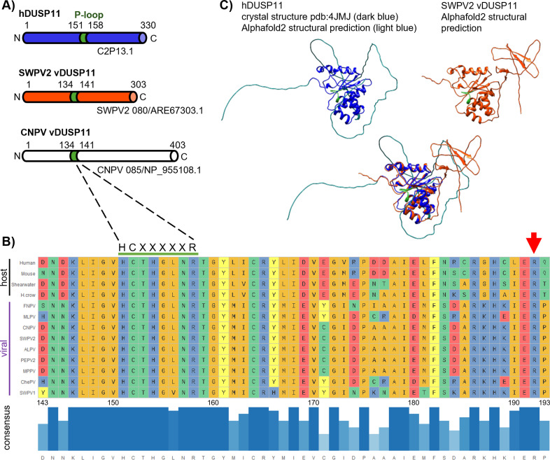
Fig 1 Avipoxviruses and related unclassified avipox-related poxviruses encode putative homologs of DUSP11. (A) Schematic of human DUSP11 (hDUSP11) and select putative viral DUSP11s with predicted domains indicated. The shared colored domain (green) indicates hDUSP11 P-loop with corresponding identical sequences present in putative viral DUSP11 homologs (vDUSP11). Putative vDUSP11 includes shearwaterpox virus 2 vDUSP11 (SWPV2 vDUSP11) and canarypox virus vDUSP11 (CNPV vDUSP11). (B) Key catalytic residues are conserved between hDUSP11 and putative avipox vDUSP11. Multiple sequence comparison (Clustal Omega) of amino acid (AA) sequences for human DUSP11, mouse DUSP11, C. borealis (Shearwater), and putative vDUSP11s from shearwaterpox virus 1 (SWPV1), finchpox virus (FNPV), mudlarkpox virus (MLPV), cheloniidpox virus (ChePV), canarypox virus (CNPV), penguinpox virus 2 (PEPV2), albatrosspox virus (ALPV), magpiepox virus (MPPV), and shearwaterpox virus 2 (SWPV2). A subset of AAs surrounding the hDUSP11 P-loop are shown. Amino acids are colored based on side-chain chemistry. Bar graph indicating relative sequence conservation with consensus amino acid sequence determined from listed sequences. Taller bar height and darker blue color correspond to relative correlation to consensus sequence. The numbering below the consensus sequence corresponds to hDUSP11 amino acid numbering. hDUSP11 residue R192 is indicated by a red arrow. (C) Partial crystal structure of hDUSP11 (PDB:4JMJ, dark blue) with unsolved regions predicted using Alphafold2 (light blue) and Alphafold2 structural prediction shearwaterpox virus 2 vDUSP11 (orange) with structural overlay. Key catalytic residues (P-loop) present in hDUSP11 are indicated in green, and corresponding residues are also indicated on the putative SWPV2 vDUSP11 structural prediction.
Fig 1 Avipoxviruses and related unclassified avipox-related poxviruses encode putative homologs of DUSP11. (A) Schematic of human DUSP11 (hDUSP11) and select putative viral DUSP11s with predicted domains indicated. The shared colored domain (green) indicates hDUSP11 P-loop with corresponding identical sequences present in putative viral DUSP11 homologs (vDUSP11). Putative vDUSP11 includes shearwaterpox virus 2 vDUSP11 (SWPV2 vDUSP11) and canarypox virus vDUSP11 (CNPV vDUSP11). (B) Key catalytic residues are conserved between hDUSP11 and putative avipox vDUSP11. Multiple sequence comparison (Clustal Omega) of amino acid (AA) sequences for human DUSP11, mouse DUSP11, C. borealis (Shearwater), and putative vDUSP11s from shearwaterpox virus 1 (SWPV1), finchpox virus (FNPV), mudlarkpox virus (MLPV), cheloniidpox virus (ChePV), canarypox virus (CNPV), penguinpox virus 2 (PEPV2), albatrosspox virus (ALPV), magpiepox virus (MPPV), and shearwaterpox virus 2 (SWPV2). A subset of AAs surrounding the hDUSP11 P-loop are shown. Amino acids are colored based on side-chain chemistry. Bar graph indicating relative sequence conservation with consensus amino acid sequence determined from listed sequences. Taller bar height and darker blue color correspond to relative correlation to consensus sequence. The numbering below the consensus sequence corresponds to hDUSP11 amino acid numbering. hDUSP11 residue R192 is indicated by a red arrow. (C) Partial crystal structure of hDUSP11 (PDB:4JMJ, dark blue) with unsolved regions predicted using Alphafold2 (light blue) and Alphafold2 structural prediction shearwaterpox virus 2 vDUSP11 (orange) with structural overlay. Key catalytic residues (P-loop) present in hDUSP11 are indicated in green, and corresponding residues are also indicated on the putative SWPV2 vDUSP11 structural prediction.CC BY · PubMed Central
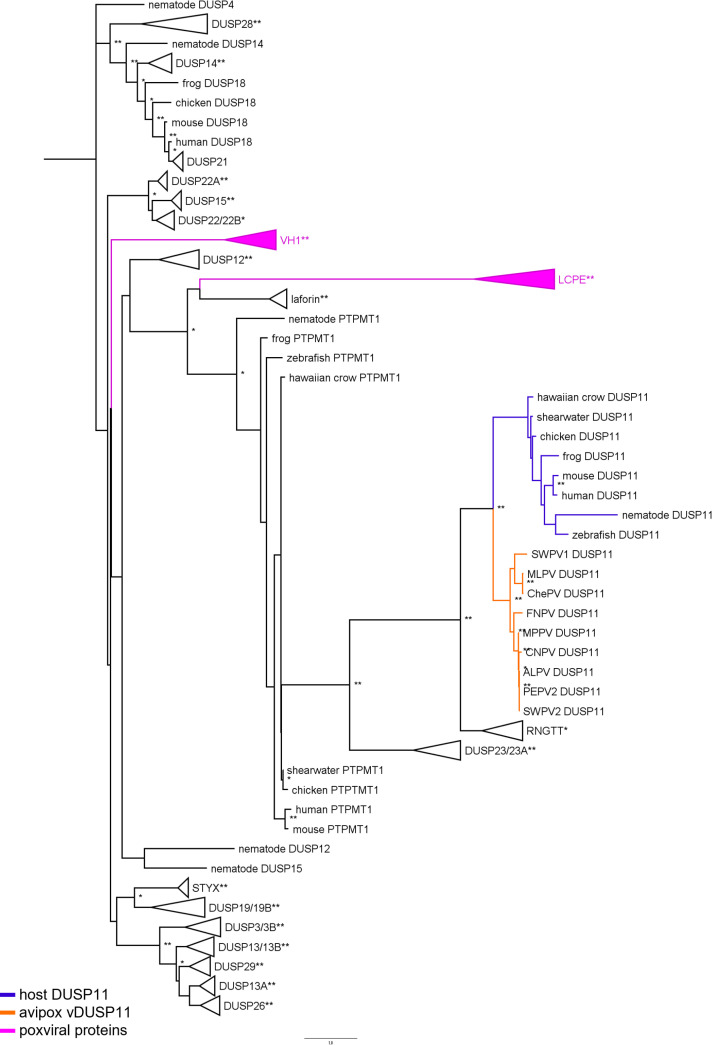
Fig 2 Phylogenetic analysis supports putative vDUSP11 acquisition from host DUSP11 sequence. An inferred tree built using 160 DUSP amino acid (AA) sequences by maximum-likelihood analysis using PhyML [42]. AA sequences for host atypical DUSPs, poxviral putative vDUSP11s, vaccinia virus DUSP H1L protein phosphatase and avipox homologs, and the large subunit the capping enzyme from poxviruses were aligned using Clustal Omega (S9 Fig) [46]. Clustal alignment was used to run PhyML analysis with LG + R model selected by SMS [53]. Sequences were retrieved from the HGNC sequence database, NCBI sequence database [41] and Uniprot (www.uniprot.org/) (S4 Table). Putative APV/AdjPV vDUSP11s (orange) cluster with host DUSP11s (blue) and not with other host protein DUSPs or other poxviral proteins (pink). Collapsed branches are used to represent proteins with homology that cluster together across numerous species. 100 bootstrap replicates were performed; branch support ≥50% (*) or ≥ 70% (**) are indicated. Nematode DUSP4, a member of the DUSP-family but not an atypical DUSP, was specified as the outgroup.
Fig 2 Phylogenetic analysis supports putative vDUSP11 acquisition from host DUSP11 sequence. An inferred tree built using 160 DUSP amino acid (AA) sequences by maximum-likelihood analysis using PhyML [42]. AA sequences for host atypical DUSPs, poxviral putative vDUSP11s, vaccinia virus DUSP H1L protein phosphatase and avipox homologs, and the large subunit the capping enzyme from poxviruses were aligned using Clustal Omega (S9 Fig) [46]. Clustal alignment was used to run PhyML analysis with LG + R model selected by SMS [53]. Sequences were retrieved from the HGNC sequence database, NCBI sequence database [41] and Uniprot (www.uniprot.org/) (S4 Table). Putative APV/AdjPV vDUSP11s (orange) cluster with host DUSP11s (blue) and not with other host protein DUSPs or other poxviral proteins (pink). Collapsed branches are used to represent proteins with homology that cluster together across numerous species. 100 bootstrap replicates were performed; branch support ≥50% (*) or ≥ 70% (**) are indicated. Nematode DUSP4, a member of the DUSP-family but not an atypical DUSP, was specified as the outgroup.CC BY · PubMed Central
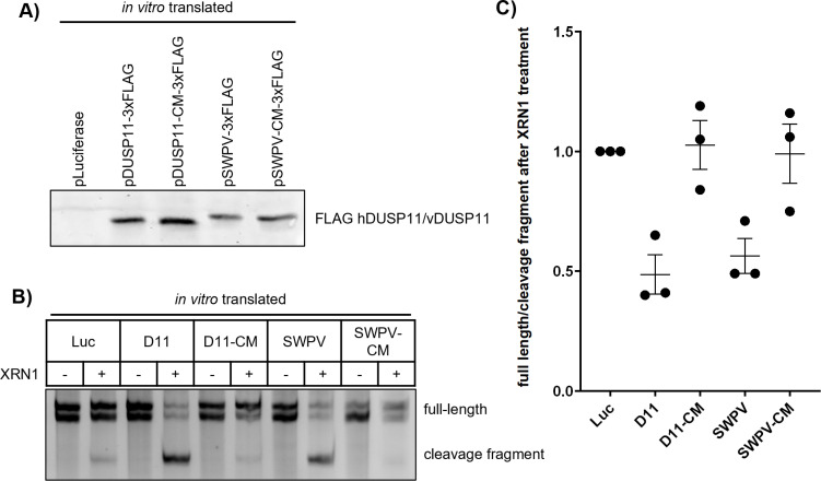
Fig 3 vDUSP11 sensitizes HCV 5’ UTR RNA to XRN-mediated degradation. (A) Confirmation of in vitro translated constructs via immunoblot analysis. Membrane was probed for FLAG-tagged proteins. Plasmid encoding luciferase was used for negative control reactions. (B) In vitro XRN susceptibility assay. In vitro transcribed HCV 5’ UTR RNA was incubated with in vitro translated products from (A) luciferase (Luc), hDUSP11 (D11), hDUSP11 catalytic mutant (D11-CM), shearwaterpox virus 2 vDUSP11 (SWPV) or shearwaterpox virus 2 vDUSP11 catalytic mutant (SWPV-CM)) and RNA was purified. Purified RNA was then subjected to treatment ± recombinant XRN1. Products were separated using urea PAGE and stained with EtBr. Migration of full-length HCV 5’ UTR is indicated, which for unknown reasons migrates as a doublet, the position of the faster-migrating cleavage fragment is indicated. (C) Graphical representation of (B), displaying ratio of band intensity of the ratio of HCV 5’ UTR full-length doublet bands to cleavage fragment band from the XRN-treated to the -XRN1 treatment reactions. Values are represented relative to the negative control Luc treatment. Data are derived from n = 3 independent replicates. In all panels, data are represented as mean ± SEM.
Fig 3 vDUSP11 sensitizes HCV 5’ UTR RNA to XRN-mediated degradation. (A) Confirmation of in vitro translated constructs via immunoblot analysis. Membrane was probed for FLAG-tagged proteins. Plasmid encoding luciferase was used for negative control reactions. (B) In vitro XRN susceptibility assay. In vitro transcribed HCV 5’ UTR RNA was incubated with in vitro translated products from (A) luciferase (Luc), hDUSP11 (D11), hDUSP11 catalytic mutant (D11-CM), shearwaterpox virus 2 vDUSP11 (SWPV) or shearwaterpox virus 2 vDUSP11 catalytic mutant (SWPV-CM)) and RNA was purified. Purified RNA was then subjected to treatment ± recombinant XRN1. Products were separated using urea PAGE and stained with EtBr. Migration of full-length HCV 5’ UTR is indicated, which for unknown reasons migrates as a doublet, the position of the faster-migrating cleavage fragment is indicated. (C) Graphical representation of (B), displaying ratio of band intensity of the ratio of HCV 5’ UTR full-length doublet bands to cleavage fragment band from the XRN-treated to the -XRN1 treatment reactions. Values are represented relative to the negative control Luc treatment. Data are derived from n = 3 independent replicates. In all panels, data are represented as mean ± SEM.CC BY · PubMed Central
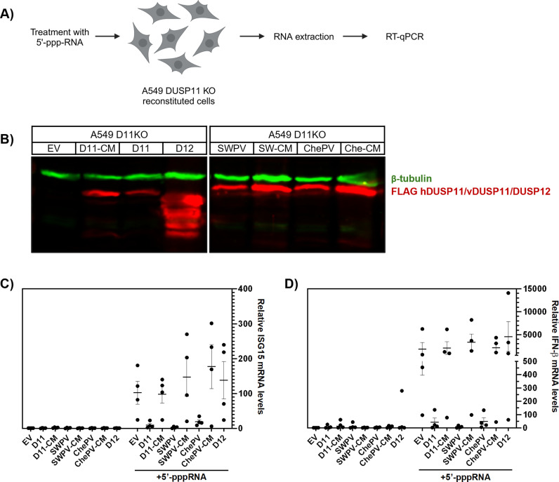
Fig 4 vDUSP11 modulates immune activation in response to liposomal 5’-ppp-RNAs. (A) Schematic diagram of the 5’-triphosphate RNA (5’-ppp-RNA) transfection assay. A549 DUSP11 knockout (KO) reconstituted cells (12-well) were transfected with 5–10 ng of in vitro transcribed 5’-ppp-RNA for 18 hours followed by RT-qPCR to assay induction of ISGs. (B) Immunoblot analysis of A549 DUSP11 knockout (KO) cells transduced with pLenti empty vector (EV), pLenti hDUSP11-3xFLAG (D11), pLenti hDUSP11 catalytic mutant-3xFLAG (D11-CM), pLenti shearwaterpox virus 2 vDUSP11-3xFLAG (SWPV), pLenti shearwaterpox virus 2 vDUSP11 catalytic mutant-3xFLAG (SW-CM/SWPV-CM), pLenti cheloniidpox virus 1 vDUSP11-3xFLAG (ChePV), pLenti 3xFLAG-cheloniidpox virus 1 vDUSP11 catalytic mutant-3xFLAG (Che-CM/ChePV-CM), or negative control pLenti DUSP12-3xFLAG (D12). (C) RT-qPCR analysis of ISG15 and (D) IFNB1 mRNA levels in A549 DUSP11 KO reconstituted cells following transfection with either lipofectamine alone or 5’-ppp-RNA, normalized to GAPDH mRNA levels. Results are represented relative to those of empty vector expressing cells. Data are derived from n = 4 independent replicates and are represented as mean ± SEM. Schematic figures in Fig 4A were created with BioRender.com.
Fig 4 vDUSP11 modulates immune activation in response to liposomal 5’-ppp-RNAs. (A) Schematic diagram of the 5’-triphosphate RNA (5’-ppp-RNA) transfection assay. A549 DUSP11 knockout (KO) reconstituted cells (12-well) were transfected with 5–10 ng of in vitro transcribed 5’-ppp-RNA for 18 hours followed by RT-qPCR to assay induction of ISGs. (B) Immunoblot analysis of A549 DUSP11 knockout (KO) cells transduced with pLenti empty vector (EV), pLenti hDUSP11-3xFLAG (D11), pLenti hDUSP11 catalytic mutant-3xFLAG (D11-CM), pLenti shearwaterpox virus 2 vDUSP11-3xFLAG (SWPV), pLenti shearwaterpox virus 2 vDUSP11 catalytic mutant-3xFLAG (SW-CM/SWPV-CM), pLenti cheloniidpox virus 1 vDUSP11-3xFLAG (ChePV), pLenti 3xFLAG-cheloniidpox virus 1 vDUSP11 catalytic mutant-3xFLAG (Che-CM/ChePV-CM), or negative control pLenti DUSP12-3xFLAG (D12). (C) RT-qPCR analysis of ISG15 and (D) IFNB1 mRNA levels in A549 DUSP11 KO reconstituted cells following transfection with either lipofectamine alone or 5’-ppp-RNA, normalized to GAPDH mRNA levels. Results are represented relative to those of empty vector expressing cells. Data are derived from n = 4 independent replicates and are represented as mean ± SEM. Schematic figures in Fig 4A were created with BioRender.com.CC BY · PubMed Central
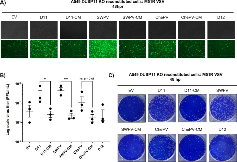
Fig 5 vDUSP11 catalytic activity promotes VSV virus replication. (A) Representative GFP images of A549 DUSP11 knockout (KO) cells stably reconstituted with either empty vector (EV) plasmid, hDUSP11 (D11), hDUSP11 catalytic mutant (D11-CM), shearwaterpox virus 2 vDUSP11 (SWPV), shearwaterpox virus 2 vDUSP11 catalytic mutant (SWPV-CM), cheloniidpox virus vDUSP11 (ChePV), cheloniidpox virus vDUSP11 catalytic mutant (ChePV-CM), or protein phosphatase DUSP12 (D12) after infection with GFP-M51R VSV at an MOI of 0.01 PFU/cell at 48 hours post-infection (hpi). (B) VSV viral titer of indicated A549 DUSP11 KO reconstituted cells determined by plaque assay analysis at 48 hpi. (C) Representative images of plaque assay analysis of virus supernatant at 48 hpi. Data are derived from n = 3 independent replicates. Data are presented as mean ± SEM. (*) P < 0.05; (**) P < 0.01 (two-tailed Student’s t-test).
Fig 5 vDUSP11 catalytic activity promotes VSV virus replication. (A) Representative GFP images of A549 DUSP11 knockout (KO) cells stably reconstituted with either empty vector (EV) plasmid, hDUSP11 (D11), hDUSP11 catalytic mutant (D11-CM), shearwaterpox virus 2 vDUSP11 (SWPV), shearwaterpox virus 2 vDUSP11 catalytic mutant (SWPV-CM), cheloniidpox virus vDUSP11 (ChePV), cheloniidpox virus vDUSP11 catalytic mutant (ChePV-CM), or protein phosphatase DUSP12 (D12) after infection with GFP-M51R VSV at an MOI of 0.01 PFU/cell at 48 hours post-infection (hpi). (B) VSV viral titer of indicated A549 DUSP11 KO reconstituted cells determined by plaque assay analysis at 48 hpi. (C) Representative images of plaque assay analysis of virus supernatant at 48 hpi. Data are derived from n = 3 independent replicates. Data are presented as mean ± SEM. (*) P < 0.05; (**) P < 0.01 (two-tailed Student’s t-test).CC BY · PubMed Central
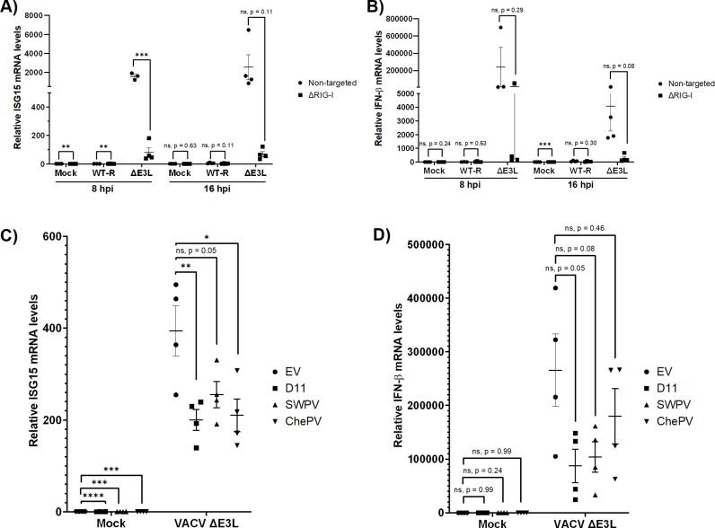
Fig 6 DUSP11 reduces immune signaling in response to infection by VACV ∆E3L. Previously characterized A549NT (non-targeted) and A549 RIG-I knock out cells (∆RIG-I) [34] were either mock infected (Mock), infected with wild-type recombinant vaccinia virus (VACV) (WT-R), or infected with an E3L deficient VACV (∆E3L/VACV ∆E3L). (A). RT-qPCR analysis of ISG15 and (B) IFNB1 mRNA normalized to GAPDH mRNA levels. Results are represented relative to those of non-targeted cells. (C) RT-qPCR analysis of ISG15 and (D) IFNB1 mRNA normalized to GAPDH mRNA of A549 DUSP11 KO cells expressing either empty vector (EV), hDUSP11 (D11), shearwaterpox virus vDUSP11 (SWPV), or cheloniidpox virus vDUSP11 (ChePV) at 16 hpi following either mock infection or infection with VACV ∆E3L. Results are represented relative to mock infected A549 DUSP11 KO cells reconstituted with EV. For panels A and B, non-targeted cells infected with either WT-R or VACV ∆E3L at 8 hpi, data are derived from n = 3 independent replicates. For all other time points, data are derived from n = 4 independent replicates. All data are represented as mean ± SEM. (*) P < 0.05; (**) P < 0.01; (***) P < 0.001; (****); P < 0.0001. Statistical analysis for panels A and B included two-tailed students t-tests; C and D included one-way ANOVAs followed by Dunnett’s multiple comparison test.
Fig 6 DUSP11 reduces immune signaling in response to infection by VACV ∆E3L. Previously characterized A549NT (non-targeted) and A549 RIG-I knock out cells (∆RIG-I) [34] were either mock infected (Mock), infected with wild-type recombinant vaccinia virus (VACV) (WT-R), or infected with an E3L deficient VACV (∆E3L/VACV ∆E3L). (A). RT-qPCR analysis of ISG15 and (B) IFNB1 mRNA normalized to GAPDH mRNA levels. Results are represented relative to those of non-targeted cells. (C) RT-qPCR analysis of ISG15 and (D) IFNB1 mRNA normalized to GAPDH mRNA of A549 DUSP11 KO cells expressing either empty vector (EV), hDUSP11 (D11), shearwaterpox virus vDUSP11 (SWPV), or cheloniidpox virus vDUSP11 (ChePV) at 16 hpi following either mock infection or infection with VACV ∆E3L. Results are represented relative to mock infected A549 DUSP11 KO cells reconstituted with EV. For panels A and B, non-targeted cells infected with either WT-R or VACV ∆E3L at 8 hpi, data are derived from n = 3 independent replicates. For all other time points, data are derived from n = 4 independent replicates. All data are represented as mean ± SEM. (*) P < 0.05; (**) P < 0.01; (***) P < 0.001; (****); P < 0.0001. Statistical analysis for panels A and B included two-tailed students t-tests; C and D included one-way ANOVAs followed by Dunnett’s multiple comparison test.CC BY · PubMed Central
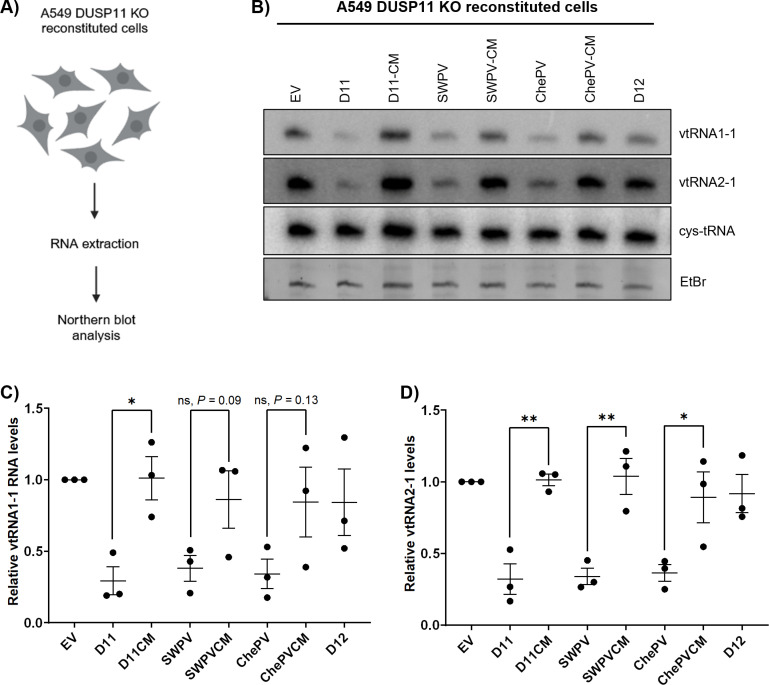
Fig 7 vDUSP11 modulates steady-state RNA levels of endogenous RNAP III transcripts. (A) Schematic diagram of vtRNA northern blot. RNA was collected from resting A549 DUSP11 KO cells stably reconstituted with empty vector (EV) plasmid, hDUSP11 (D11), hDUSP11 catalytic mutant (D11-CM), shearwaterpox virus 2 vDUSP11 (SWPV), shearwaterpox virus 2 vDUSP11 catalytic mutant (SWPV-CM), cheloniidpox virus vDUSP11 (ChePV), cheloniidpox virus vDUSP11 catalytic mutant (ChePV-CM), or negative control protein phosphatase DUSP12 (D12). Purified RNA was then subjected to northern blot analysis. (B) Northern blot analysis of vtRNA1–1 and vtRNA2–1 using RNA from A549 DUSP11 KO reconstituted cells. (C) Graphical representation of relative band intensity of vtRNA1–1 and (D) vtRNA2–1 normalized to the relative band intensity of the 5’ monophosphate control cysteine-tRNA. Values are represented relative to the A549 DUSP11 KO + EV cell line. Data are derived from n = 3 independent replicates. In all panels, data are represented as mean ± SEM. (*) P < 0.05; (**) P < 0.01 (two-tailed Student’s t-test). Schematic figures in Fig 7A were created with BioRender.com.
Fig 7 vDUSP11 modulates steady-state RNA levels of endogenous RNAP III transcripts. (A) Schematic diagram of vtRNA northern blot. RNA was collected from resting A549 DUSP11 KO cells stably reconstituted with empty vector (EV) plasmid, hDUSP11 (D11), hDUSP11 catalytic mutant (D11-CM), shearwaterpox virus 2 vDUSP11 (SWPV), shearwaterpox virus 2 vDUSP11 catalytic mutant (SWPV-CM), cheloniidpox virus vDUSP11 (ChePV), cheloniidpox virus vDUSP11 catalytic mutant (ChePV-CM), or negative control protein phosphatase DUSP12 (D12). Purified RNA was then subjected to northern blot analysis. (B) Northern blot analysis of vtRNA1–1 and vtRNA2–1 using RNA from A549 DUSP11 KO reconstituted cells. (C) Graphical representation of relative band intensity of vtRNA1–1 and (D) vtRNA2–1 normalized to the relative band intensity of the 5’ monophosphate control cysteine-tRNA. Values are represented relative to the A549 DUSP11 KO + EV cell line. Data are derived from n = 3 independent replicates. In all panels, data are represented as mean ± SEM. (*) P < 0.05; (**) P < 0.01 (two-tailed Student’s t-test). Schematic figures in Fig 7A were created with BioRender.com.CC BY · PubMed Central
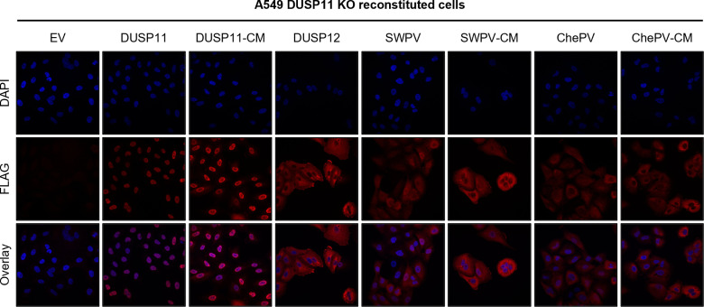
Fig 8 vDUSP11s differ in subcellular localization compared to hDUSP11. Representative confocal images of A549 DUSP11 KO cells stably expressing either an empty vector (EV) plasmid or individual 3xFLAG tagged proteins including hDUSP11 (D11), hDUSP11 catalytic mutant (D11-CM), SWPV2 vDUSP11 (SWPV), SWPV2-CM (SWPV-CM), ChePV1 vDUSP11 (ChePV), ChePV1 vDUSP11-CM (ChePV-CM), or negative control protein phosphatase DUSP12 (D12). Prolong Gold Antifade Mountant (Thermo Fisher Scientific) was used in slide preparation to visualize nuclei.
Fig 8 vDUSP11s differ in subcellular localization compared to hDUSP11. Representative confocal images of A549 DUSP11 KO cells stably expressing either an empty vector (EV) plasmid or individual 3xFLAG tagged proteins including hDUSP11 (D11), hDUSP11 catalytic mutant (D11-CM), SWPV2 vDUSP11 (SWPV), SWPV2-CM (SWPV-CM), ChePV1 vDUSP11 (ChePV), ChePV1 vDUSP11-CM (ChePV-CM), or negative control protein phosphatase DUSP12 (D12). Prolong Gold Antifade Mountant (Thermo Fisher Scientific) was used in slide preparation to visualize nuclei.CC BY · PubMed Central
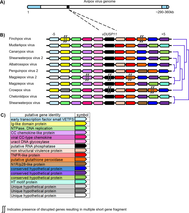
Fig 9 A single acquisition event for vDUSP11 is indicated by synteny analysis. (A) Graphical representation of avipoxvirus genome using published information from the canarypox virus genome as a model [62]. Inverted terminal repeats (ITRs) are indicated in light blue, relative genome position of vDUSP11 indicated in black. (B) Analysis of upstream and downstream flanking genes indicated vDUSP11s (black) reside at similar genomic locations across poxvirus genomes. Protein homologs were determined using reciprocal BLAST hits for the 5 genes upstream (left of vDUSP11) and 5 genes downstream (right of vDUSP11) of vDUSP11. Homologous genes are indicated by shared color. Genes oriented to the right indicate ORFs on the top strand while genes oriented to the left indicate genes on the bottom strand. Cladogram indicating virus relatedness from published data [63] to the right of each gene diagram. (C) Corresponding functional annotation for genes indicated in (B). Predicted functional annotations were determined using NCBI database genome annotations [41]. For genes lacking descriptive annotations, more detailed annotations based on identified homologous genes were used (S5 Table).
Fig 9 A single acquisition event for vDUSP11 is indicated by synteny analysis. (A) Graphical representation of avipoxvirus genome using published information from the canarypox virus genome as a model [62]. Inverted terminal repeats (ITRs) are indicated in light blue, relative genome position of vDUSP11 indicated in black. (B) Analysis of upstream and downstream flanking genes indicated vDUSP11s (black) reside at similar genomic locations across poxvirus genomes. Protein homologs were determined using reciprocal BLAST hits for the 5 genes upstream (left of vDUSP11) and 5 genes downstream (right of vDUSP11) of vDUSP11. Homologous genes are indicated by shared color. Genes oriented to the right indicate ORFs on the top strand while genes oriented to the left indicate genes on the bottom strand. Cladogram indicating virus relatedness from published data [63] to the right of each gene diagram. (C) Corresponding functional annotation for genes indicated in (B). Predicted functional annotations were determined using NCBI database genome annotations [41]. For genes lacking descriptive annotations, more detailed annotations based on identified homologous genes were used (S5 Table).CC BY · PubMed Central

Verification

Claims the paper makes

The claims and statistics of this paper haven’t been checked yet.

Run the check — free, no account needed. It reads the open-access text and checks the sources it cites.

A few free checks a day · papers already verified open instantly · results are public

Self-check

Percentages, sums and reported differences re-computed from the paper’s own numbers.

The self-check needs the paper’s full text.

Citations

Cites

1 retracted

Show all 66 references

Research works printed but not linked: 1

  1. T Deshpande, 1999, Human PIR1 of the protein-tyrosine phosphatase superfamily has RNA 5’-triphosphatase and diphosphatase activities, J Biol Chem, 274, 16590, 10.1074/jbc.274.23.16590 · DOI

Metadata

Authors
Szymanik, K. H., Rex, E. A., Pothireddy, V. R., Gammon, D. B., Hancks, D. C., & Sullivan, C. S.
Affiliations
  • The University of Texas at Austin (US)
  • The University of Texas Southwestern Medical Center (US)
Corresponding author
  • Hancks, Dustin C.
  • Sullivan, Christopher S.
Venue
PLOS Pathogens
Published
2025-04-21
Volume
21
Issue
4
Pages
e1013101
ISSN
  • 1553-7366 (ISSN-L)
  • 1553-7374
OpenAlex
W4409640449
Cited by
0
References
66
FWCI
0.00
Type
journal article
Language
en
Open access
Locations
  • PLoS Pathogens (publishedVersion, cc-by)
  • bioRxiv (Cold Spring Harbor Laboratory) (acceptedVersion)
  • PubMed Central (publishedVersion, cc-by)
  • PubMed (publishedVersion)
  • PubMed Central (submittedVersion)
  • DOAJ (DOAJ: Directory of Open Access Journals) (submittedVersion, cc-by-sa)
Funders
  • NIH (1R35GM142689-04, 1R01AI134980, 1R35GM137978, T32 AI007520)
  • Burroughs Wellcome Fund (1011070)
Topics
  • Life Sciences › Immunology and Microbiology › Immunology › interferon and immune responses
  • Health Sciences › Medicine › Epidemiology › Virology and Viral Diseases
  • Life Sciences › Immunology and Microbiology › Immunology › Immune Response and Inflammation
Keywords
RIG-I, innate immune evasion, RNA triphosphatase, RNA polymerase III transcripts, viral RNA-dependent RNA polymerase, RNA metabolism

About these checks

Registry, retraction, Cites and statistics results are computed, not judged, with one exception: a printed reference that no index lists is read once by a model before it counts as a citation error. Claims are extracted by a model and kept only when their quote appears verbatim in the text; each cited source is then checked, with a model’s help, for whether it says what the claim relies on. Those verdicts can be wrong — read the claim’s quote and the cited source before relying on one.

Statistics and arithmetic are recomputed, not judged.

Confidence bands are shown only for fields calibrated on our annotated test set. None is calibrated yet, so no band is shown.

What counts as a claim ›How we measure confidence ›How we check this ›

Are you an author, or do you see a problem with this page? Request a review

How to cite

Cite CoChat for the checks on this page, and the paper for its own findings.

This page
CoChat. (2026). Citation check: Viral piracy of host RNA phosphatase DUSP11 by avipoxviruses. https://app.cochat.ai/papers/10.1371/journal.ppat.1013101
The paper
Szymanik, K. H., Rex, E. A., Pothireddy, V. R., Gammon, D. B., Hancks, D. C., & Sullivan, C. S. (2025). Viral piracy of host RNA phosphatase DUSP11 by avipoxviruses. PLOS Pathogens. https://doi.org/10.1371/journal.ppat.1013101

Data sources

Bibliographic data: Semantic Scholar, OpenAlex, Crossref.

Contains information from the Semantic Scholar Academic Graph Datasets, made available under the ODC Attribution License (ODC-BY 1.0).

Semantic Scholar’s data is described in: Kinney, R., et al. (2023). The Semantic Scholar Open Data Platform. arXiv:2301.10140.