

The DOI matches its Crossref record; 303 of the 303 references it cites were found in a registry or an index.
Microglia and Astrocytes in Alzheimer’s Disease: Significance and Summary of Recent Advances
Open accessAbstract
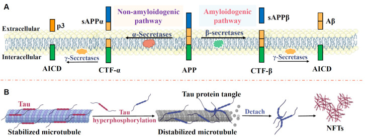
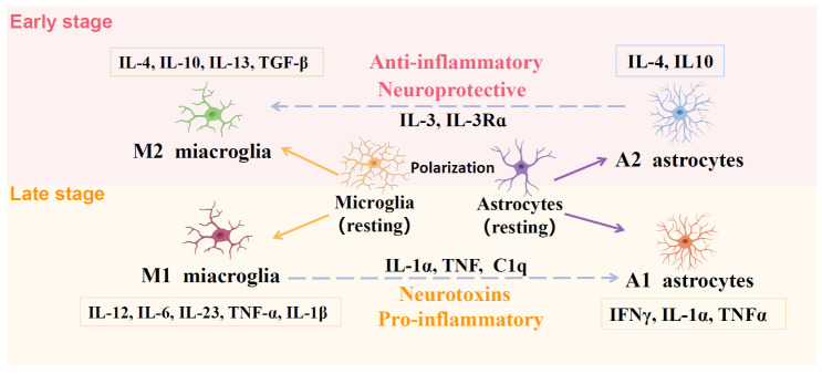
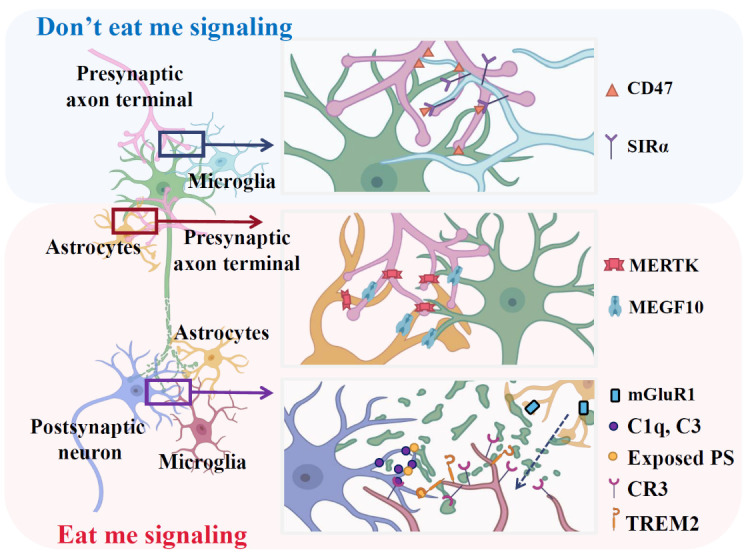
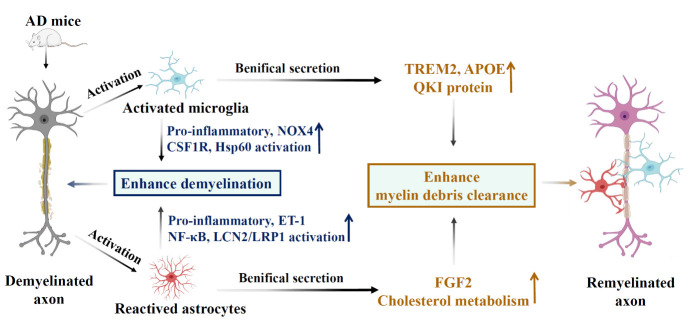
Alzheimer's disease, one of the most common forms of dementia, is characterized by a slow progression of cognitive impairment and neuronal loss. Currently, approved treatments for AD are hindered by various side effects and limited efficacy. Despite considerable research, practical treatments for AD have not been developed. Increasing evidence shows that glial cells, especially microglia and astrocytes, are essential in the initiation and progression of AD. During AD progression, activated resident microglia increases the ability of resting astrocytes to transform into reactive astrocytes, promoting neurodegeneration. Extensive clinical and molecular studies show the involvement of microglia and astrocyte-mediated neuroinflammation in AD pathology, indicating that microglia and astrocytes may be potential therapeutic targets for AD. This review will summarize the significant and recent advances of microglia and astrocytes in the pathogenesis of AD in three parts. First, we will review the typical pathological changes of AD and discuss microglia and astrocytes in terms of function and phenotypic changes. Second, we will describe microglia and astrocytes' physiological and pathological role in AD. These roles include the inflammatory response, "eat me" and "don't eat me" signals, Aβ seeding, propagation, clearance, synapse loss, synaptic pruning, remyelination, and demyelination. Last, we will review the pharmacological and non-pharmacological therapies targeting microglia and astrocytes in AD. We conclude that microglia and astrocytes are essential in the initiation and development of AD. Therefore, understanding the new role of microglia and astrocytes in AD progression is critical for future AD studies and clinical trials. Moreover, pharmacological, and non-pharmacological therapies targeting microglia and astrocytes, with specific studies investigating microglia and astrocyte-mediated neuronal damage and repair, may be a promising research direction for future studies regarding AD treatment and prevention.
Overview
The claims and statistics of this paper haven’t been checked yet.
Run the check — free, no account needed. It reads the open-access text and checks the sources it cites.
Integrity signals
Registry
Cites
Statistics
Figures
Verification
Claims the paper makes
The claims and statistics of this paper haven’t been checked yet.
Run the check — free, no account needed. It reads the open-access text and checks the sources it cites.
Claims other papers make about this one
Each is checked against this paper’s own text: does it say what the citing paper says it does?
The source backs part of the claim, not all of it — or backs it for a different population, condition or comparison.
In the citing paper
“For example, misidentifying disease-associated microglia (DAMs), reactive astrocytes, or rare cell subpopulations can hinder efforts to characterize disease progression and cellular dysfunction in AD[58].”
From Comparative analysis of nuclei isolation methods for brain single-nucleus RNA sequencing (2026) · cites this paper as [58]
Experimental — accuracy not yet measured
Self-check
Percentages, sums and reported differences re-computed from the paper’s own numbers.
The self-check needs the paper’s full text.
Citations
Cites
Polazzi, E., Mengoni, I., Caprini, M., Peña‐Altamira, E., Kurtys, E., & Monti, B. (2012) · Neurosignals · doi:10.1159/000337115 · Cited by 9,023
Neurotoxic reactive astrocytes are induced by activated microglia
Liddelow, S. A., Guttenplan, K. A., Clarke, L. E., Bennett, F. C., Bohlen, C. J., Schirmer, L., Bennett, M. L., Münch, A. E., Chung, W.-S., Peterson, T. C., et al. (2017) · Nature · doi:10.1038/nature21029 · Cited by 7,971
Alzheimer's disease: the amyloid cascade hypothesis
Hardy, J. A., & Higgins, G. A. (1992) · Science · doi:10.1126/science.1566067 · Cited by 7,484
Astrocyte-endothelial interactions at the blood-brain barrier
Abbott, N. J., Rönnbäck, L., & Hansson, E. (2006) · Nature Reviews Neuroscience · doi:10.1038/nrn1824 · Cited by 5,595
Microglia: Active sensor and versatile effector cells in the normal and pathologic brain
Hanisch, U.-K., & Kettenmann, H. (2007) · Nature Neuroscience · doi:10.1038/nn1997 · Cited by 3,676
Complement and microglia mediate early synapse loss in Alzheimer mouse models
Hong, S., Beja-Glasser, V. F., Nfonoyim, B. M., Frouin, A., Li, S., Ramakrishnan, S., Merry, K. M., Shi, Q., Rosenthal, A., Barres, B. A., et al. (2016) · Science · doi:10.1126/science.aad8373 · Cited by 3,343 · cited 2×
“Previous research suggested that knocking out complement systems such as C1q, C3, or CR3 disrupted microglia-specific phagocytic pathways, consequently impairing synaptic connectivity [158, 159].”
BackgroundToll-Like Receptor Signaling Pathways
Kawasaki, T., & Kawai, T. (2014) · Frontiers in Immunology · doi:10.3389/fimmu.2014.00461 · Cited by 3,245
Neuroinflammation and microglial activation in Alzheimer disease: where do we go from here?
Leng, F., & Edison, P. (2021) · Nature Reviews Neurology · doi:10.1038/s41582-020-00435-y · Cited by 3,179
NLRP3 is activated in Alzheimer’s disease and contributes to pathology in APP/PS1 mice
Heneka, M. T., Kummer, M. P., Stutz, A., Delekate, A., Schwartz, S., Vieira-Saecker, A., Griep, A., Axt, D., Remus, A., Tzeng, T.-C., et al. (2013) · Nature · doi:10.1038/nature11729 · Cited by 2,898
Neuroinflammation in neurodegenerative disorders: the roles of microglia and astrocytes
Kwon, H. S., & Koh, S.-H. (2020) · Translational Neurodegeneration · doi:10.1186/s40035-020-00221-2 · Cited by 2,595
Cited by
Sorted by citations
Targeting Microglia in Alzheimer’s Disease: Pathogenesis and Potential Therapeutic Strategies
Sun, Z., Zhang, X., So, K.-F., Jiang, W., & Chiu, K. (2024) · Biomolecules · doi:10.3390/biom14070833 · Cited by 71
Unveiling the role of astrocytes in postoperative cognitive dysfunction
He, L., Duan, X., Li, S., Zhang, R., Dai, X., & Lu, M. (2024) · Ageing Research Reviews · doi:10.1016/j.arr.2024.102223 · Cited by 66
Research progress in Alzheimer's disease and bone-brain axis
Zhang, F., & Zhang, W. (2024) · Ageing Research Reviews · doi:10.1016/j.arr.2024.102341 · Cited by 62
Voicu, V. A., Toader, C., Șerban, M., Covache-Busuioc, R.-A., & Ciurea, A. V. (2025) · Biomedicines · doi:10.3390/biomedicines13082025 · Cited by 59
Alzheimer’s disease pathogenesis: standing at the crossroad of lipid metabolism and immune response
Wang, Z., Zhang, L., & Qin, C. (2025) · Molecular Neurodegeneration · doi:10.1186/s13024-025-00857-6 · Cited by 60
Wahl, D., Risen, S. J., Osburn, S. C., Emge, T., Sharma, S., Gilberto, V. S., Chatterjee, A., Nagpal, P., Moreno, J. A., & LaRocca, T. J. (2024) · Journal of Neuroinflammation · doi:10.1186/s12974-024-03182-9 · Cited by 41
Boccardi, V., Mancinetti, F., & Mecocci, P. (2025) · Antioxidants · doi:10.3390/antiox14091044 · Cited by 40
The role of estrogen in Alzheimer’s disease pathogenesis and therapeutic potential in women
Wang, X., Feng, S., Deng, Q., Wu, C., Duan, R., & Yang, L. (2024) · Molecular and Cellular Biochemistry · doi:10.1007/s11010-024-05071-4 · Cited by 40
Self-citationYan, Q., Qin, Q., Zhang, S., Chen, F., Ru, Y., Zhong, Y., & Wu, G. (2025) · Critical Reviews in Food Science and Nutrition · doi:10.1080/10408398.2025.2568606 · Cited by 28
Han, J., Zhang, X., Kang, L., & Guan, J. (2025) · Journal of Neuroinflammation · doi:10.1186/s12974-025-03443-1 · Cited by 26
Sugandhi, V. V., Gadhave, D. G., Ugale, A. R., Kulkarni, N. N., Nangare, S., Patil, H. P., Rath, S., Saxena, R., Lavate, A., Patel, A. T., et al. (2025) · Ageing Research Reviews · doi:10.1016/j.arr.2025.102775 · Cited by 32
Han, X., Zhang, Y., Zhang, L., Zhang, L., & Wang, Y. (2024) · European Journal of Medicinal Chemistry · doi:10.1016/j.ejmech.2024.117144 · Cited by 24
Wu, Y., Yang, L., Jiang, W., Zhang, X., & Yao, Z. (2024) · Neural Regeneration Research · doi:10.4103/nrr.nrr-d-24-00190 · Cited by 19
Self-citationJin, Z., Lu, Y., Tang, H., & Cui, H. (2025) · Alzheimer s & Dementia · doi:10.1002/alz.70472 · Cited by 24
Beyond the blood–brain barrier: unraveling T cell subsets in CNS immunity and disease
Shokr, M. M. (2025) · Inflammopharmacology · doi:10.1007/s10787-025-01955-z · Cited by 21
Dastan, M., Rajaei, Z., Sharifi, M., & Salehi, H. (2024) · Metabolic Brain Disease · doi:10.1007/s11011-024-01441-5 · Cited by 19
Tolédano, A., Rodríguez‐Casado, A., Álvarez, M., & Toledano‐Díaz, A. (2024) · Brain Sciences · doi:10.3390/brainsci14111101 · Cited by 19
Wu, J., Xu, W., Su, Y., Wang, G., & Ma, J. (2024) · Acta Pharmacologica Sinica · doi:10.1038/s41401-024-01416-3 · Cited by 21
The Role of Astrocytes in Synaptic Dysfunction and Memory Deficits in Alzheimer’s Disease
de León-López, C. A. M., Navarro‐Lobato, I., & Khan, Z. U. (2025) · Biomolecules · doi:10.3390/biom15070910 · Cited by 19
Role of MARK2 in the nervous system and cancer
Lei, Y., Zhang, R., & Cai, F. (2024) · Cancer Gene Therapy · doi:10.1038/s41417-024-00737-z · Cited by 15
Refining the interactions between microglia and astrocytes in Alzheimer’s disease pathology
Chen, J., Xu, S., Wang, L., Liu, X., Liu, G., Tan, Q., Li, W., Zhang, S., & Du, Y. (2025) · Neuroscience · doi:10.1016/j.neuroscience.2025.03.033 · Cited by 17
The role of peripheral innate immune cells in Alzheimer’s disease progression
Cao, Y., Tang, K., Ma, P., Zhang, R., Yang, Y., Li, T., Zhang, Y., & Peng, X. (2025) · Frontiers in Immunology · doi:10.3389/fimmu.2025.1616939 · Cited by 15
Njoku, G. C., & Kanmogne, G. D. (2025) · International Journal of Molecular Sciences · doi:10.3390/ijms26189069 · Cited by 15
Das, D., Sonthalia, S., Stein-O.’Brien, G., Wahbeh, M., Feuer, K. L., Goff, L. A., Colantuoni, C., Mahairaki, V., & Avramopoulos, D. (2024) · BMC Biology · doi:10.1186/s12915-024-01867-4 · Cited by 14
Hao, F., Zeng, M., Cao, B., Liang, X., Ye, K., Jiao, X., Feng, W., & Zheng, X. (2024) · CNS Neuroscience & Therapeutics · doi:10.1111/cns.70068 · Cited by 12
Targeting autophagy in astrocytes: a potential for neurodegenerative disease intervention
Potokar, M., & Jorgačevski, J. (2025) · Frontiers in Cellular Neuroscience · doi:10.3389/fncel.2025.1584767 · Cited by 14
Vascular Contributions to Healthy Aging and Dementia
Ge, Y. (2024) · Aging and Disease · doi:10.14336/ad.2023.1719 · Cited by 10
Brezáni, V., Joshi, R., Ortega‐Ribera, M., Nagesh, P. T., Brezáni, V., Zivny, A., Kurt‐Jones, E. A., Golenbock, D. T., & Szabó, G. (2025) · Journal of Neuroinflammation · doi:10.1186/s12974-025-03501-8 · Cited by 12
Beyond amyloid: nanobody-mediated neuroinflammatory therapy for Alzheimer’s disease
Amniouel, S., Suh, J., Zheng, W. H., & Zhang, Q. (2025) · Translational Neurodegeneration · doi:10.1186/s40035-025-00513-5 · Cited by 11
Deng, Q., Parker, E., Wu, C., Zhu, L., Liu, T. C., Duan, R., & Yang, L. (2024) · Aging and Disease · doi:10.14336/ad.2024.0239 · Cited by 10
Self-citationExercise-induced upregulation of TRIM9 attenuates neuroinflammation in Alzheimer’s disease-like rat
Zhang, X.-Y., Zhang, J., Li, X.-C., Lu, H., & Liu, T. C. (2024) · International Immunopharmacology · doi:10.1016/j.intimp.2024.113676 · Cited by 9
Self-citationThe aggregation propensity of Tau and amyloid-β in Alzheimer’s disease
Chinnathambi, S., Kumarappan, M., Chandrshekar, M., & Malik, S. (2025) · Advances in protein chemistry and structural biology · doi:10.1016/bs.apcsb.2024.10.004 · Cited by 9
Wu, Z., Zhai, M., Wang, Y., Ren, L., Pan, J., Xiao, L., & Liu, Y. (2025) · Neurobiology of Disease · doi:10.1016/j.nbd.2025.107117 · Cited by 13
Comparative analysis of nuclei isolation methods for brain single-nucleus RNA sequencing
Kersey, H. N., Acri, D. J., Dabin, L. C., Hartigan, K. A., Mustaklem, R., Park, J. H., & Kim, J. (2026) · Cell Reports Methods · doi:10.1016/j.crmeth.2026.101337 · Cited by 8
Que, X., Zhang, T., Liu, X. Y., Yin, Y., Xia, X., Gong, P., Song, W., Qin, Q., Xu, Q., & Tang, Y. (2025) · Progress in Neurobiology · doi:10.1016/j.pneurobio.2025.102732 · Cited by 8
Neuroimmune Dysregulation and AI-Driven Therapeutic Strategies in Alzheimer’s Disease
Ghosh, S., Bhaskar, R., Singh, D. K. K., Sharma, S., Yarlagadda, B., Sinha, J. K., & Han, S. S. (2025) · Cellular and Molecular Neurobiology · doi:10.1007/s10571-025-01651-0 · Cited by 11
Alzheimer’s Disease and Other Dementias
Brugulat‐Serrat, A., Shaaban, C. E., Emrani, S., & Sundermann, E. E. (2025) · Springer eBooks · doi:10.1007/978-3-031-87228-0_5 · Cited by 7
Moraitis, S., & Piperi, C. (2025) · Biology · doi:10.3390/biology14070749 · Cited by 7
Radi, R. A., Kandeil, M. A., Mohammed, E. T., Ibrahim, M. A., Fouad, A. G., Abdel‐Razik, A. H., Khalıl, F., & Sabry, D. (2025) · Scientific Reports · doi:10.1038/s41598-025-25892-7 · Cited by 7
Targeting Microglial Activation to Modulate Neuroinflammation in Alzheimer’s Disease
Patil, V., Sharma, A., Parekh, B. K., Farah, H., Jyothi, S. R., Mishra, S. S., Nanda, A., Al-Hasnaawei, S., & Mishra, M. K. (2025) · NeuroMolecular Medicine · doi:10.1007/s12017-025-08898-2 · Cited by 9
Xu, J., Zhao, Y., Wang, G., & Tian, W. (2025) · International Immunopharmacology · doi:10.1016/j.intimp.2025.114281 · Cited by 7
Magnolol acts as a neurorestorative agent in an Aβ1‑42‑induced mouse model of Alzheimer's disease
Qian, Y., Shi, Y., Wu, Y., Liu, R., Zhang, H., Wu, L., & Ding, M. (2024) · Experimental and Therapeutic Medicine · doi:10.3892/etm.2024.12762 · Cited by 5
Xu, Z., Li, J., Liu, X., Liu, L., Lin, W., Sun, D., & Zeng, Y. (2025) · Journal of Pharmacy and Pharmacology · doi:10.1093/jpp/rgaf034 · Cited by 5
Role of astrocytes in Alzheimer's disease pathogenesis and the impact of exercise-induced remodeling
Shao, J., Deng, Q., Feng, S., Wu, C., Liu, X., & Yang, L. (2024) · Biochemical and Biophysical Research Communications · doi:10.1016/j.bbrc.2024.150418 · Cited by 5
Self-citationBoosting Neurogenesis as a Strategy in Treating Alzheimer’s Disease
Dwamena, A., Arachchi, R. Y. B., & Wang, H. (2025) · International Journal of Molecular Sciences · doi:10.3390/ijms26188926 · Cited by 4
Correale, J. D. (2026) · Journal of Neurochemistry · doi:10.1111/jnc.70414 · Cited by 4
Katola, F. O., Adana, M. Y., & Olajide, O. A. (2024) · Inflammation Research · doi:10.1007/s00011-024-01952-y · Cited by 4
Wu, L., Zhang, R., Wang, Y., Dai, S., & Yang, N. (2025) · Frontiers in Aging Neuroscience · doi:10.3389/fnagi.2025.1571783 · Cited by 4
Tacrolimus mitigates pathological patterns in mouse models of Alzheimer’s disease
Gomes, G. F., Batista, C. R. Á., da Silva, M. C. M., Ferrari, C. Z., Koerich, S., Brant, M. L., Saliba, S. W., Perez, V. G., Lima, A. L. D., Ribeiro, R., et al. (2025) · Biomedicine & Pharmacotherapy · doi:10.1016/j.biopha.2025.118168 · Cited by 5
Li, P., Zhang, H., Ma, J., Wang, J., Lv, S., Zhao, X., Yang, X., Luo, Y., Guo, X., Tao, L., et al. (2025) · Diabetology & Metabolic Syndrome · doi:10.1186/s13098-025-01805-6 · Cited by 3
Gurikar, S., Ahmed, S. S., & Raghunatahanaidu, B. D. (2025) · Journal of Molecular Neuroscience · doi:10.1007/s12031-025-02433-5 · Cited by 3
Wu, Z., Yu, S., Tian, D., Li, C., & Jing, J. (2025) · Frontiers in Cellular Neuroscience · doi:10.3389/fncel.2025.1705069 · Cited by 3
The dual effects of anesthetics on glial cells: a review of neuroprotection and neurotoxicity
Wang, X., Zhao, M., & Liu, Z. (2026) · Frontiers in Pharmacology · doi:10.3389/fphar.2026.1724695 · Cited by 3
Glial cells in dementia: From cellular dysfunction to therapeutic frontiers
Jubair, H. (2026) · Cell Transplantation · doi:10.1177/09636897251414216 · Cited by 3
Yang, W., Yu, Q., Wang, N., Lam, K. K. Y., Lin, Z., & Xian, Y. (2025) · NeuroMolecular Medicine · doi:10.1007/s12017-025-08860-2 · Cited by 4
Mechanistic Role of Astrocytic Rac1 Protein in Alzheimer's Disease
Jiang, F., Wang, N., & Meng, Q. (2025) · Aging and Disease · doi:10.14336/ad.2025.0502 · Cited by 2
Pratt, T., Lee, A., Zheng, J., Tavacoli, F., Ho, H., Liu, T., Zhan, H., Chen, J., & Initiative, A. D. N. (2025) · Journal of Alzheimer s Disease · doi:10.1177/13872877251362258 · Cited by 2
Self-citationFunctions of the basal lamina and ApoE in cerebral amyloid angiopathy
Juan-Palencia, A., & Yao, Y. (2025) · Fluids and Barriers of the CNS · doi:10.1186/s12987-025-00734-w · Cited by 2
Li, X., Xie, M., & Ibáñez, C. F. (2026) · Journal of Neuroscience · doi:10.1523/jneurosci.1939-25.2026 · Cited by 1
de Souza, M. R. M., Lindner, M. R., Mladinich, M. C., Gorbunova, E. E., Kirillov, V., Byrne, A. G., Laird, A. Y., Finnerty, C. E., Ikeda, P., Vorkas, C. K., et al. (2026) · mBio · doi:10.1128/mbio.01294-26 · Cited by 2
Gong, M., Lan, J. X., & Miao, J. (2026) · Frontiers in Aging Neuroscience · doi:10.3389/fnagi.2026.1908574 · Cited by 3
3-N-Butylphthalide alleviate Aβ-induced cellular senescence through the CDK2-pRB1-Caspase3 axis
Tian, Y., Li, W., & Zhang, Y. (2024) · Brain Research · doi:10.1016/j.brainres.2024.149435 · Cited by 1
Simula, E. R., Ercoli, T., Ruiu, E., Fais, M., Noli, M., Solla, P., & Sechi, L. A. (2026) · Frontiers in Cellular and Infection Microbiology · doi:10.3389/fcimb.2026.1777560 · Cited by 1
Natale, F., Dellaria, A., Sarrapochiello, I. N., Leone, L., Spinelli, M., Rinaudo, M., Garofalo, N., Grassi, C., & Fusco, S. (2026) · Alzheimer s Research & Therapy · doi:10.1186/s13195-026-02014-7 · Cited by 1
Xie, Y., Tian, J., Li, H., Peng, Y., Li, M., & Wu, Y. H. (2026) · Frontiers in Neuroscience · doi:10.3389/fnins.2026.1771310 · Cited by 1
Mechanisms and Therapeutic Targeting of the Gut Microbiota-Immune-Brain Axis in Alzheimer’s Disease
Wang, S., Yuan, X., Wang, T., Yang, M., Dong, P., & Han, H. (2026) · Immunological Investigations · doi:10.1080/08820139.2026.2669375 · Cited by 1
Li, Y., Zhang, L., Lin, J., Yang, L., & Duan, R. (2025) · Brain Research · doi:10.1016/j.brainres.2025.149981 · Cited by 1
Self-citationSORLA upregulation suppresses global pathological effects in aged tauopathy mouse brainPreprint
Huang, H., Shi, C. H., Yang, W., Piña-Crespo, J., Bhatnagar, J., Curatolo, J., Murad, R., Shah, P. M., Campos, A. R., Houser, A. K., et al. (2025) · bioRxiv (Cold Spring Harbor Laboratory) · doi:10.1101/2025.06.25.661599 · Cited by 1
Kaur, B., Nada, H., Zhang, L., & Gabr, M. T. (2026) · bioRxiv (Cold Spring Harbor Laboratory) · doi:10.64898/2026.01.13.699206 · Cited by 1
Wang, Q. L., Jiang, M., Luna, A., Niu, X., Yuan, N., Sun, Q., Perrin, R. J., Franklin, E. E., Cruchaga, C., Flores, S. E., et al. (2026) · medRxiv · doi:10.64898/2026.06.11.26354743 · Cited by 1
SORLA up-regulation suppresses pathological effects in aged tauopathy mouse brain
Huang, H., Shi, C. H., Yang, W., Piña-Crespo, J. C., Bhatnagar, J., Curatolo, J., Murad, R., Shah, P., Campos, A., Houser, A., et al. (2026) · Science Advances · doi:10.1126/sciadv.aed6825 · Cited by 1
Immune cells and Alzheimer's disease: a bidirectional two-sample Mendelian randomization studyPreprint
Zhang, J., Zhang, Y., Wang, H., Huang, Z., Xie, S., Fan, D., Chen, Y., Zhong, C., Chen, L., Kong, D., et al. (2024) · Research Square · doi:10.21203/rs.3.rs-3831902/v1
Raghuvanshi, S., Mazumder, A., & Das, S. (2025) · CNS & Neurological Disorders - Drug Targets · doi:10.2174/0118715273378092250623064748
GSDME unlocks astrocyte-driven neurotoxicity in Alzheimer’s DiseasePreprint
Xie, X., Ji, C., Xu, J., Lu, X., Guo, G., Wu, X., Liu, W., Chen, Y., Zhang, Y., Wang, J., et al. (2025) · bioRxiv (Cold Spring Harbor Laboratory) · doi:10.1101/2025.08.28.672784
Okhalnikov, A. D., Anastasia, M., Ermoshina, A. A., & Maria, G. B. (2025) · International Conference Neurotechnologies and Neurointerfaces (CNN) · doi:10.1109/cnn67635.2025.11177480
Asrij/OCIAD1 contributes to age-associated microglial activation and neuroinflammation in mice
Dongre, P., Ramesh, M., Govindaraju, T., & Inamdar, M. S. (2025) · Frontiers in Aging Neuroscience · doi:10.3389/fnagi.2025.1674136
Long-term locus coeruleus stimulation exacerbates tau pathology in PS19 mice
Nong, Y., Wellman, S. M., Zhang, H., Liu, Y., Argyrousi, E. K., Arancio, O., & Wang, Q. (2026) · Journal of Alzheimer s Disease · doi:10.1177/13872877261468577
Neural Stem Cell-conditioned Medium Protected Against Cognitive Dysfunction in APP/PS1 Mice
Zheng, X., Xiang, Q., Dong, X., Shen, Y., Qu, T., Fang, W., & Yang, H. (2025) · Molecular Neurobiology · doi:10.1007/s12035-025-05282-w · Cited by 2
Garcia, M. L., Denton, A. R., Jackson, N. L., Scofield, M. D., & McMahon, L. L. (2025) · Frontiers in Aging Neuroscience · doi:10.3389/fnagi.2025.1690410
Neuroinflammatory Modulation and Potential Photocompounds
Anuradha, K., Perera, D., & Gunasekara, P. (2026) · Springer eBooks · doi:10.1007/978-981-95-4751-7_3
Huang, Z., Liu, W., Zhang, Y., Zha, B., Wang, P., & Yang, J. (2026) · Frontiers in Aging Neuroscience · doi:10.3389/fnagi.2025.1708008
A Cross Talk Between Natural Biomolecules and Neuroglia
Iman, T., Akram, R., Tehreem, A., Asghar, A., Mustafa, M., & Iqbal, J. (2026) · IGI Global eBooks · doi:10.4018/979-8-3373-4139-2.ch001
Wang, Y., Zhu, M., Park, J., Kim, H. K., Ge, X., Chen, X., Chen, B., Ding, S., Lin, W., & Guo, F. (2026) · bioRxiv (Cold Spring Harbor Laboratory) · doi:10.64898/2026.01.29.702669
Chenghan, M., Zhong, Q., Xiaojie, L., Yao, H., Zhang, T., Zhao, B., Jiangshang, L., & Zhihui, Z. (2026) · bioRxiv (Cold Spring Harbor Laboratory) · doi:10.64898/2026.02.17.706258
Zafar, S., Rasheed, U., & Zerr, I. (2026) · IntechOpen eBooks · doi:10.5772/intechopen.1013648 · Cited by 1
Liu, A., Xing, L., Li, J., Yao, M., Song, J., Guo, W., Duan, P., & Li, H. (2026) · Drug Design Development and Therapy · doi:10.2147/dddt.s590868
Benhora-Chabeaux, G., MORISSET, C., NICOD, T., Mor, D., DELABROUILLE-CAULIEZ, S., Cabeza, L., Etiévant, A., & Bourasset, F. (2026) · bioRxiv (Cold Spring Harbor Laboratory) · doi:10.64898/2026.03.13.711598
BDCD: a comprehensive Brain Disease Cell-cell communication Database
Liu, X., Citu, C., Qu, G., Liu, W., Enduru, N., Liu, A., Tung, C.-H., & Zhao, Z. (2026) · Database · doi:10.1093/database/baag017
Xu, J.-J., Xu, T., Gan, L., Deng, S., Lian, Q., Ye, J., Guo, Y., Li, H., Geng, Y., Yao, H., et al. (2026) · Glia · doi:10.1002/glia.70159
Kaur, B., Nada, H., Zhang, L., & Gabr, M. T. (2026) · ACS Bio & Med Chem Au · doi:10.1021/acsbiomedchemau.5c00279 · Cited by 1
Mao, Y., Sui, Z., Zhang, M., & M, Y. (2026) · Frontiers in Aging Neuroscience · doi:10.3389/fnagi.2026.1799025
Ansari, S., Khali, A., Joshi, B. C., & Uniyal, S. (2026) · Physiological Mini Reviews · doi:10.2174/0116695410415209251114033740
Predicting the risk of neurodegenerative diseases based on clinical microsignals
Bondarenko, Y. D., & Kauk, O. I. (2026) · Ukrainian Neurosurgical Journal · doi:10.25305/unj.341180
Meng, J., Liang, H., Wu, X.-D., Liu, Q., & Zhu, A. (2026) · Frontiers in Immunology · doi:10.3389/fimmu.2026.1872294
Clinical Indication of the Role of Inflammation in Alzheimer’s Disease
Kumar, A., Sarkar, N., Kundu, M., Singh, V., & Dasgupta, D. (2026) · BENTHAM SCIENCE PUBLISHERS eBooks · doi:10.2174/9798898815646126010010
Li, Z., Ming, Q., Fu, L., Yang, J., Lv, Y., Chen, B., Qiu, X., & Lu, Z. (2026) · Frontiers in Nutrition · doi:10.3389/fnut.2026.1897378
OmniBioTwin: A System-of-Twinned-Systems Framework for Health Digital Twins
Wang, Z., Huang, Y., & Bian, J. (2026) · IEEE International Conference on Healthcare Informatics (ICHI) · doi:10.1109/ichi69079.2026.00266
Li, Y., Liu, S., & 蓝娇 (2026) · Frontiers in Aging Neuroscience · doi:10.3389/fnagi.2026.1915244
Peng, W., Wu, X., & Sun, L. (2026) · Research Square · doi:10.21203/rs.3.rs-10351234/v1
Kim, T. Y., Bhalla, M., Park, U., Hyeon, S. J., Hwang, I., Seo, Y., Youn, W., Lee, J.-A., Lee, J.-H., Lee, B., et al. (2026) · bioRxiv (Cold Spring Harbor Laboratory) · doi:10.64898/2026.08.23.746508
Metadata
- Authors
- Deng, Q., Wu, C., Parker, E., Liu, T. C.-Y., Duan, R., & Yang, L.
- Affiliations
- South China Normal University (CN)
- Augusta University (US)
- Corresponding author
- Duan, Rui
- Yang, Luodan
- Venue
- Aging and disease
- Publisher
- Aging and Disease
- Published
- 2023
- Volume
- 15
- Issue
- 4
- Pages
- 0
- ISSN
- 2152-5250 (ISSN-L)
- PMC
- PMC11272214
- OpenAlex
- W4387196264
- Cited by
- 102
- References
- 302
- Citations by year
- 2026: 38 · 2025: 43 · 2024: 21
- FWCI
- 2.66
- Type
- journal article
- Language
- en
- Open access
- gold
- doi.org
- Licence
- cc-by
- Locations
- Aging and Disease (publishedVersion, cc-by)
- PubMed Central (publishedVersion, cc-by)
- PubMed (publishedVersion)
- Funders
- National Natural Science Foundation of China (32100918, 31971096, 31771256)
- China Postdoctoral Science Foundation (2022 T150227, SL2022B04J00013)
- National Key Research and Development Program of China (2017YFB0403801)
- Topics
- Life Sciences › Neuroscience › Neurology › Neuroinflammation and Neurodegeneration Mechanisms
- Life Sciences › Neuroscience › Cellular and Molecular Neuroscience › Alzheimer's disease research and treatments
- Life Sciences › Neuroscience › Neurology › Neurological Disease Mechanisms and Treatments
- Keywords
- Alzheimer's disease, microglia, astrocytes, neuroinflammation, reactive astrocytes, microglial activation, amyloid-beta, synaptic pruning, synaptic loss, remyelination, demyelination, neurodegeneration, therapeutic target
- MeSH terms
- Alzheimer Disease, Animals, Astrocytes, Humans, Microglia
About these checks
Registry, retraction, Cites and statistics results are computed, not judged, with one exception: a printed reference that no index lists is read once by a model before it counts as a citation error. Claims are extracted by a model and kept only when their quote appears verbatim in the text; each cited source is then checked, with a model’s help, for whether it says what the claim relies on. Those verdicts can be wrong — read the claim’s quote and the cited source before relying on one.
Statistics and arithmetic are recomputed, not judged.
Confidence bands are shown only for fields calibrated on our annotated test set. None is calibrated yet, so no band is shown.
What counts as a claim ›How we measure confidence ›How we check this ›
Are you an author, or do you see a problem with this page? Request a review
