

The DOI matches its Crossref record; 65 of the 65 references it cites were found in a registry or an index; none of the 39 sources its claims cite is checked for support yet.
Whole-Brain Wiring Diagram of Oxytocin System in Adult Mice
Open accessPublished version of a preprint
Abstract
Abstract Oxytocin (OT) neurons regulate diverse physiological responses via direct connections with different neural circuits. However, the lack of comprehensive input-output wiring diagrams of OT neurons and their quantitative relationship with OT receptor (OTR) expression presents challenges to understanding circuit specific OT functions. Here, we establish a whole-brain distribution and anatomical connectivity map of OT neurons, and their relationship with OTR expression using cell type specific viral tools and high-resolution 3D mapping methods. We utilize a flatmap to describe OT neuronal expression in four hypothalamic domains including under-characterized OT neurons in the tuberal nucleus. OT neurons in the paraventricular hypothalamus (PVH) broadly project to nine functional circuits that control cognition, brain state, and somatic visceral response. In contrast, OT neurons in the supraoptic (SO) and accessory nuclei have limited central projection to a small subset of the nine circuits. Surprisingly, quantitative comparison between OT output and OTR expression showed no significant correlation across the whole brain, suggesting abundant indirect OT signaling in OTR expressing areas. Unlike output, OT neurons in both the PVH and SO receive similar mono-synaptic inputs from a subset of the nine circuits mainly in the thalamic, hypothalamic, and cerebral nuclei areas. Our results suggest that PVH-OT neurons serve as a central modulator to integrate external and internal information via largely reciprocal connection with the nine circuits while the SO-OT neurons act mainly as unidirectional OT hormonal output. In summary, our OT wiring diagram provides anatomical insights about distinct behavioral functions of OT signaling in the brain. Significance Statement Oxytocin (OT) neurons regulate diverse physiological functions from pro-social behavior to pain sensation via central projection in the brain. Thus, understanding detailed anatomical connectivity of OT neurons can provide insight on circuit specific roles of OT signaling in regulating different physiological functions. Here, we utilize high resolution mapping methods to describe the 3D distribution, mono-synaptic input and long-range output of OT neurons, and their relationship with OT receptor (OTR) expression across the entire mouse brain. We found OT connections with nine functional circuits controlling cognition, brain state, and somatic visceral response. Furthermore, we identified a quantitatively unmatched OT-OTR relationship, suggesting broad indirect OT signaling. Together, our comprehensive OT wiring diagram advances our understanding of circuit specific roles of OT neurons.
Overview
AI snapshot
Written from the paper’s full text.
TL;DR
This paper provides a whole-brain wiring diagram of oxytocin (Oxt) neurons in adult mice, mapping their distribution, long-range outputs, monosynaptic inputs, and relationship to Oxt receptor (Oxtr) expression. It finds broad PVH-Oxt projections to nine functional circuits, limited SO/AN projections, and a quantitative mismatch between Oxt output and Oxtr expression across the brain.
Key findings
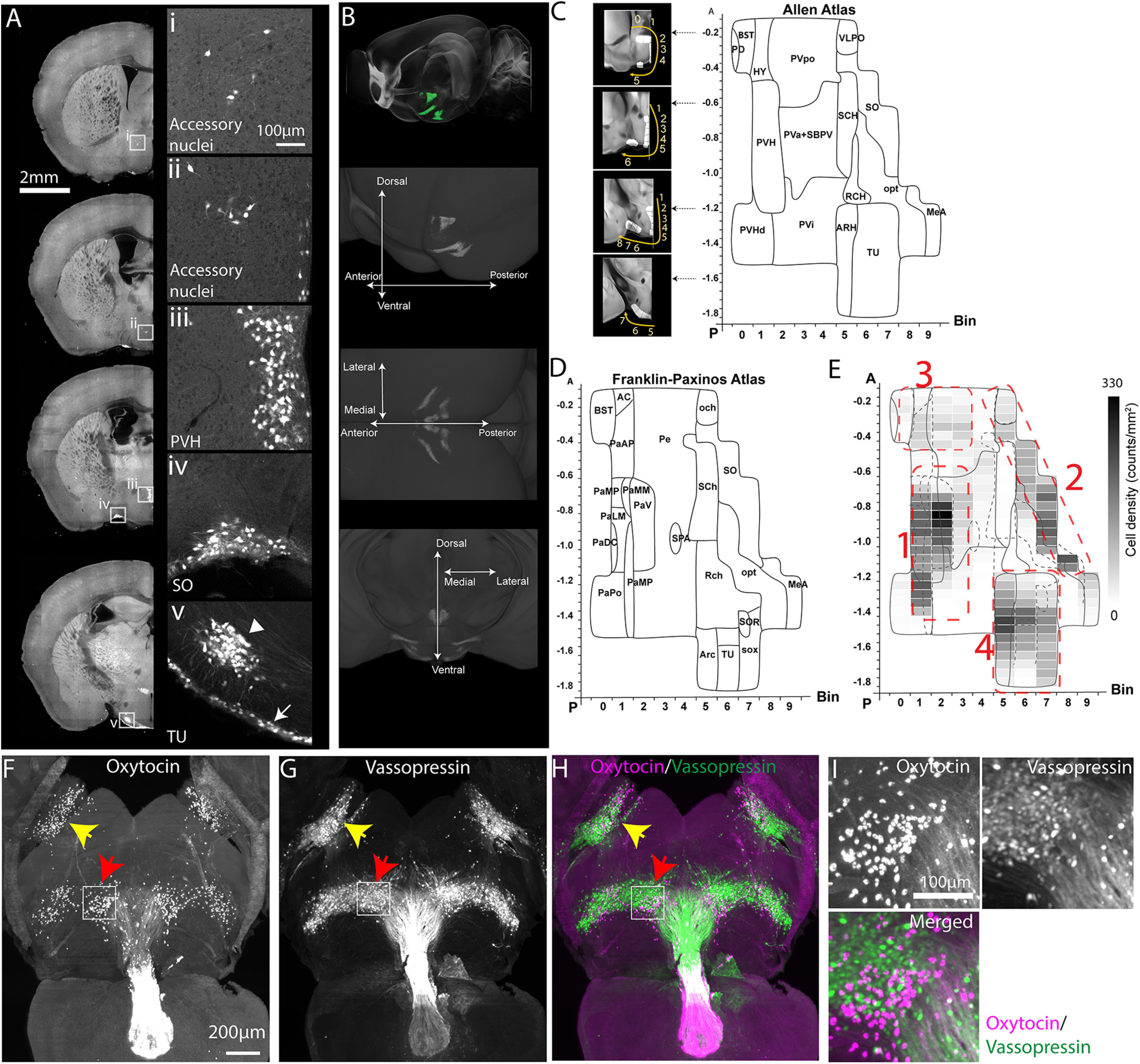
- Oxt neurons form four hypothalamic clusters: PVH, SO, AN, and TU. PVH had the highest transgenic count (511.5 ± 147.1 cells) and TU was nearly as dense (472.9 ± 65.2 cells); immunolabeling gave PVH 818 ± 169 and TU 393 ± 121 cells. PVH regions contained ∼39% of transgenic Oxt cells (742 of 1,899).
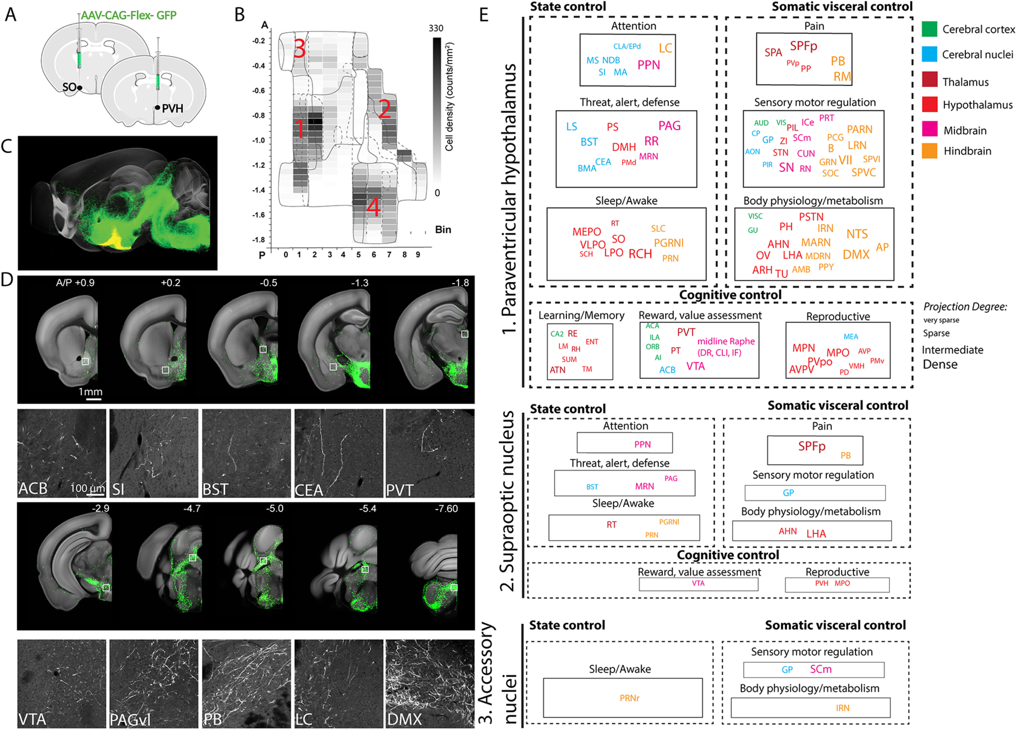
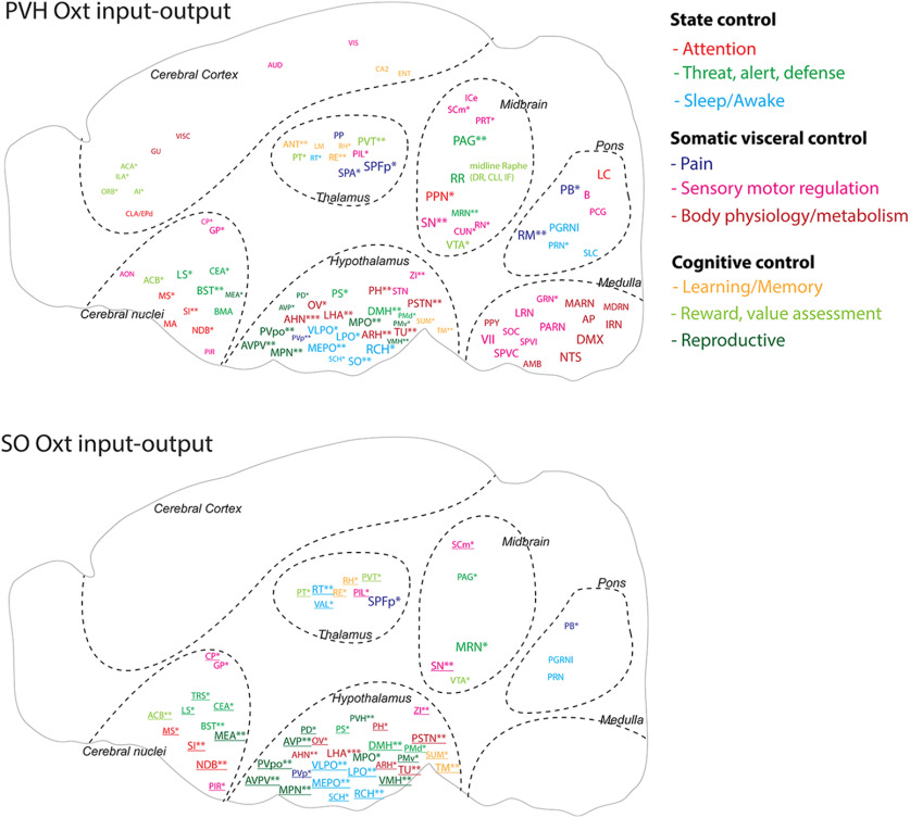
- PVH-Oxt neurons broadly project to nine functional circuits controlling attention, threat/alert/defense, sleep/awake, pain, sensory motor, body physiology/metabolism, learning/memory, reward/value assessment, and reproduction. SO and AN project to only a small subset, and TU-Oxt neurons showed no long-range projections.
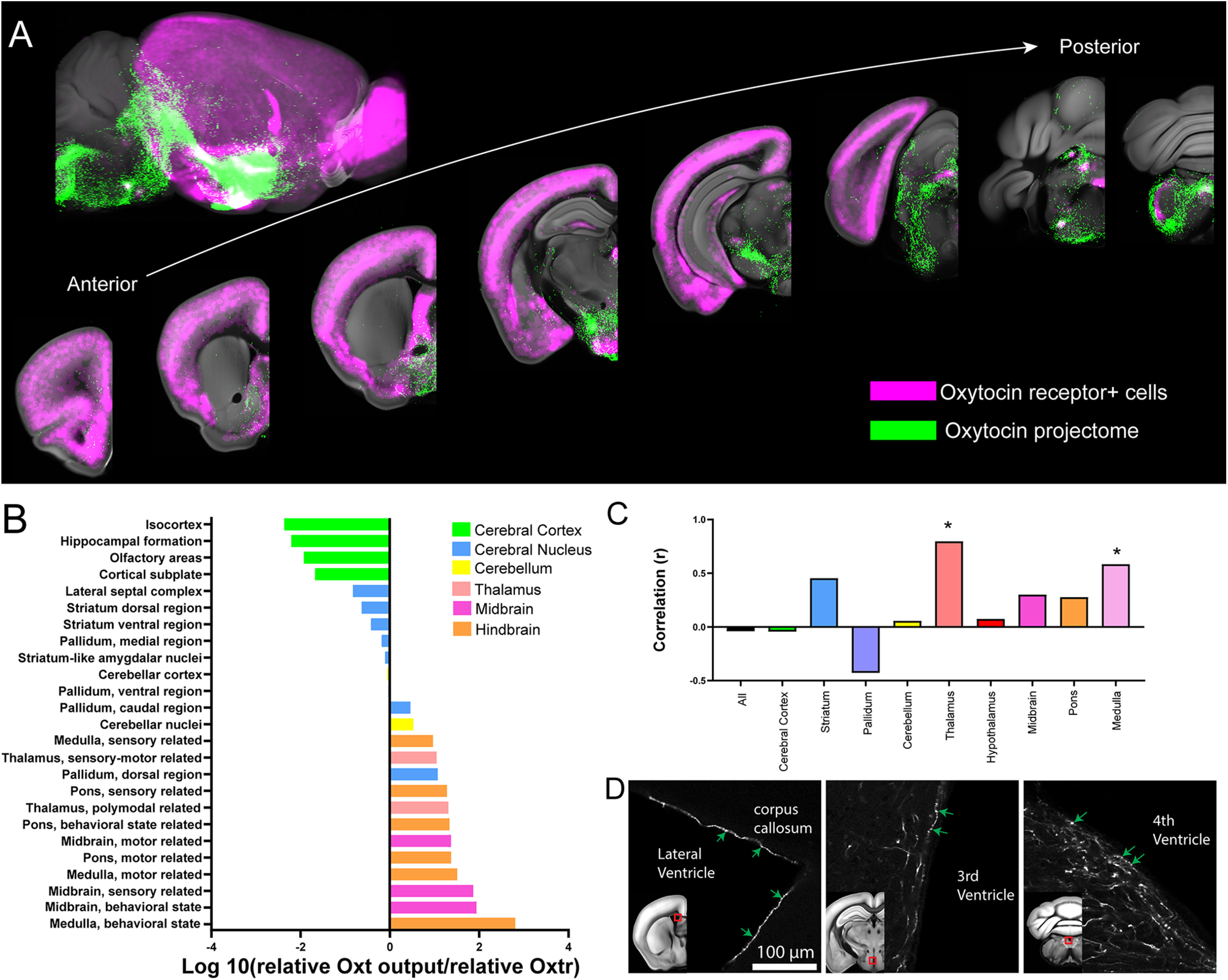
- Oxt output and Oxtr expression showed no significant correlation across the whole brain, except in the thalamus and medulla (Spearman nonparametric correlation, *p < 0.05), suggesting abundant indirect or non-synaptic Oxt signaling; Oxt fibers were found contacting lateral, third, and fourth ventricle surfaces.
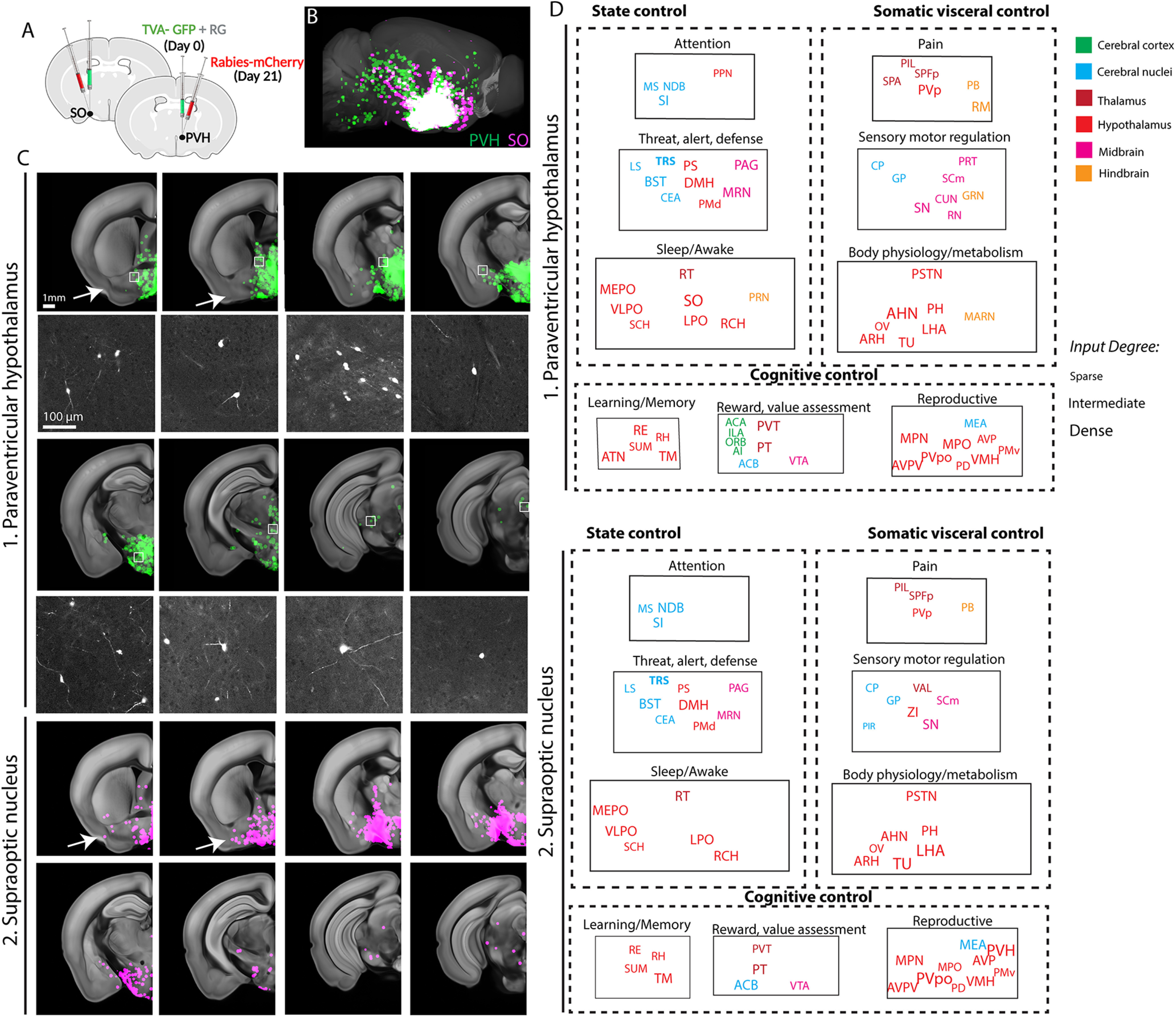
- PVH and SO Oxt neurons receive similar monosynaptic inputs mainly from thalamus, hypothalamus, and cerebral nuclei, with little hindbrain or cortical input. PVH connections are largely reciprocal except for unidirectional output to hindbrain, while SO-Oxt neurons have limited central projection and are proposed to act mainly as hormonal output.
Methods
- Used Oxt-Cre;Ai14, Oxtr-Venus, and C57Bl/6 J mice with serial two-photon tomography (STPT), tissue clearing, light sheet fluorescence microscopy (LSFM), 3D immunolabeling, and registration to the Allen Common Coordinate Framework.
- Anterograde tracing used AAV2-CAG-Flex-EGFP injections into PVH (N = 15), SO (N = 10), AN (N = 2), and TU (N = 5); Oxtr mapping used Oxtr-Venus mice (6 males, 8 females); rabies-based monosynaptic input mapping used N = 6 for PVH and N = 4 for SO.
- Cell counting used machine-learning detection and 3D conversion; projection strength was quantified per anatomically defined area, and Oxt output–Oxtr density relationships were tested with Spearman correlation after D’Agostino–Pearson normality testing.
Limitations
- Paper-stated: TU-Oxt monosynaptic input was not examined, and future ablation studies are needed to determine TU-Oxt function; noncanonical Oxt signaling pathways are described as under-explored.
- Evident from the text: the study is primarily anatomic and correlational, with no behavioral, optogenetic, or causal functional manipulations to test circuit-specific Oxt roles.
- Evident from the text: male, virgin female, and lactating female data were merged after no significant differences were observed, and sample sizes for some tracing conditions were modest; the whole-brain Oxt–Oxtr correlation was not significant except in thalamus and medulla.
Model-written summary. Check it against the paper before relying on it.
Integrity signals
Registry
Cites
Statistics
Figures
Verification
Claims the paper makes
46 Own result · 31 Own interpretation · 23 Not checked yet · 7 Own method · 3 No basis given
A result the paper reports from its own data — it cites no other source for it, so there is none to check. The quote shows it is in the paper.
In the paper
“Surprisingly, quantitative comparison between Oxt output and Oxtr expression showed no significant correlation across the whole brain, suggesting abundant indirect Oxt signaling in Oxtr-expressing areas.”
- Also stated in: § Introduction · plain
“we found lack of quantitative correlation between Oxt output and Oxtr expression across the whole brain”
§ Whole-Brain Wiring Diagram of Oxytocin System in Adult Mice > Abstract
plain- Asserted: passes — The authors put it forward as true.
- Checkable: passes — It can be checked against evidence.
- Load-bearing: passes — The paper’s conclusions depend on it.
- Based: passes — The paper gives a reason why it holds.
- Also stated in: § Introduction · plain
A result the paper reports from its own data — it cites no other source for it, so there is none to check. The quote shows it is in the paper.
In the paper
“Oxt neurons in the paraventricular hypothalamus (PVH) broadly project to nine functional circuits that control cognition, brain state, and somatic visceral response.”
- Also stated in: § Abstract · plain
“We found Oxt connections with nine functional circuits controlling cognition, brain state, and somatic visceral response.”
§ Whole-Brain Wiring Diagram of Oxytocin System in Adult Mice > Abstract
plaindirect- Asserted: passes — The authors put it forward as true.
- Checkable: passes — It can be checked against evidence.
- Load-bearing: passes — The paper’s conclusions depend on it.
- Based: passes — The paper gives a reason why it holds.
- Also stated in: § Abstract · plain
A result the paper reports from its own data — it cites no other source for it, so there is none to check. The quote shows it is in the paper.
In the paper
“In contrast, the cerebrum (the cerebral nuclei and cerebral cortex) that works as high cognitive controller receives more sparse projection (Fig. 2E).”
§ Whole-Brain Wiring Diagram of Oxytocin System in Adult Mice > Results > Quantitative whole-brain projection mapping of Oxt neurons reveals broad long-range projections in nine functional circuits
plain- Asserted: passes — The authors put it forward as true.
- Checkable: passes — It can be checked against evidence.
- Load-bearing: passes — The paper’s conclusions depend on it.
- Based: passes — The paper gives a reason why it holds.
A result the paper reports from its own data — it cites no other source for it, so there is none to check. The quote shows it is in the paper.
In the paper
“Importantly, we confirmed the robust Oxt expression in the TU area that was not colocalized with vasopressin staining (Fig. 1I).”
§ Whole-Brain Wiring Diagram of Oxytocin System in Adult Mice > Results > Quantitative density mapping of Oxt neurons reveals four clusters in the adult mouse brain
boostedn = 7- Asserted: passes — The authors put it forward as true.
- Checkable: passes — It can be checked against evidence.
- Load-bearing: unclear — It is unclear whether the paper’s conclusions depend on it.
- Based: passes — The paper gives a reason why it holds.
A result the paper reports from its own data — it cites no other source for it, so there is none to check. The quote shows it is in the paper.
In the paper
“The PVH regions (PVH, descending division of PVH, anterior, intermediate, and subparaventricular zone) contain the highest density of Oxt neurons (∼39%, 742 out of total 1899 cells) followed by the tuberal nucleus (TU), SO, and other areas (Table 1).”
§ Whole-Brain Wiring Diagram of Oxytocin System in Adult Mice > Results > Quantitative density mapping of Oxt neurons reveals four clusters in the adult mouse brain
plainn = 8- Asserted: passes — The authors put it forward as true.
- Checkable: passes — It can be checked against evidence.
- Load-bearing: passes — The paper’s conclusions depend on it.
- Based: passes — The paper gives a reason why it holds.
A result the paper reports from its own data — it cites no other source for it, so there is none to check. The quote shows it is in the paper.
In the paper
“Notably, the largely overlooked TU area contains almost as high density of Oxt neurons as the PVH area (Fig. 1E).”
§ Whole-Brain Wiring Diagram of Oxytocin System in Adult Mice > Results > Quantitative density mapping of Oxt neurons reveals four clusters in the adult mouse brain
plain- Asserted: passes — The authors put it forward as true.
- Checkable: passes — It can be checked against evidence.
- Load-bearing: passes — The paper’s conclusions depend on it.
- Based: passes — The paper gives a reason why it holds.
The paper states this without a source or data behind it here, so there is nothing to check it against. The quote shows it is in the paper.
In the paper
“The somatic visceral module includes pain, sensory motor, and body physiology/metabolism circuits (Fig. 2E).”
§ Whole-Brain Wiring Diagram of Oxytocin System in Adult Mice > Results > Quantitative whole-brain projection mapping of Oxt neurons reveals broad long-range projections in nine functional circuits
plain- Asserted: passes — The authors put it forward as true.
- Checkable: passes — It can be checked against evidence.
- Load-bearing: unclear — It is unclear whether the paper’s conclusions depend on it.
- Based: passes — The paper gives a reason why it holds.
A result the paper reports from its own data — it cites no other source for it, so there is none to check. The quote shows it is in the paper.
In the paper
“Dense projection occurs largely onto the hindbrain (e.g., the dorsal motor nucleus of the vagus nerve; DMX, the PB), the midbrain (the substantia nigra compacta; SNc), and the hypothalamus (the medial preoptic nucleus; MPN) to directly modulate motor output and sensory input (Fig. 2E).”
§ Whole-Brain Wiring Diagram of Oxytocin System in Adult Mice > Results > Quantitative whole-brain projection mapping of Oxt neurons reveals broad long-range projections in nine functional circuits
plaindirect- Asserted: passes — The authors put it forward as true.
- Checkable: passes — It can be checked against evidence.
- Load-bearing: passes — The paper’s conclusions depend on it.
- Based: passes — The paper gives a reason why it holds.
A result the paper reports from its own data — it cites no other source for it, so there is none to check. The quote shows it is in the paper.
In the paper
“For example, the estimated number of Oxt neurons in the PVH with the immunostaining was 1095 cells out of total 3149 cells (∼35%), which is higher than our transgenic based estimate mostly likely because of sensitive labeling based on antibody detection.”
§ Whole-Brain Wiring Diagram of Oxytocin System in Adult Mice > Results > Quantitative density mapping of Oxt neurons reveals four clusters in the adult mouse brain
hedgedn = 7- Asserted: passes — The authors put it forward as true.
- Checkable: passes — It can be checked against evidence.
- Load-bearing: unclear — It is unclear whether the paper’s conclusions depend on it.
- Based: passes — The paper gives a reason why it holds.
A result the paper reports from its own data — it cites no other source for it, so there is none to check. The quote shows it is in the paper.
In the paper
“When we examined whether relative projection of Oxt neurons is correlated with relative Oxtr density across the entire brain, we found no significant correlation across the whole brain and major brain areas, except for the thalamus and the medulla (Fig. 3C).”
§ Whole-Brain Wiring Diagram of Oxytocin System in Adult Mice > Results > Oxtr expression showed quantitative mismatch with Oxt neuronal output
plain- Asserted: passes — The authors put it forward as true.
- Checkable: passes — It can be checked against evidence.
- Load-bearing: passes — The paper’s conclusions depend on it.
- Based: passes — The paper gives a reason why it holds.
- Rests on an assumption: The quantitative relationship between Oxt projection and Oxtr expression across the whole brain is currently lacking.
- It may state two separate claims as one. 21
- Probably not a substantial claim. 12
A result the paper reports from its own data — it cites no other source for it, so there is none to check. The quote shows it is in the paper.
In the paper
“The TU (in v panel) contains two subpopulations of Oxt neurons with a small cluster (arrowhead) and a thin layer along the bottom edge of the brain (arrow).”
§ Whole-Brain Wiring Diagram of Oxytocin System in Adult Mice > Results > Quantitative density mapping of Oxt neurons reveals four clusters in the adult mouse brain
plain- Asserted: passes — The authors put it forward as true.
- Checkable: passes — It can be checked against evidence.
- Load-bearing: unclear — It is unclear whether the paper’s conclusions depend on it.
- Based: passes — The paper gives a reason why it holds.
It may state two separate claims as one.
In the paper
“The regional boundaries of the two labeling systems generally agreed with each other in the major Oxt-expressing regions (e.g., the PVH and the SO) despite noticeable discrepancies in the caudal hypothalamic area (e.g., the TU; Fig. 1C,D; Chon et al., 2019).”
§ Whole-Brain Wiring Diagram of Oxytocin System in Adult Mice > Results > Quantitative density mapping of Oxt neurons reveals four clusters in the adult mouse brain
What the sources say
- Not checked yetcites Chon et al., 2019
plain- Asserted: passes — The authors put it forward as true.
- Checkable: passes — It can be checked against evidence.
- Load-bearing: unclear — It is unclear whether the paper’s conclusions depend on it.
- Based: passes — The paper gives a reason why it holds.
It may state two separate claims as one.
A result the paper reports from its own data — it cites no other source for it, so there is none to check. The quote shows it is in the paper.
In the paper
“The internal state module contains attention, threat/alert/defense, and sleep/awake circuits (Fig. 2E).”
§ Whole-Brain Wiring Diagram of Oxytocin System in Adult Mice > Results > Quantitative whole-brain projection mapping of Oxt neurons reveals broad long-range projections in nine functional circuits
plain- Asserted: passes — The authors put it forward as true.
- Checkable: passes — It can be checked against evidence.
- Load-bearing: passes — The paper’s conclusions depend on it.
- Based: passes — The paper gives a reason why it holds.
It may state two separate claims as one.
A result the paper reports from its own data — it cites no other source for it, so there is none to check. The quote shows it is in the paper.
In the paper
“Lastly, the cognitive control module has learning/memory, reward/value assessment, and reproduction circuits (Fig. 2E).”
§ Whole-Brain Wiring Diagram of Oxytocin System in Adult Mice > Results > Quantitative whole-brain projection mapping of Oxt neurons reveals broad long-range projections in nine functional circuits
plain- Asserted: passes — The authors put it forward as true.
- Checkable: passes — It can be checked against evidence.
- Load-bearing: passes — The paper’s conclusions depend on it.
- Based: passes — The paper gives a reason why it holds.
It may state two separate claims as one.
A result the paper reports from its own data — it cites no other source for it, so there is none to check. The quote shows it is in the paper.
In the paper
“Overall, the Oxtr showed high expression in the cortical area with minimal Oxt projection, while many midbrain and hindbrain regions have strong Oxt with little Oxtr expression (Fig. 3A,B; Movie 3).”
§ Whole-Brain Wiring Diagram of Oxytocin System in Adult Mice > Results > Oxtr expression showed quantitative mismatch with Oxt neuronal output
plaincorrelational- Asserted: passes — The authors put it forward as true.
- Checkable: passes — It can be checked against evidence.
- Load-bearing: passes — The paper’s conclusions depend on it.
- Based: passes — The paper gives a reason why it holds.
It may state two separate claims as one.
- Rests on an assumption: The quantitative relationship between Oxt projection and Oxtr expression across the whole brain is currently lacking.
A result the paper reports from its own data — it cites no other source for it, so there is none to check. The quote shows it is in the paper.
In the paper
“We observed similar Oxt staining distribution and overall slightly higher counting compared with our transgenic based mapping results (Table 1).”
§ Whole-Brain Wiring Diagram of Oxytocin System in Adult Mice > Results > Quantitative density mapping of Oxt neurons reveals four clusters in the adult mouse brain
plaincorrelationaln = 7- Asserted: passes — The authors put it forward as true.
- Checkable: passes — It can be checked against evidence.
- Load-bearing: passes — The paper’s conclusions depend on it.
- Based: passes — The paper gives a reason why it holds.
It may state two separate claims as one.
A result the paper reports from its own data — it cites no other source for it, so there is none to check. The quote shows it is in the paper.
In the paper
“We found abundant projections from Oxt neurons in the PVH to the midbrain and hindbrain areas while relatively sparse projection to the diencephalon and telencephalon areas (Fig. 2D)”
§ Whole-Brain Wiring Diagram of Oxytocin System in Adult Mice > Results > Quantitative whole-brain projection mapping of Oxt neurons reveals broad long-range projections in nine functional circuits
plainn = 15- Asserted: passes — The authors put it forward as true.
- Checkable: passes — It can be checked against evidence.
- Load-bearing: passes — The paper’s conclusions depend on it.
- Based: passes — The paper gives a reason why it holds.
It may state two separate claims as one.
A result the paper reports from its own data — it cites no other source for it, so there is none to check. The quote shows it is in the paper.
In the paper
“SO-Oxt neurons receive similar mid-level circuit input compared with the PVH-Oxt neurons while showing limited central projection to the midbrain and pons (Fig. 5).”
§ Whole-Brain Wiring Diagram of Oxytocin System in Adult Mice > Results > Input-output wiring diagrams of PVH-Oxt and SO-Oxt neurons provide overall neural circuit control patterns
plain- Asserted: passes — The authors put it forward as true.
- Checkable: passes — It can be checked against evidence.
- Load-bearing: passes — The paper’s conclusions depend on it.
- Based: passes — The paper gives a reason why it holds.
It may state two separate claims as one.
A result the paper reports from its own data — it cites no other source for it, so there is none to check. The quote shows it is in the paper.
In the paper
“For example, the cerebral cortical area contains abundant Oxtr with little to no Oxt axons.”
§ Whole-Brain Wiring Diagram of Oxytocin System in Adult Mice > Discussion
plain- Asserted: passes — The authors put it forward as true.
- Checkable: passes — It can be checked against evidence.
- Load-bearing: unclear — It is unclear whether the paper’s conclusions depend on it.
- Based: passes — The paper gives a reason why it holds.
It may state two separate claims as one.
None of the sources cited for this claim have been checked yet.
In the paper
“Oxytocin (Oxt) is a highly conserved neuropeptide, playing key roles in regulating social behavior and other physiological functions (Althammer et al., 2018; Jurek and Neumann, 2018; Quintana and Guastella, 2020).”
§ Whole-Brain Wiring Diagram of Oxytocin System in Adult Mice > Introduction
What the sources say
- Not checked yetcites Althammer et al., 2018
- Not checked yetcites Jurek and Neumann, 2018
- Not checked yetcites Quintana and Guastella, 2020
plaindirect- Asserted: passes — The authors put it forward as true.
- Checkable: passes — It can be checked against evidence.
- Load-bearing: fails — The paper’s conclusions may not depend on it.
- Based: passes — The paper gives a reason why it holds.
Probably not a substantial claim. The paper’s conclusions may not depend on it. It may state two separate claims as one.
None of the sources cited for this claim have been checked yet.
In the paper
“Oxt neurons receive input from distinct brain regions (e.g., the thalamus) and integrate sensory input with internal information to release Oxt in a context dependent manner to modulate specific downstream circuitry (Tang et al., 2020; Grinevich and Neumann, 2021).”
§ Whole-Brain Wiring Diagram of Oxytocin System in Adult Mice > Introduction
What the sources say
- Not checked yetcites Tang et al., 2020
- Not checked yetcites Grinevich and Neumann, 2021
plaindirect- Asserted: passes — The authors put it forward as true.
- Checkable: passes — It can be checked against evidence.
- Load-bearing: fails — The paper’s conclusions may not depend on it.
- Based: passes — The paper gives a reason why it holds.
Probably not a substantial claim. The paper’s conclusions may not depend on it. It may state two separate claims as one.
None of the sources cited for this claim have been checked yet.
In the paper
“The majority of Oxt producing neurons are located in the paraventricular nucleus of the hypothalamus (PVH) and the supraoptic nucleus (SO) while fewer Oxt neurons reside in the extended amygdala (Biag et al., 2012; Madrigal and Jurado, 2021).”
§ Whole-Brain Wiring Diagram of Oxytocin System in Adult Mice > Introduction
What the sources say
- Not checked yetcites Biag et al., 2012
- Not checked yetcites Madrigal and Jurado, 2021
plain- Asserted: passes — The authors put it forward as true.
- Checkable: passes — It can be checked against evidence.
- Load-bearing: fails — The paper’s conclusions may not depend on it.
- Based: passes — The paper gives a reason why it holds.
Probably not a substantial claim. The paper’s conclusions may not depend on it. It may state two separate claims as one.
None of the sources cited for this claim have been checked yet.
In the paper
“In addition to the well-known peripheral release of Oxt as a hormone via the posterior pituitary, Oxt neurons send direct projections to specific brain areas that frequently express Oxtr, thereby modulating circuit-specific functions (Grinevich et al., 2016; Liao et al., 2020).”
§ Whole-Brain Wiring Diagram of Oxytocin System in Adult Mice > Introduction
What the sources say
- Not checked yetcites Grinevich et al., 2016
- Not checked yetcites Liao et al., 2020
boosteddirect- Asserted: passes — The authors put it forward as true.
- Checkable: passes — It can be checked against evidence.
- Load-bearing: passes — The paper’s conclusions depend on it.
- Based: passes — The paper gives a reason why it holds.
It may state two separate claims as one.
In the paper
“Although most Oxt projecting areas are known to contain Oxtr expression (Grinevich et al., 2016), the quantitative relationship between Oxt projection and Oxtr expression across the whole brain is currently lacking.”
§ Whole-Brain Wiring Diagram of Oxytocin System in Adult Mice > Results > Oxtr expression showed quantitative mismatch with Oxt neuronal output
What the sources say
- Not checked yetcites Grinevich et al., 2016
boostedcorrelational- Asserted: passes — The authors put it forward as true.
- Checkable: passes — It can be checked against evidence.
- Load-bearing: passes — The paper’s conclusions depend on it.
- Based: passes — The paper gives a reason why it holds.
Probably not a substantial claim.
In the paper
“Since Oxt can be released via axons, dendrites, and even neuronal processes (Jurek and Neumann, 2018), we injected a Cre-dependent adeno associated virus 2”
§ Whole-Brain Wiring Diagram of Oxytocin System in Adult Mice > Results > Quantitative whole-brain projection mapping of Oxt neurons reveals broad long-range projections in nine functional circuits
What the sources say
- Not checked yetcites Jurek and Neumann, 2018
plaindirect- Asserted: passes — The authors put it forward as true.
- Checkable: passes — It can be checked against evidence.
- Load-bearing: passes — The paper’s conclusions depend on it.
- Based: passes — The paper gives a reason why it holds.
Probably not a substantial claim. It may state two separate claims as one.
What the paper did — it cites no source for this, so there is none to check. The quote shows it is in the paper.
In the paper
“We included male, virgin female, and lactating female mice in our study (see Table 2 and Materials and Methods for more detail), and observed no significant difference between sex or lactating state. Thus, we merged all data from the same anatomic areas.”
§ Whole-Brain Wiring Diagram of Oxytocin System in Adult Mice > Results > Quantitative whole-brain projection mapping of Oxt neurons reveals broad long-range projections in nine functional circuits
plain- Asserted: passes — The authors put it forward as true.
- Checkable: passes — It can be checked against evidence.
- Load-bearing: passes — The paper’s conclusions depend on it.
- Based: passes — The paper gives a reason why it holds.
It may state two separate claims as one.
What the paper did — it cites no source for this, so there is none to check. The quote shows it is in the paper.
In the paper
“Since we did not observe significant difference in Oxt neuronal number, we combined data from both sexes to generate representative cell counting (Table 1; Extended Data Table 1-1).”
§ Whole-Brain Wiring Diagram of Oxytocin System in Adult Mice > Materials and Methods > Experimental design and statistical analyses
plain- Asserted: passes — The authors put it forward as true.
- Checkable: passes — It can be checked against evidence.
- Load-bearing: passes — The paper’s conclusions depend on it.
- Based: passes — The paper gives a reason why it holds.
It may state two separate claims as one.
A conclusion the paper draws from its own results — it cites no other source for it, so there is none to check. The quote shows it is in the paper.
In the paper
“PVH-Oxt neurons project broadly to all nine identified functional circuits throughout the brain, indicating that Oxt neurons can modulate information processing at different level of circuits with overall stronger influence in the midbrain and hindbrain circuits (Fig. 5).”
§ Whole-Brain Wiring Diagram of Oxytocin System in Adult Mice > Results > Input-output wiring diagrams of PVH-Oxt and SO-Oxt neurons provide overall neural circuit control patterns
hedgeddirect- Asserted: passes — The authors put it forward as true.
- Checkable: passes — It can be checked against evidence.
- Load-bearing: passes — The paper’s conclusions depend on it.
- Based: passes — The paper gives a reason why it holds.
It may state two separate claims as one.
None of the sources cited for this claim have been checked yet.
In the paper
“The most well-established role of Oxt signaling is to promote social behavior (Kemp and Guastella, 2010; Shamay-Tsoory and Abu-Akel, 2016).”
§ Whole-Brain Wiring Diagram of Oxytocin System in Adult Mice > Discussion
What the sources say
- Not checked yetcites Kemp and Guastella, 2010
- Not checked yetcites Shamay-Tsoory and Abu-Akel, 2016
boosteddirect- Asserted: passes — The authors put it forward as true.
- Checkable: passes — It can be checked against evidence.
- Load-bearing: fails — The paper’s conclusions may not depend on it.
- Based: passes — The paper gives a reason why it holds.
Probably not a substantial claim. The paper’s conclusions may not depend on it.
In the paper
“A recent single cell reconstruction study demonstrated that even a single magnocellular Oxt neuron can make multiple collateral projections to extrahypothalamic areas to coordinate neuromodulation across functionally related brain circuits (Zhang et al., 2021).”
§ Whole-Brain Wiring Diagram of Oxytocin System in Adult Mice > Discussion
What the sources say
- Not checked yetcites Zhang et al., 2021
boosteddirect- Asserted: passes — The authors put it forward as true.
- Checkable: passes — It can be checked against evidence.
- Load-bearing: fails — The paper’s conclusions may not depend on it.
- Based: passes — The paper gives a reason why it holds.
Probably not a substantial claim. The paper’s conclusions may not depend on it.
None of the sources cited for this claim have been checked yet.
In the paper
“Oxt signaling is known to modulate many distinct brain functions such as anxiolytic effect, social memory, and attention (Lee et al., 2009; Marlin et al., 2015; Grinevich and Stoop, 2018; Schiavo et al., 2020).”
§ Whole-Brain Wiring Diagram of Oxytocin System in Adult Mice > Discussion
What the sources say
- Not checked yetcites Lee et al., 2009
- Not checked yetcites Marlin et al., 2015
- Not checked yetcites Grinevich and Stoop, 2018
boosteddirect- Asserted: passes — The authors put it forward as true.
- Checkable: passes — It can be checked against evidence.
- Load-bearing: fails — The paper’s conclusions may not depend on it.
- Based: passes — The paper gives a reason why it holds.
Probably not a substantial claim. The paper’s conclusions may not depend on it.
None of the sources cited for this claim have been checked yet.
In the paper
“Oxt neurons are mostly located in hypothalamic nuclei with a complex 3D shape (Biag et al., 2012; Madrigal and Jurado, 2021).”
§ Whole-Brain Wiring Diagram of Oxytocin System in Adult Mice > Discussion
What the sources say
- Not checked yetcites Biag et al., 2012
- Not checked yetcites Madrigal and Jurado, 2021
plain- Asserted: passes — The authors put it forward as true.
- Checkable: passes — It can be checked against evidence.
- Load-bearing: fails — The paper’s conclusions may not depend on it.
- Based: passes — The paper gives a reason why it holds.
Probably not a substantial claim. The paper’s conclusions may not depend on it. It may state two separate claims as one.
What the paper did — it cites no source for this, so there is none to check. The quote shows it is in the paper.
In the paper
“None of the TVA infected neurons were AVP positive and were largely Oxt positive (Extended Data Fig. 4-1).”
§ Whole-Brain Wiring Diagram of Oxytocin System in Adult Mice > Results > Oxt neurons mainly receive monosynaptic inputs from the thalamus, hypothalamus, and cerebral nuclei
plain- Asserted: passes — The authors put it forward as true.
- Checkable: passes — It can be checked against evidence.
- Load-bearing: passes — The paper’s conclusions depend on it.
- Based: passes — The paper gives a reason why it holds.
It may state two separate claims as one.
What the paper did — it cites no source for this, so there is none to check. The quote shows it is in the paper.
In the paper
“All the neurons observed were confined to the injection site and both samples were devoid of any long-range input cells.”
§ Whole-Brain Wiring Diagram of Oxytocin System in Adult Mice > Results > Oxt neurons mainly receive monosynaptic inputs from the thalamus, hypothalamus, and cerebral nuclei
plainSingle case- Asserted: passes — The authors put it forward as true.
- Checkable: passes — It can be checked against evidence.
- Load-bearing: passes — The paper’s conclusions depend on it.
- Based: passes — The paper gives a reason why it holds.
It may state two separate claims as one.
None of the sources cited for this claim have been checked yet.
In the paper
“However, Oxt can still mediate sensory stimuli in the cortex to modify mouse behavior (Marlin et al., 2015; Schiavo et al., 2020).”
§ Whole-Brain Wiring Diagram of Oxytocin System in Adult Mice > Discussion
What the sources say
- Not checked yetcites Marlin et al., 2015
- Not checked yetcites Schiavo et al., 2020
plainconditional- Asserted: passes — The authors put it forward as true.
- Checkable: passes — It can be checked against evidence.
- Load-bearing: fails — The paper’s conclusions may not depend on it.
- Based: passes — The paper gives a reason why it holds.
Probably not a substantial claim. The paper’s conclusions may not depend on it.
A conclusion the paper draws from its own results — it cites no other source for it, so there is none to check. The quote shows it is in the paper.
In the paper
“Particularly, almost all afferent brain regions to PVH-Oxt neurons also receive efferent projections, suggesting strong reciprocal control of target regions by PVH-Oxt neurons except the hindbrain for unidirectional output.”
§ Whole-Brain Wiring Diagram of Oxytocin System in Adult Mice > Discussion
hedgeddirect- Asserted: passes — The authors put it forward as true.
- Checkable: passes — It can be checked against evidence.
- Load-bearing: passes — The paper’s conclusions depend on it.
- Based: passes — The paper gives a reason why it holds.
It may state two separate claims as one.
None of the sources cited for this claim have been checked yet.
In the paper
“Previous studies suggest that Oxtr neurons in the isocortex may receive Oxt signals indirectly from ventricular pathways via cerebral spinal fluid with delayed and long-lasting effects (Mens et al., 1983; Zheng et al., 2014).”
§ Whole-Brain Wiring Diagram of Oxytocin System in Adult Mice > Discussion
What the sources say
- Not checked yetcites Mens et al., 1983
- Not checked yetcites Zheng et al., 2014
hedgedconditional- Asserted: passes — The authors put it forward as true.
- Checkable: passes — It can be checked against evidence.
- Load-bearing: fails — The paper’s conclusions may not depend on it.
- Based: passes — The paper gives a reason why it holds.
Probably not a substantial claim. The paper’s conclusions may not depend on it.
In the paper
“For example, Oxt can elicit TRPV1 activity in the spinal cord to modulate nociception (Nersesyan et al., 2017).”
§ Whole-Brain Wiring Diagram of Oxytocin System in Adult Mice > Discussion
What the sources say
- Not checked yetcites Nersesyan et al., 2017
plaindirect- Asserted: passes — The authors put it forward as true.
- Checkable: passes — It can be checked against evidence.
- Load-bearing: fails — The paper’s conclusions may not depend on it.
- Based: passes — The paper gives a reason why it holds.
Probably not a substantial claim. The paper’s conclusions may not depend on it.
The paper relies on this without support.
A comprehensive input-output wiring diagram of Oxt neurons and their quantitative relationship with Oxtr expression is lacking, which limits understanding of circuit-specific Oxt functions.
“However, the lack of comprehensive input-output wiring diagrams of Oxt neurons and their quantitative relationship with Oxt receptor (Oxtr) expression presents challenges to understanding circuit-specific Oxt functions.”
The paper relies on this without support.
A quantitative and comprehensive wiring diagram of Oxt neurons in a standard 3D reference brain is still lacking.
“Despite these prior studies, we still lack a quantitative and comprehensive wiring diagram of the Oxt neurons in a standard 3D reference brain.”
The paper relies on this without support.
Limited neural circuit-based understanding of Oxt signaling hampers development of targeted therapeutic approaches aimed at altering specific Oxt functions without affecting other biological pathways.
“However, our limited neural circuit based understanding of Oxt signaling in the brain hampers the development of targeted therapeutic approaches aimed at altering specific Oxt functions without affecting other biological pathways.”
The paper relies on this without support.
The quantitative relationship between Oxt projection and Oxtr expression across the whole brain is currently lacking.
“Although most Oxt projecting areas are known to contain Oxtr expression (Grinevich et al., 2016), the quantitative relationship between Oxt projection and Oxtr expression across the whole brain is currently lacking.”
The paper relies on this without support.
Oxt neurons integrate external stimuli and internal state.
“Since Oxt neurons are known to integrate external stimuli and internal state, we ask whether Oxt neurons in the PVH and the SO receive monosynaptic input from sensory and integrative information processing brain areas.”
Claims other papers make about this one
Each is checked against this paper’s own text: does it say what the citing paper says it does?
In the citing paper
“Recently, Son et al (59) found no correlation between OXT neuronal projections and OXTR expression, consistent with paracrine signaling.”
From Oxytocin, Vasopressin, and Social Behavior: From Neural Circuits to Clinical Opportunities (2022) · cites this paper as [59]
Self-check
Percentages, sums and reported differences re-computed from the paper’s own numbers.
Each contradiction between two passages listed here was confirmed by an independent check.
Rated minor: it would not touch a result — a value no result rests on, a p-value that stays on the same side of the significance level, or a conflict that is clerical, resolved elsewhere in the paper, or outside its own findings.
In the paper
§ Whole-Brain Wiring Diagram of Oxytocin System in Adult Mice > Results > Oxtr expression showed quantitative mismatch with Oxt neuronal output
“These mapped Oxtr positive neurons (Fig. 3A, magenta) were registered onto the same reference brain along with Oxt-projections (Fig. 3A, green).”
§ Whole-Brain Wiring Diagram of Oxytocin System in Adult Mice > Results > Oxtr expression showed quantitative mismatch with Oxt neuronal output
“A, Composite images of representative Oxt neuronal projection outputs (red: combined from both the PVH and the SO) and Oxtr expression (green) across the mouse brain.”
Citations
Cites
The mouse brain in stereotaxic coordinates
Konsman, J. (2003) · Psychoneuroendocrinology · doi:10.1016/s0306-4530(03)00088-x · Cited by 16,796
Elastix: a toolbox for intensity-based medical image registration
Klein, S., Staring, M., Murphy, K., Viergever, M. A., & Pluim, J. (2010) · IEEE Transactions on Medical Imaging · doi:10.1109/tmi.2009.2035616 · Cited by 4,311
The oxytocin receptor system: structure, function, and regulation
Gimpl, G., & Fahrenholz, F. (2001) · Physiological Reviews · doi:10.1152/physrev.2001.81.2.629 · Cited by 3,089
The Allen Mouse Brain Common Coordinate Framework: A 3D Reference Atlas
Wang, Q., Ding, S., Li, Y., Royall, J. J., Feng, D. H., Lesnar, P., Graddis, N., Naeemi, M., Facer, B. A. C., Ho, A., et al. (2020) · Cell · doi:10.1016/j.cell.2020.04.007 · Cited by 1,480
Monosynaptic Restriction of Transsynaptic Tracing from Single, Genetically Targeted Neurons
Wickersham, I. R., Lyon, D. C., Barnard, R. J. O., Mori, T., Finke, S., Conzelmann, K., Young, J. A. T., & Callaway, E. M. (2007) · Neuron · doi:10.1016/j.neuron.2007.01.033 · Cited by 1,176
Social reward requires coordinated activity of nucleus accumbens oxytocin and serotonin
Dölen, G., Darvishzadeh, A., Huang, K. W., & Malenka, R. C. (2013) · Nature · doi:10.1038/nature12518 · Cited by 1,161
Evoked axonal oxytocin release in the central amygdala attenuates fear response
Knobloch, H. S., Charlet, A., Hoffmann, L. C., Eliava, M., Khrulev, S., Cetin, A. H., Osten, P., Schwarz, M. K., Seeburg, P. H., Stoop, R., et al. (2012) · Neuron · doi:10.1016/j.neuron.2011.11.030 · Cited by 1,049
Oxytocin: The Great Facilitator of Life
Lee, H.-J., Macbeth, A. H., Pagani, J., & Young, W. S. (2009) · Progress in Neurobiology · doi:10.1016/j.pneurobio.2009.04.001 · Cited by 1,037
The oxytocin receptor: from intracellular signaling to behavior
Jurek, B., & Neumann, I. D. (2018) · Physiological Reviews · doi:10.1152/physrev.00031.2017 · Cited by 999
Mapping of Brain Activity by Automated Volume Analysis of Immediate Early Genes
Renier, N., Adams, E. L., Kirst, C., Wu, Z., Azevedo, R., Kohl, J., Autry, A. E., Kadiri, L., Umadevi Venkataraju, K., Zhou, Y., et al. (2016) · Cell · doi:10.1016/j.cell.2016.05.007 · Cited by 918
Cited by
Sorted by citations
Items 101–125 of 125
顾, 文., 郝, 振., 毛, 俊., & 田, 莹. (2025) · Research Square · doi:10.21203/rs.3.rs-6817321/v1
Vandendoren, M., Landen, J. G., Rogers, J. F., Killmer, S., Alamiri, B., Pohlman, C., Tattersall, G. J., Bedford, N. L., & Nelson, A. C. (2025) · eLife · doi:10.7554/elife.108212
Numan, M. (2025) · Hormones and Behavior · doi:10.1016/j.yhbeh.2025.105856
Hu, S., Wang, Y., Han, X., Dai, M., Zhang, Y., Ma, Y., Weng, S., & Xiao, L. (2022) · Figshare · doi:10.6084/m9.figshare.21175657
Hu, S., Wang, Y., Han, X., Dai, M., Zhang, Y., Ma, Y., Weng, S., & Xiao, L. (2022) · Figshare · doi:10.6084/m9.figshare.21175651
Knoop, M. S., Possovre, M.-L., Trak, E., Pitetti, J.-L., van de Looij, Y., Sanches, E. F., Tsartsalis, S., Pansiot, J., Schirmbeck, G. H., & Baud, O. (2025) · Neurobiology of Disease · doi:10.1016/j.nbd.2025.107250
Oxytocin mediates the acquisition and strategy formation of cooperation in ratsPreprint
Lin, Y., Wei, L., Wang, Q., & Wang, Z. (2026) · bioRxiv (Cold Spring Harbor Laboratory) · doi:10.64898/2026.03.02.708935
Gu, W., Mao, J., Wang, X., Lü, F., He, P., Cai, L., Dai, L., Jia, M., Tian, Y., & Hao, Z. (2026) · Journal of Oceanology and Limnology · doi:10.1007/s00343-026-5280-8
Narattil, N. R., Chandran, S. K., & Maroun, M. (2026) · Communications Biology · doi:10.1038/s42003-026-10088-0
Eramo, B., Sepe, C., Friuli, M., Campagna, A., Romano, A., & Gaetani, S. (2026) · Pharmacological Research · doi:10.1016/j.phrs.2026.108279
Choi, U.-S., Kiyama, S., Sung, Y., Fukunaga, M., & Ogawa, S. (2026) · Discover Mental Health · doi:10.1007/s44192-026-00521-5
Experimental quality control induces changes in Allen mouse brain connectomes
Nathan, V., Tullo, S., Portillo, L. H., Devenyi, G. A., Yee, Y., & Chakravarty, M. M. (2026) · Imaging Neuroscience · doi:10.1162/imag.a.1310
Judd, E. N., Pauli, J. L., Kenmochi, S. J., Bruchas, M. R., & Palmiter, R. D. (2026) · bioRxiv (Cold Spring Harbor Laboratory) · doi:10.64898/2026.08.06.743398
Hypothalamic supraoptic and paraventricular nuclei
Krabichler, Q., & Grinevich, V. (2026) · Elsevier eBooks · doi:10.1016/b978-0-443-24078-2.00012-7
Yue, Z., Zhao, D., Chen, X., Zhang, Y., Zhang, H., Yinchang, W., Liu, Y., Yang, Y., Geng, C., Song, Y., et al. (2026) · CNS Neuroscience & Therapeutics · doi:10.1002/cns.71127
Comparative atlas of PVN oxytocin and vasopressin systems reveals sex-conserved input–output wiring
Freda, S. N., Lowe, Y. F., Priest, M. F., Badong, D., Liu, Y., Xiao, L., & Kozorovitskiy, Y. (2026) · Proceedings of the National Academy of Sciences · doi:10.1073/pnas.2536750123
Luis, J., & Romero‐Morales, L. (2026) · Elsevier eBooks · doi:10.1016/b978-0-443-33657-7.00015-4
Vandendoren, M., Landen, J. G., Rogers, J. F., Killmer, S., Alamiri, B., Pohlman, C., Tattersall, G. J., Bedford, N. L., & Nelson, A. C. (2026) · eLife · doi:10.7554/elife.108212.3
Iba, K., Masuda, Y., Kitano, R., Ohbayashi, K., Ago, Y., Nagai, H., Furuyashiki, T., Abe, C., & Iwasaki, Y. (2026) · iScience · doi:10.1016/j.isci.2026.117713
Ameloot, A., Disarbois, É., Elseedy, H., Duperron, C., Grinevich, V., Sirigu, A., & Duhamel, J. (2026) · bioRxiv (Cold Spring Harbor Laboratory) · doi:10.64898/2026.09.17.751377
Endogenous oxytocin reconfigures brainwide network dynamics toward salience-related statesPreprint
de Guzman, E. A., Montani, C., Hayward, A., Migliarini, S., Morelli, G., Gutierrez‐Barragan, D., Galbusera, A., Alvino, F. G., Coletta, L., Kim, Y., et al. (2026) · bioRxiv (Cold Spring Harbor Laboratory) · doi:10.64898/2026.09.22.753403
Self-citationDeciphering nervous systems wiring: Tract-tracing and complementary methods
Schmitt, O., Eipert, P., Maestro, R. T., Bellmann, M., Haase, S., Morawska, P., & Prathapan, V. (2026) · Neuroscience & Biobehavioral Reviews · doi:10.1016/j.neubiorev.2026.106943
Metadata
- Authors
- Son, S., Manjila, S. B., Newmaster, K. T., Wu, Y.-T., Vanselow, D. J., Ciarletta, M., Anthony, T. E., Cheng, K. C., & Kim, Y.
- Affiliations
- Pennsylvania State University (US)
- Harvard University (US)
- Corresponding author
- Kim, Yongsoo
- ORCID
- Venue
- The Journal of Neuroscience
- Publisher
- Society for Neuroscience
- Published
- 2022-05-23
- Volume
- 42
- Issue
- 25
- Pages
- 5021-5033
- ISSN
- 0270-6474 (ISSN-L)
- 1529-2401
- PMC
- PMC9233446
- OpenAlex
- W4281401274
- Semantic Scholar
- c94fb30f0763f603278c5485b90f905de9f79b7a
- Cited by
- 125
- References
- 62
- Citations by year
- 2026: 29 · 2025: 32 · 2024: 34 · 2023: 17 · 2022: 12
- FWCI
- 22.41
- Type
- journal article
- Language
- en
- Open access
- hybrid
- doi.org
- Licence
- cc-by
- Locations
- Journal of Neuroscience (publishedVersion, cc-by)
- bioRxiv (Cold Spring Harbor Laboratory) (acceptedVersion, cc-by-nc-nd)
- PubMed Central (publishedVersion, cc-by)
- PubMed (publishedVersion)
- Funders
- University of Pennsylvania
- Pennsylvania State University
- National Institutes of Health (R24OD018559)
- Penn State College of Medicine
- National Institute of Mental Health (R01MH116176)
- Eunice Kennedy Shriver National Institute of Child Health and Human Development (P50HD105351)
- Topics
- Life Sciences › Neuroscience › Endocrine and Autonomic Systems › Neuroendocrine regulation and behavior
- Life Sciences › Neuroscience › Cellular and Molecular Neuroscience › Neuroscience and Neuropharmacology Research
- Life Sciences › Neuroscience › Cellular and Molecular Neuroscience › Neuropeptides and Animal Physiology
- Keywords
- oxytocin neurons, oxytocin receptor, whole-brain connectivity, paraventricular hypothalamus, supraoptic nucleus, accessory olfactory bulb, monosynaptic input, brain circuits, sex differences, mice
- MeSH terms
- Animals, Brain, Female, Male, Neurons, Oxytocin, Signal Transduction, Receptors, Oxytocin, Mice
About these checks
Registry, retraction, Cites and statistics results are computed, not judged, with one exception: a printed reference that no index lists is read once by a model before it counts as a citation error. Claims are extracted by a model and kept only when their quote appears verbatim in the text; each cited source is then checked, with a model’s help, for whether it says what the claim relies on. Those verdicts can be wrong — read the claim’s quote and the cited source before relying on one.
A language model drew the claims from the paper on 2026-10-09, under version 3 of the claim definition (model deepseek-v4.1-flash, prompt extract-v3).
A second model labelled each claim on 2026-10-09 (model jev-1.13-20260917, definition revision gold-guideline-v0.2).
Statistics and arithmetic are recomputed, not judged.
Confidence bands are shown only for fields calibrated on our annotated test set. None is calibrated yet, so no band is shown.
What counts as a claim ›How we measure confidence ›How we check this ›
Are you an author, or do you see a problem with this page? Request a review
