

The DOI matches its Crossref record; 35 of the 35 references it cites were found in a registry or an index.
Neurological outcomes after hematopoietic stem cell transplantation for cerebral X-linked adrenoleukodystrophy, late onset metachromatic leukodystrophy and Hurler syndrome
Abstract
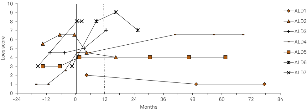
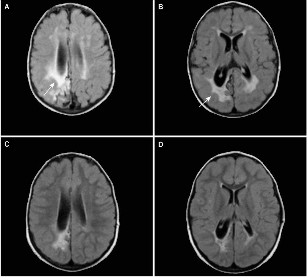
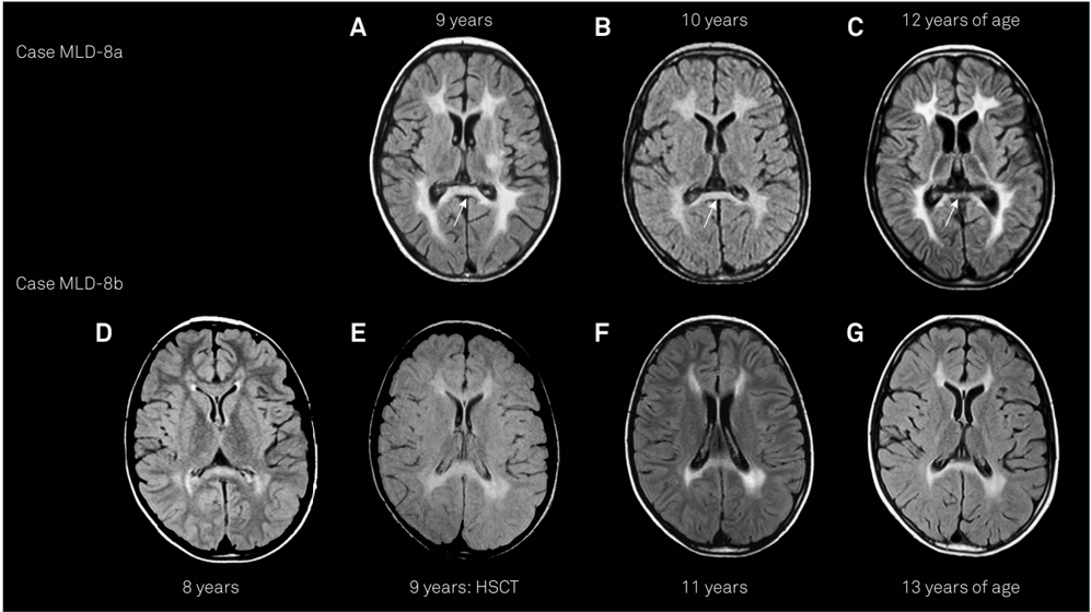
ABSTRACT Hematopoietic stem cell transplantation (HSCT) is the only available treatment for the neurological involvement of disorders such as late-onset metachromatic leukodystrophy (MLD), mucopolysaccharidosis type I-Hurler (MPS-IH), and X-linked cerebral adrenoleukodystrophy (CALD). Objective To describe survival and neurological outcomes after HSCT for these disorders. Methods Seven CALD, 2 MLD and 2 MPS-IH patients underwent HSCT between 2007 and 2014. Neurological examinations, magnetic resonance imaging, molecular and biochemical studies were obtained at baseline and repeated when appropriated. Results Favorable outcomes were obtained with 4/5 related and 3/6 unrelated donors. Two patients died from procedure-related complications. Nine transplanted patients were alive after a median of 3.7 years: neurological stabilization was obtained in 5/6 CALD, 1/2 MLD, and one MPS-IH patient. Brain lesions of the MPS-IH patient were reduced four years after HSCT. Conclusion Good outcomes were obtained when HSCT was performed before adulthood, early in the clinical course, and/or from a related donor.
Overview
The claims and statistics of this paper haven’t been checked yet.
Run the check — free, no account needed. It reads the open-access text and checks the sources it cites.
Integrity signals
Registry
Cites
Statistics
Figures
Verification
Claims the paper makes
The claims and statistics of this paper haven’t been checked yet.
Run the check — free, no account needed. It reads the open-access text and checks the sources it cites.
Claims other papers make about this one
Each is checked against this paper’s own text: does it say what the citing paper says it does?
In the citing paper
“Allogeneic transplantation has been shown to eventually halt disease progression in many patients, although some white-matter disease progression on MRI is commonly observed during the first 12 to 18 months after transplantation.6,7,18”
From Hematopoietic stem-cell gene therapy for cerebral adrenoleukodystrophy (2017) · cites this paper as [18]
Self-check
Percentages, sums and reported differences re-computed from the paper’s own numbers.
The self-check needs the paper’s full text.
Citations
Cites
Engelen, M., Kemp, S., de Visser, M., van Geel, B. M., Wanders, R. J., Aubourg, P., & Poll-The, B. (2012) · Orphanet Journal of Rare Diseases · doi:10.1186/1750-1172-7-51 · Cited by 562
Bone marrow transplantation for leukemia following a new busulfan and cyclophosphamide regimen
Tutschka, P. J., Copelan, E., & Klein, J. (1987) · Blood · doi:10.1182/blood.v70.5.1382.1382 · Cited by 540
Peters, C., Charnas, L. R., Tan, Y., Ziegler, R. S., Shapiro, E. G., DeFor, T., Grewal, S. S., Orchard, P. J., Abel, S. L., Goldman, A. I., et al. (2004) · Blood · doi:10.1182/blood-2003-10-3402 · Cited by 374
Techniques in diagnostic human biochemical genetics
Shapira, E. (1991) · Europe PMC (PubMed Central) · Cited by 351
Peters, C., Shapiro, E., Anderson, J. R., Henslee‐Downey, P. J., Klemperer, M. R., Cowan, M. J., Saunders, E. F., deAlarcon, P. A., Twist, C. J., Nachman, J. B., et al. (1998) · Blood · doi:10.1182/blood.v91.7.2601 · Cited by 337
Adrenoleukodystrophy: a scoring method for brain MR observations.
(1994) · PubMed Central · Cited by 321
Miller, W. P., Rothman, S. M., Nascene, D., Kivisto, T., DeFor, T. E., Ziegler, R. S., Eisengart, J., Leiser, K., Raymond, G., Lund, T. C., et al. (2011) · Blood · doi:10.1182/blood-2011-01-329235 · Cited by 290
de Ru, M. H., Boelens, J. J., Das, A. M., Jones, S. A., van der Lee, J. H., Mahlaoui, N., Mengel, E., Offringa, M., O’Meara, A., Parini, R., et al. (2011) · Orphanet Journal of Rare Diseases · doi:10.1186/1750-1172-6-55 · Cited by 238
Boelens, J. J., Aldenhoven, M., Purtill, D., Ruggeri, A., DeFor, T. E., Wynn, R. F., Wraith, E., Cavazzana, M., Rovelli, A., Fischer, A., et al. (2013) · Blood · doi:10.1182/blood-2012-09-455238 · Cited by 205
The Clinical Outcome of Hurler Syndrome after Stem Cell Transplantation
Aldenhoven, M., Boelens, J. J., & de Koning, T. J. (2008) · Transplantation and Cellular Therapy · doi:10.1016/j.bbmt.2008.01.009 · Cited by 180
Research works printed but not linked: 1
- Moser HW, 1991, Measurement of saturated very long chain fatty acid in plasma, 177
Cited by
Hematopoietic stem-cell gene therapy for cerebral adrenoleukodystrophy
Eichler, F., Duncan, C., Musolino, P. L., Orchard, P. J., De Oliveira, S., Thrasher, A. J., Armant, M., Dansereau, C., Lund, T. C., Miller, W. P., et al. (2017) · New England Journal of Medicine · doi:10.1056/nejmoa1700554 · Cited by 604
Clinical efficacy of haematopoietic stem cell transplantation for adult adrenoleukodystrophy
Matsukawa, T., Yamamoto, T., Honda, A., Toya, T., Ishiura, H., Mitsui, J., Tanaka, M., Hao, A., Shinohara, A., Ogura, M., et al. (2020) · Brain Communications · doi:10.1093/braincomms/fcz048 · Cited by 26
Metadata
- Authors
- Saute, J. A. M., Souza, C. F. M. D., Poswar, F. D. O., Donis, K. C., Campos, L. G., Deyl, A. V. S., Burin, M. G., Vargas, C. R., Matte, U. D. S., Giugliani, R., Saraiva-Pereira, M. L., Vedolin, L. M., Gregianin, L. J., & Jardim, L. B.
- Affiliations
- Hospital de Clínicas de Porto Alegre, Brasil; Hospital de Clínicas de Porto Alegre, Brasil; Universidade Federal do Rio Grande do Sul, Brasil
- Hospital de Clínicas de Porto Alegre, Brasil
- Universidade Federal do Rio Grande do Sul, Brasil
- Hospital de Clínicas de Porto Alegre, Brasil; Universidade Federal do Rio Grande do Sul, Brasil
- Universidade Federal do Rio Grande do Sul, Brasil; Hospital de Clínicas de Porto Alegre, Brasil; Universidade Federal do Rio Grande do Sul, Brasil
- Hospital de Clínicas de Porto Alegre, Brasil; Universidade Federal do Rio Grande do Sul, Brasil; Universidade Federal do Rio Grande do Sul, Brasil; Universidade Federal do Rio Grande do Sul, Brasil; Hospital de Clínicas de Porto Alegre, Brasil; Universidade Federal do Rio Grande do Sul, Brasil
- Hospital de Clínicas de Porto Alegre, Brasil; Hospital de Clínicas de Porto Alegre, Brasil; Universidade Federal do Rio Grande do Sul, Brasil; Universidade Federal do Rio Grande do Sul, Brasil
- Hospital de Clínicas de Porto Alegre, Brasil; Hospital de Clínicas de Porto Alegre, Brasil; Universidade Federal do Rio Grande do Sul, Brasil; Universidade Federal do Rio Grande do Sul, Brasil; Universidade Federal do Rio Grande do Sul, Brasil; Universidade Federal do Rio Grande do Sul, Brasil
- Venue
- Arquivos de Neuro-Psiquiatria
- Publisher
- FapUNIFESP (SciELO)
- Published
- 2016-12
- Volume
- 74
- Issue
- 12
- Pages
- 953-966
- ISSN
- 0004-282X
- Cited by
- 16
- References
- 35
About these checks
Registry, retraction, Cites and statistics results are computed, not judged, with one exception: a printed reference that no index lists is read once by a model before it counts as a citation error. Claims are extracted by a model and kept only when their quote appears verbatim in the text; each cited source is then checked, with a model’s help, for whether it says what the claim relies on. Those verdicts can be wrong — read the claim’s quote and the cited source before relying on one.
Statistics and arithmetic are recomputed, not judged.
Confidence bands are shown only for fields calibrated on our annotated test set. None is calibrated yet, so no band is shown.
What counts as a claim ›How we measure confidence ›How we check this ›
Are you an author, or do you see a problem with this page? Request a review
Data sources
Bibliographic data: Semantic Scholar, OpenAlex, Crossref.
Contains information from the Semantic Scholar Academic Graph Datasets, made available under the ODC Attribution License (ODC-BY 1.0).
Semantic Scholar’s data is described in: Kinney, R., et al. (2023). The Semantic Scholar Open Data Platform. arXiv:2301.10140.
