

The DOI matches its Crossref record; 2 of the 2 references it cites were found in a registry or an index.
Studies on Cross-linking of succinic acid with chitosan/collagen
Open accessAbstract
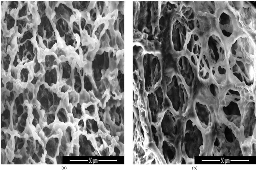
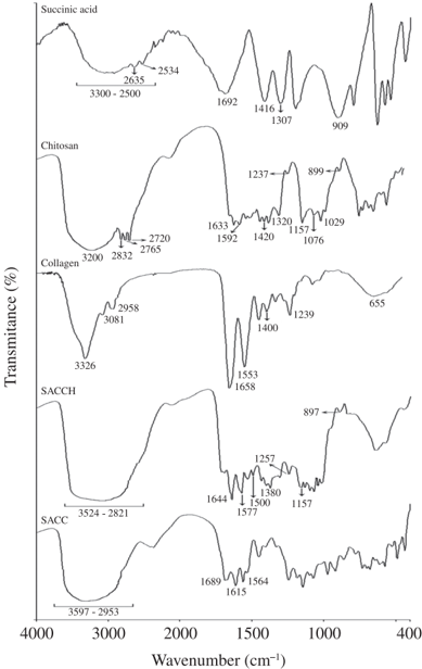
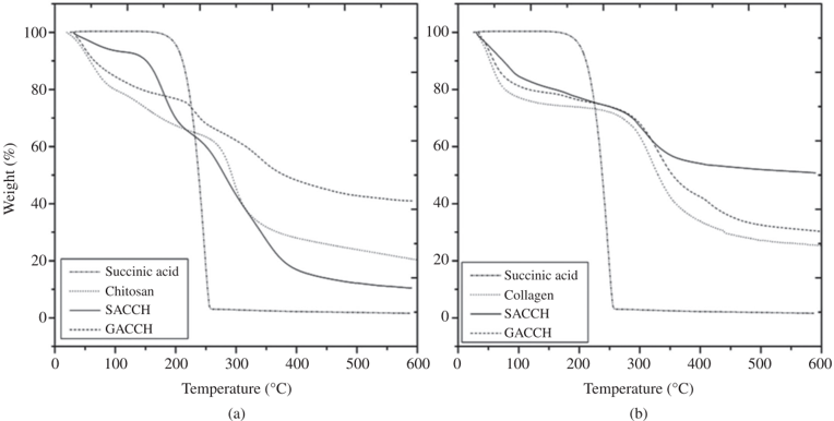
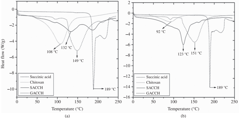
The present study summarizes the cross-linking property of succinic acid with chitosan /collagen. In detail, the chemistry behind the cross-linking and the improvement in mechanical and thermal properties of the cross-linked material were discussed with suitable instruments and bioinformatics tools. The concentration of succinic acid with reference to the chosen polymers was optimized. A 3D scaffold prepared using an optimized concentration of succinic acid (0.2% (w/v)) with chitosan (1.0% (w/v)) and similarly with collagen (0.5% (w/v)), was subjected to surface morphology, FT-IR analysis, tensile strength assessment, thermal stability and biocompatibility. Results revealed, cross-linking with succinic acid impart appreciable mechanical strength to the scaffold material. In silico analysis suggested the prevalence of non-covalent interactions, which played a crucial role in improving the mechanical and thermal properties of the cross-linked scaffold. The resultant 3D scaffold may find application as wound dressing material, as an implant in clinical applications and as a tissue engineering material.
Overview
The claims and statistics of this paper haven’t been checked yet.
Run the check — free, no account needed. It reads the open-access text and checks the sources it cites.
Integrity signals
Registry
Cites
Statistics
Figures
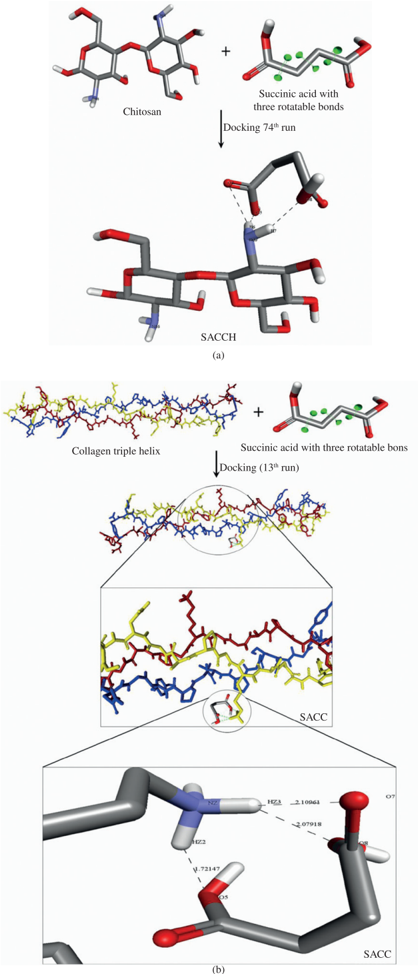
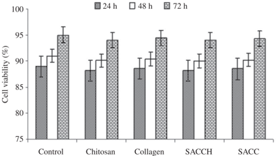
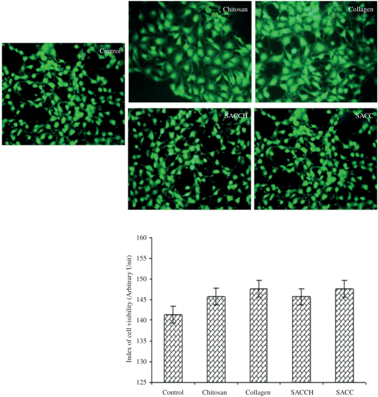
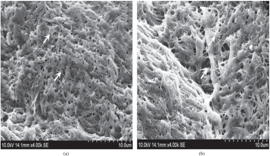
Verification
Claims the paper makes
The claims and statistics of this paper haven’t been checked yet.
Run the check — free, no account needed. It reads the open-access text and checks the sources it cites.
Claims other papers make about this one
Each is checked against this paper’s own text: does it say what the citing paper says it does?
The source backs part of the claim, not all of it — or backs it for a different population, condition or comparison.
In the citing paper
“The second mass loss event of Col, with a T onset of 266.0 °C and a T max of 312.0 °C (60 %), corresponds to the decomposition of the macromolecular structure of Col (Figure 4A and B) [53].”
From Montmorillonite-reinforced methacrylated collagen/methacrylated silk fibroin hydrogel-based inks: rheological and physicochemical characterization for bone scaffold applications (2026) · cites this paper as [53]
Experimental — accuracy not yet measured
Self-check
Percentages, sums and reported differences re-computed from the paper’s own numbers.
The self-check needs the paper’s full text.
Citations
Cites
A tough biodegradable elastomer
Wang, Y., Ameer, G. A., Sheppard, B. J., & Langer, R. (2002) · Nature Biotechnology · doi:10.1038/nbt0602-602 · Cited by 1,314
Bubnis, W. A., & Ofner, C. M. (1992) · Analytical Biochemistry · doi:10.1016/0003-2697(92)90513-7 · Cited by 265
Research works printed but not linked: 28
- Hashimoto K, 2004, Synthesis of novel block copolymers containing polyamide4 segments and control of their biodegradability, Journal of Applied Polymer Science, 92, 3492, 10.1002/app.20348 · DOI
- Liu Y, 2003, Preparation and Properties of a Biodegradable Polymer as a Novel Drug Delivery System, Journal of Applied Polymer Science, 90, 3150, 10.1002/app.13061 · DOI
- Engelmayr GC, 2003, A novel bioreactor for the dynamic flexural stimulation of tissue engineered heart valve biomaterials, Biomaterials, 24, 2523, 10.1016/S0142-9612(03)00051-6 · DOI
- Ligler FS, 2001, Development of uniform chitosan thin-film layers on silicon chips, Langmuir, 17, 5082, 10.1021/la010148b · DOI
- Schauer CL, 2003, Color changes in chitosan and poly (allyl amine) films upon metal binding, Thin Solid Films, 434, 250, 10.1016/S0040-6090(03)00055-5 · DOI
- Jin J, 2004, Chitosan-Based Films Cross-Linked by Genipin with improved Physical Properties, Biomacromolecules, 5, 162, 10.1021/bm034286m · DOI
- Tual C, 2000, Transport properties of chitosan membranes: influence of crosslinking, Journal of Polymer Science Part B: Polymer Physics, 38, 1521, 10.1002/(SICI)1099-0488(20000601)38:11<1521::AID-POLB120>3.0.CO;2-# · DOI
- Usha R, 2000, Effect of crosslinking agents (basic chromium sulfate and formaldehyde) on the thermal and thermomechanical stability of rat tail tendon type-I collagen fibre, Thermochim Acta, 356, 59, 10.1016/S0040-6031(00)00518-9 · DOI
- Sheu MT, 2001, Characterization of type-I collagen gel solutions and type-I collagen matrices for cell culture, Biomaterials, 22, 1713, 10.1016/S0142-9612(00)00315-X · DOI
- Miles CA, 2005, The increase in denaturation temperature following cross-linking of type-I collagen is caused by dehydration of the fibres, Journal of Molecular Biology, 346, 551, 10.1016/j.jmb.2004.12.001 · DOI
- Nam K, 2008, Controlling coupling reaction of EDC and NHS for preparation of type-I collagen gels using ethanol/water co-solvents, Macromolecular Bioscience, 8, 32, 10.1002/mabi.200700206 · DOI
- Song TY, 2010, Electrospinning of zein/chitosan composite fibrous membranes, Chinese Journal of Polymer Science, 28, 171, 10.1007/s10118-010-8239-2 · DOI
- Saito H, 2008, Characterization of alkali-treated type-I collagen gels prepared by different crosslinkers, Journal Materials Science: Materials in Medicine, 19, 1297
- Charulatha V, 2003, Influence of different crosslinking treatments on the physical properties of collagen membranes, Biomaterials, 24, 759, 10.1016/S0142-9612(02)00412-X · DOI
- Leung HW, 2001, Ecotoxicology of glutaraldehyde: review of environmental fate and effects studies, Ecotoxicology and Environmental Safety, 49, 26, 10.1006/eesa.2000.2031 · DOI
- Ballantyne B, 2001, Toxicological, medical and industrial hygiene aspects of glutaraldehyde with particular reference to its biocidal use in cold sterilization procedures, Journal of applied toxicology, 21, 131, 10.1002/jat.741 · DOI
- Horn HJ, 1957, Food additives, safety of adipic acid as compares with citric and tartaric acid, Journal of Agricultural and Food Chemistry, 5, 759, 10.1021/jf60080a007 · DOI
- Mitra T, 2011, Preparation and characterization of a thermostable and biodegradable biopolymers using natural cross-linker, International Journal of Biological Macromolecules, 48, 276, 10.1016/j.ijbiomac.2010.11.011 · DOI
- Peng L, 2006, Scaffolds for Periodontal Tissue Engineering, Journal of Bioactive and Compatible Polymers, 21, 207, 10.1177/0883911506065100 · DOI
- Morris GM, 2009, Autodock4 and AutoDockTools4: automated docking with selective receptor flexibility, Journal of Computational Chemistry, 30, 2785, 10.1002/jcc.21256 · DOI
- Trentani L, 2002, Evaluation of the TiMo12Zr6Fe2 alloy for orthopaedic implants: vitro biocompatibility study by using primary human fibroblasts and osteoblasts, Biomaterials, 23, 2863, 10.1016/S0142-9612(01)00413-6 · DOI
- Mossmann T, 1983, Rapid colorimetric assay for cellular growth and survival: application to proliferation and cytotoxicity assays, Journal of Immunological Methods, 65, 55, 10.1016/0022-1759(83)90303-4 · DOI · Paper page
- Majumder S, 2011, Simulated Microgravity Promoted Differentiation of Bipotential Murine Oval Liver Stem Cells by Modulating BMP4/Notch1 Signaling, Journal of Cellular Biochemistry, 112, 1898, 10.1002/jcb.23110 · DOI
- Ohkawa K, 2004, Electrospinning of chitosan, Macromolecular Rapid Communication, 25, 1600, 10.1002/marc.200400253 · DOI
- El-Tahlawy K, 2006, Novel method for preparation of cyclodextrin/grafted chitosan and its application, Carbohydrate Polymers, 63, 385, 10.1016/j.carbpol.2005.08.057 · DOI
- Bhumkar DR, 2006, Studies on effect of pH on crosslinking of chitosan with sodium tripolyphosphate: a technical note, Journal of the American Association of Pharmaceutical Scientists, 7, 1
- Schiffman JD, 2007, Cross-linking chitosan nanofibers, Biomacromolecules, 8, 594, 10.1021/bm060804s · DOI
- Nunes PS, 2010, Thermal characterization of usnic acid/collagen-based films, Journal of Thermal Analysis and Calorimetry, 99, 1011, 10.1007/s10973-009-0661-8 · DOI
Cited by
Sorted by citations
Collagen and its modifications—crucial aspects with concern to its processing and analysis
Rýglová, Š., Braun, M., & Suchý, T. (2017) · Macromolecular Materials and Engineering · doi:10.1002/mame.201600460 · Cited by 106
Antunes, B. S. L., Pedrini, F., Komatsu, D., Asami, J., Dicko, C., & Duek, E. A. R. (2026) · International Journal of Biological Macromolecules · doi:10.1016/j.ijbiomac.2026.154129
Metadata
- Authors
- Mitra, T., Sailakshmi, G., Gnanamani, A., & Mandal, A. B.
- Affiliations
- Central Leather Research Institute (IN)
- Venue
- Materials Research
- Publisher
- FapUNIFESP (SciELO)
- Published
- 2013-04-23
- Volume
- 16
- Issue
- 4
- Pages
- 755-765
- ISSN
- 1516-1439 (ISSN-L)
- 1980-5373
- OpenAlex
- W2042567183
- Cited by
- 104
- References
- 2
- Citations by year
- 2026: 5 · 2025: 7 · 2024: 8 · 2023: 8 · 2022: 6 · 2021: 10 · 2020: 11 · 2019: 9 · 2018: 8 · 2017: 13 · 2016: 8 · 2015: 8 · 2014: 3
- FWCI
- 2.42
- Type
- journal article
- Language
- en
- Open access
- diamond
- doi.org
- Licence
- cc-by-nc
- Locations
- Materials Research (publishedVersion, cc-by-nc)
- LA Referencia (Red Federada de Repositorios Institucionales de Publicaciones Científicas) (publishedVersion, other-oa)
- Americanae (AECID Library) (submittedVersion, cc-by-nc)
- DOAJ (DOAJ: Directory of Open Access Journals) (submittedVersion, cc-by-sa)
- Topics
- Physical Sciences › Materials Science › Biomaterials › Collagen: Extraction and Characterization
- Physical Sciences › Materials Science › Biomaterials › Nanocomposite Films for Food Packaging
- Physical Sciences › Materials Science › Biomaterials › Hydrogels: synthesis, properties, applications
- Keywords
- succinic acid, chitosan, collagen, crosslinking, 3D scaffolds, mechanical properties, thermal stability, biocompatibility, non-covalent interactions, wound dressings, tissue engineering
About these checks
Registry, retraction, Cites and statistics results are computed, not judged, with one exception: a printed reference that no index lists is read once by a model before it counts as a citation error. Claims are extracted by a model and kept only when their quote appears verbatim in the text; each cited source is then checked, with a model’s help, for whether it says what the claim relies on. Those verdicts can be wrong — read the claim’s quote and the cited source before relying on one.
Statistics and arithmetic are recomputed, not judged.
Confidence bands are shown only for fields calibrated on our annotated test set. None is calibrated yet, so no band is shown.
What counts as a claim ›How we measure confidence ›How we check this ›
Are you an author, or do you see a problem with this page? Request a review
