

The DOI matches its Crossref record.
Immunohistochemical analysis of RHAMM expression in normal and neoplastic human tissues: a cell cycle protein with distinctive expression in mitotic cells and testicular germ cells
Overview
The claims and statistics of this paper haven’t been checked yet.
Run the check — free, no account needed. It reads the open-access text and checks the sources it cites.
Integrity signals
Registry
Cites
Statistics
Figures
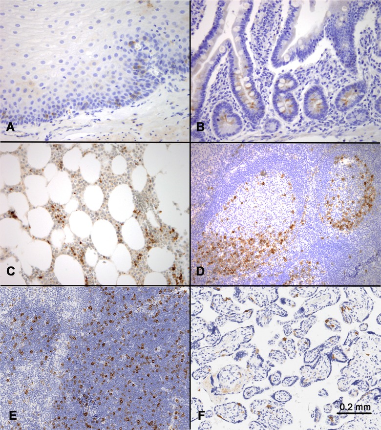
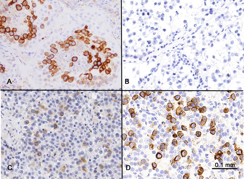
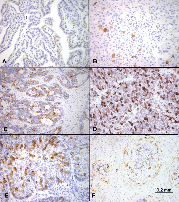
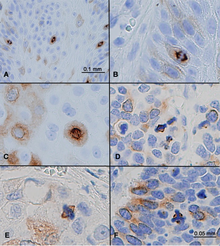
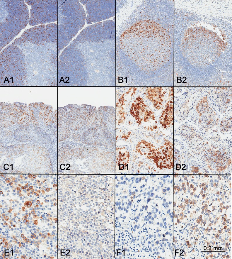
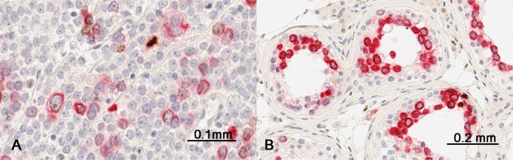
Verification
Claims the paper makes
The claims and statistics of this paper haven’t been checked yet.
Run the check — free, no account needed. It reads the open-access text and checks the sources it cites.
Claims other papers make about this one
Each is checked against this paper’s own text: does it say what the citing paper says it does?
In the citing paper
“Akin to CD44, there have been studies that have shown that RHAMM protein expression is found in organs with high cell turnover, such as the small intestine and colon, in particular the base of the intestinal crypts.”
From Hyaluronic a cid–doxorubicin nanoparticles for targeted treatment of colorectal cancer (2021) · cites this paper as [89]
Self-check
Percentages, sums and reported differences re-computed from the paper’s own numbers.
The self-check needs the paper’s full text.
Citations
Cites
No references could be resolved yet.
Research works printed but not linked: 26
- Turley, 1982, Purification of a hyaluronate-binding protein fraction that modifies cell social behavior, Biochem Biophys Res Commun, 108, 1016, 10.1016/0006-291X(82)92101-5 · DOI
- Knudson, 2001, Cartilage proteoglycans, Semin Cell Dev Biol, 12, 69, 10.1006/scdb.2000.0243 · DOI
- Hart, 1999, The intracellular hyaluronan receptor RHAMM/IHABP interacts with microtubules and actin filaments, J Cell Sci, 112, 3943, 10.1242/jcs.112.22.3943 · DOI
- Turley, 1996, HA receptors: regulators of signalling to the cytoskeleton, J Cell Biochem, 61, 569, 10.1002/(SICI)1097-4644(19960616)61:4<569::AID-JCB10>3.0.CO;2-B · DOI
- Lugli, 2017, The hyaluronan-mediated motility receptor RHAMM promotes growth, invasiveness and dissemination of colorectal cancer, Oncotarget, 8, 70617, 10.18632/oncotarget.19904 · DOI
- Varmus, 2011, Receptor for hyaluronan-mediated motility isoform B promotes liver metastasis in a mouse model of multistep tumorigenesis and a tail vein assay for metastasis, Proc Natl Acad Sci U S A, 108, 16753, 10.1073/pnas.1114022108 · DOI
- Du, 2016, Expression of the receptor for hyaluronic acid mediated motility (RHAMM) is associated with poor prognosis and metastasis in non-small cell lung carcinoma, Oncotarget, 7, 39957, 10.18632/oncotarget.9554 · DOI
- Tzankov, 2015, Receptor for hyaluronic acid-mediated motility (RHAMM, CD168) expression is prognostically important in both nodal negative and nodal positive large cell lung cancer, J Clin Pathol, 68, 368, 10.1136/jclinpath-2014-202819 · DOI
- Hirata, 2015, Receptor for Hyaluronic Acid-Mediated Motility is Associated with Poor Survival in Pancreatic Ductal Adenocarcinoma, J Cancer, 6, 1093, 10.7150/jca.12990 · DOI
- Natsugoe, 2011, Prognostic impact of CD168 expression in gastric cancer, BMC Cancer, 11, 106, 10.1186/1471-2407-11-106 · DOI
- Lugli, 2015, Expression of the hyaluronan-mediated motility receptor RHAMM in tumor budding cells identifies aggressive colorectal cancers, Hum Pathol, 46, 1573, 10.1016/j.humpath.2015.07.010 · DOI
- Milde-Langosch, 2016, RHAMM splice variants confer radiosensitivity in human breast cancer cell lines, Oncotarget, 7, 21428, 10.18632/oncotarget.7258 · DOI
- Jass, 2006, Overexpression of the receptor for hyaluronic acid mediated motility is an independent adverse prognostic factor in colorectal cancer, Mod Pathol, 19, 1302, 10.1038/modpathol.3800648 · DOI
- Dall, 2003, Expression of the hyaluronan receptor RHAMM in endometrial carcinomas suggests a role in tumour progression and metastasis, J Cancer Res Clin Oncol, 129, 161, 10.1007/s00432-003-0415-0 · DOI
- Kruk, 2017, Increased RHAMM expression relates to ovarian cancer progression, J Ovarian Res, 10, 66, 10.1186/s13048-017-0360-1 · DOI
- Chou, 2005, Integrative genomics based identification of potential human hepatocarcinogenesis-associated cell cycle regulators: RHAMM as an example, Biochem Biophys Res Commun, 330, 489, 10.1016/j.bbrc.2005.03.005 · DOI
- Schmitt, 2002, Receptor for hyaluronan acid-mediated motility (RHAMM) is a new immunogenic leukemia-associated antigen in acute and chronic myeloid leukemia, Exp Hematol, 30, 1029, 10.1016/S0301-472X(02)00874-3 · DOI
- Engeland, 2008, RHAMM is differentially expressed in the cell cycle and downregulated by the tumor suppressor p53, Cell Cycle, 7, 3448, 10.4161/cc.7.21.7014 · DOI
- Lockhart, 2001, Transcriptional regulation and function during the human cell cycle, Nat Genet, 27, 48, 10.1038/83751 · DOI · Paper page
- Botstein, 2002, Identification of genes periodically expressed in the human cell cycle and their expression in tumors, Mol Biol Cell, 13, 1977, 10.1091/mbc.02-02-0030 · DOI · Paper page
- Nussenzweig, 2012, Identification of human germinal center light and dark zone cells and their relationship to human B-cell lymphomas, Blood, 120, 2240, 10.1182/blood-2012-03-415380 · DOI
- Hua, 2010, Regulation of cyclin B2 expression and cell cycle G2/m transition by menin, J Biol Chem, 285, 18291, 10.1074/jbc.M110.106575 · DOI
- Old, 2011, Chromosome X-encoded cancer/testis antigens show distinctive expression patterns in developing gonads and in testicular seminoma, Hum Reprod, 26, 3232, 10.1093/humrep/der330 · DOI
- Lange, 2016, Impaired Planar Germ Cell Division in the Testis, Caused by Dissociation of RHAMM from the Spindle, Results in Hypofertility and Seminoma, Cancer Res, 76, 6382, 10.1158/0008-5472.CAN-16-0179 · DOI
- Frappart, 2017, HMMR acts in the PLK1-dependent spindle positioning pathway and supports neural development, Elife, 6, e28672, 10.7554/eLife.28672 · DOI
- Ringhoffer, 2009, RHAMM (CD168) is overexpressed at the protein level and may constitute an immunogenic antigen in advanced prostate cancer disease, Neoplasia, 11, 956, 10.1593/neo.09694 · DOI
Cited by
Sorted by citations
Hyaluronic a cid–doxorubicin nanoparticles for targeted treatment of colorectal cancer
Pan, D. C., Krishnan, V., Salinas, A. K., Kim, J., Sun, T., Ravid, S., Peng, K., Wu, D., Nurunnabi, M., Nelson, J. A., et al. (2021) · Bioengineering & Translational Medicine · doi:10.1002/btm2.10166 · Cited by 42
Metadata
- Authors
- Chen, Y.-T., Chen, Z., & Du, Y.-C. N.
- Affiliations
- Department of Pathology and Laboratory Medicine, Weill Cornell Medicine, New York, 10065 NY, USA
- Division of Biostatistics and Epidemiology, Department of Healthcare Policy and Research, Weill Cornell Medicine, New York, 10065 NY, USA
- Venue
- Oncotarget
- Publisher
- Impact Journals, LLC
- Published
- 2018-04-20
- Volume
- 9
- Issue
- 30
- Pages
- 20941-20952
- ISSN
- 1949-2553
- PMC
- PMC5940366
- Cited by
- 32
- References
- 0
- Language
- en
About these checks
Registry, retraction, Cites and statistics results are computed, not judged, with one exception: a printed reference that no index lists is read once by a model before it counts as a citation error. Claims are extracted by a model and kept only when their quote appears verbatim in the text; each cited source is then checked, with a model’s help, for whether it says what the claim relies on. Those verdicts can be wrong — read the claim’s quote and the cited source before relying on one.
Statistics and arithmetic are recomputed, not judged.
Confidence bands are shown only for fields calibrated on our annotated test set. None is calibrated yet, so no band is shown.
What counts as a claim ›How we measure confidence ›How we check this ›
Are you an author, or do you see a problem with this page? Request a review
