PreprintCited by 20Cancer-related gene regulationDNA Repair MechanismsEpigenetics and DNA Methylation

The DOI matches its Crossref record; 1 of the 1 references it cites were found in a registry or an index.

Structure, Activity, and Function of SETMAR protein Lysine Methyltransferase

Open access
Tellier, Michael·2021·Life

Preprint — published as 10.3390/life11121342 · Paper page

Abstract

SETMAR is a protein lysine methyltransferase that is involved in several DNA processes, including DNA repair via the non-homologous end joining (NHEJ) pathway, regulation of gene expression, illegitimate DNA integration, and DNA decatenation. However, SETMAR is an atypical protein lysine methyltransferase since in anthropoid primates, the SET domain is fused to an inactive DNA transposase. The presence of the DNA transposase domain confers to SETMAR a DNA binding activity towards the remnants of its transposable element, which has resulted in the emergence of a gene regulatory function. Both the SET and the DNA transposase domains are involved in the different cellular roles of SETMAR, indicating the presence of novel and specific functions in anthropoid primates. In addition, SETMAR is dysregulated in different types of cancer, indicating a potential pathological role. While some light has been shed on SETMAR functions, more research and new tools are needed to better understand the cellular activities of SETMAR and to investigate the therapeutic potential of SETMAR.

Overview

The claims and statistics of this paper haven’t been checked yet.

Run the check — free, no account needed. It reads the open-access text and checks the sources it cites.

A few free checks a day · papers already verified open instantly · results are public

Integrity signals

Registry

Crossref · 10.20944/preprints202111.0164.v1

Cites

1 of 1 references verified0 citation errors · 0 not checkable

Statistics

Figures

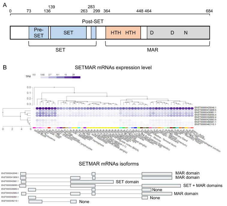
Figure 1 Protein organization and expression level of SETMAR in human. (A) SETMAR is composed of two domains, the protein methyltransferase domain (SET) that includes the pre-SET, the SET- and the post-SET subdomains, and the DNA transposase domain (MAR) that contains the DNA binding (orange) and the catalytic (gray) subdomains. (B) SETMAR encodes for eight different isoforms. One isoform encodes for the full-length SETMAR, one isoform includes only the SET domain, and three isoforms contain only the DNA transposase domain. Out of the eight mRNA isoforms, only four are expressed at a high level in human tissues (from GTEx).
Figure 1 Protein organization and expression level of SETMAR in human. (A) SETMAR is composed of two domains, the protein methyltransferase domain (SET) that includes the pre-SET, the SET- and the post-SET subdomains, and the DNA transposase domain (MAR) that contains the DNA binding (orange) and the catalytic (gray) subdomains. (B) SETMAR encodes for eight different isoforms. One isoform encodes for the full-length SETMAR, one isoform includes only the SET domain, and three isoforms contain only the DNA transposase domain. Out of the eight mRNA isoforms, only four are expressed at a high level in human tissues (from GTEx).CC BY · PubMed Central
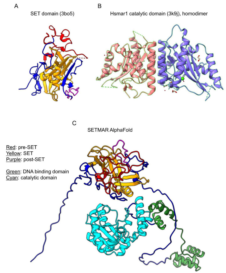
Figure 2 Crystal structures of SETMAR. (A) Structure of the SET domain of SETMAR with the pre-SET, SET, and post-SET subdomains shown in red, yellow, and purple, respectively. (B) Structure of the catalytic subdomain of the DNA transposase domain of SETMAR, shown as a homodimer. Each monomer is represented by a different color. (B,C) Proposed structure of a SETMAR monomer by AlphaFold with the pre-SET, SET, and post-SET subdomains shown in red, yellow, and purple, respectively, and the DNA binding and catalytic subdomains shown in green and cyan, respectively.
Figure 2 Crystal structures of SETMAR. (A) Structure of the SET domain of SETMAR with the pre-SET, SET, and post-SET subdomains shown in red, yellow, and purple, respectively. (B) Structure of the catalytic subdomain of the DNA transposase domain of SETMAR, shown as a homodimer. Each monomer is represented by a different color. (B,C) Proposed structure of a SETMAR monomer by AlphaFold with the pre-SET, SET, and post-SET subdomains shown in red, yellow, and purple, respectively, and the DNA binding and catalytic subdomains shown in green and cyan, respectively.CC BY · PubMed Central
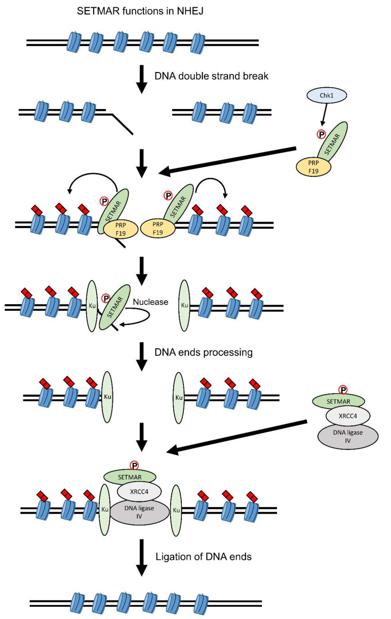
Figure 3 SETMAR functions in non-homologous end joining (NHEJ). SETMAR has been found to act on several aspects of the NHEJ pathway. Following DNA double-strand breaks (DSBs), SETMAR is phosphorylated by Chk1 on its Ser508 residue and interacts with PRPF19. The SETMAR-PRPF19 complex binds to the DSB site and SETMAR dimethylates H3K36 on the neighboring nucleosomes. H3K36me2 stabilizes the association of Ku70 and NBS1 to DSB sites. SETMAR is also involved in the processing of the DNA ends via its endonuclease activity, similarly to Artemis. Following DNA ends processing, SETMAR is also involved in the ligation of the two DNA ends as it interacts with the DNA ligase IV and XRCC4 complex. For the readability of the figure, the proteins are shown as monomer.
Figure 3 SETMAR functions in non-homologous end joining (NHEJ). SETMAR has been found to act on several aspects of the NHEJ pathway. Following DNA double-strand breaks (DSBs), SETMAR is phosphorylated by Chk1 on its Ser508 residue and interacts with PRPF19. The SETMAR-PRPF19 complex binds to the DSB site and SETMAR dimethylates H3K36 on the neighboring nucleosomes. H3K36me2 stabilizes the association of Ku70 and NBS1 to DSB sites. SETMAR is also involved in the processing of the DNA ends via its endonuclease activity, similarly to Artemis. Following DNA ends processing, SETMAR is also involved in the ligation of the two DNA ends as it interacts with the DNA ligase IV and XRCC4 complex. For the readability of the figure, the proteins are shown as monomer.CC BY · PubMed Central
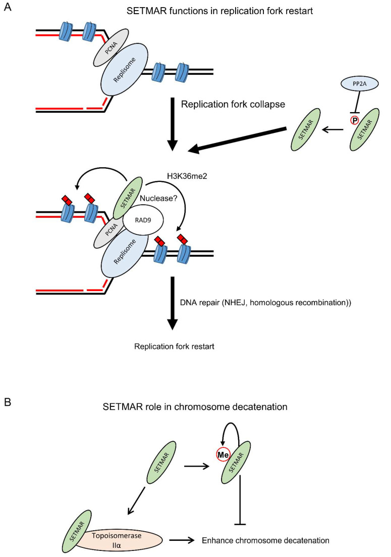
Figure 4 SETMAR functions in replication fork restart and chromosome decatenation. (A) Following replication fork collapse, the unphosphorylated form of SETMAR, mediated by the protein phosphatase PP2A, can interact with PCNA and RAD9 to promote replication fork restart via the dimethylation of H3K36, which helps recruiting DNA repair factors, and its endonuclease activity. (B) SETMAR can also enhance chromosome decatenation via its interaction with TOP2A. SETMAR activity in chromosome decatenation is regulated through its automethylation on the lysine 498 residue. For the readability of the figure, the proteins are shown as monomer.
Figure 4 SETMAR functions in replication fork restart and chromosome decatenation. (A) Following replication fork collapse, the unphosphorylated form of SETMAR, mediated by the protein phosphatase PP2A, can interact with PCNA and RAD9 to promote replication fork restart via the dimethylation of H3K36, which helps recruiting DNA repair factors, and its endonuclease activity. (B) SETMAR can also enhance chromosome decatenation via its interaction with TOP2A. SETMAR activity in chromosome decatenation is regulated through its automethylation on the lysine 498 residue. For the readability of the figure, the proteins are shown as monomer.CC BY · PubMed Central
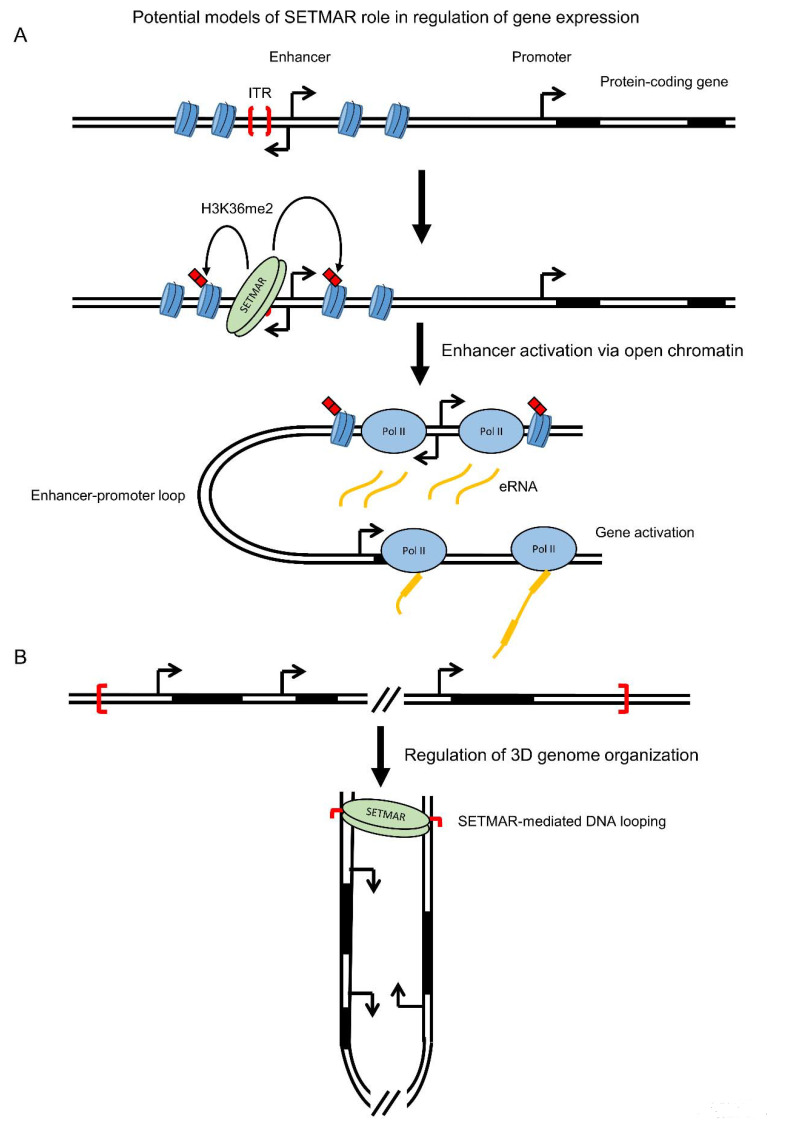
Figure 5 Potential models of SETMAR role in regulation of gene expression. (A) SETMAR could regulate gene expression via its binding to enhancer regions. Following SETMAR binding, dimethylation of H3K36 of the neighboring nucleosomes can create an open chromatin environment, via a decrease in H3K27me3, allowing the binding of transcription factors. Activation of the enhancer will then promote transcription of its associated protein-coding gene. (B) SETMAR could also regulate gene expression via an involvement in the 3D genome. Binding of SETMAR on two single ITRs can mediate the formation of a DNA loop that could regulate the expression of the genes present in the loop. Additionally, SETMAR could participate in 3D genome organization via the ITRs located near cohesin-CTCF-anchored loops.
Figure 5 Potential models of SETMAR role in regulation of gene expression. (A) SETMAR could regulate gene expression via its binding to enhancer regions. Following SETMAR binding, dimethylation of H3K36 of the neighboring nucleosomes can create an open chromatin environment, via a decrease in H3K27me3, allowing the binding of transcription factors. Activation of the enhancer will then promote transcription of its associated protein-coding gene. (B) SETMAR could also regulate gene expression via an involvement in the 3D genome. Binding of SETMAR on two single ITRs can mediate the formation of a DNA loop that could regulate the expression of the genes present in the loop. Additionally, SETMAR could participate in 3D genome organization via the ITRs located near cohesin-CTCF-anchored loops.CC BY · PubMed Central
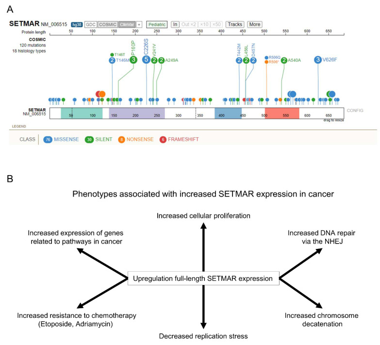
Figure 6 SETMAR roles in disease. (A) Visualization of SETMAR known mutations in cancer using ProteinPaint [68]. SETMAR major domains are shown in color at the bottom. The amino acid mutated is shown next to the circle while the number of observations of each mutation is shown within the circle. (B) Upregulation of SETMAR expression in cancer is associated with several phenotypes that can promote the tumor’s growth and resistance to treatment.
Figure 6 SETMAR roles in disease. (A) Visualization of SETMAR known mutations in cancer using ProteinPaint [68]. SETMAR major domains are shown in color at the bottom. The amino acid mutated is shown next to the circle while the number of observations of each mutation is shown within the circle. (B) Upregulation of SETMAR expression in cancer is associated with several phenotypes that can promote the tumor’s growth and resistance to treatment.CC BY · PubMed Central

Verification

Claims the paper makes

The claims and statistics of this paper haven’t been checked yet.

Run the check — free, no account needed. It reads the open-access text and checks the sources it cites.

A few free checks a day · papers already verified open instantly · results are public

Self-check

Percentages, sums and reported differences re-computed from the paper’s own numbers.

The self-check needs the paper’s full text.

Citations

Cites

Metadata

Authors
Tellier, M.
Affiliations
University of Oxford (GB)
Corresponding author
Tellier, Michael
Venue
Life
Publisher
MDPI AG
Published
2021-11-09
Volume
11
Issue
12
Pages
1342-1342
ISSN
2075-1729 (ISSN-L)
OpenAlex
W4226047180
Cited by
20
References
1
Citations by year
2026: 3 · 2025: 4 · 2024: 5 · 2023: 5 · 2022: 2 · 2021: 1
FWCI
0.39
Type
journal article
Language
en
Open access
Locations
  • Life (publishedVersion, cc-by)
  • Preprints.org (acceptedVersion, cc-by)
  • PubMed Central (publishedVersion, cc-by)
  • PubMed (publishedVersion)
  • DOAJ (DOAJ: Directory of Open Access Journals) (submittedVersion, cc-by-sa)
Funders
  • National Institutes of Health
  • National Heart, Lung, and Blood Institute
  • National Institute of Mental Health
  • National Institute on Drug Abuse
  • National Human Genome Research Institute
  • National Cancer Institute
  • National Institute of Neurological Disorders and Stroke
  • Common Fund
Topics
  • Life Sciences › Biochemistry, Genetics and Molecular Biology › Molecular Biology › Cancer-related gene regulation
  • Life Sciences › Biochemistry, Genetics and Molecular Biology › Molecular Biology › DNA Repair Mechanisms
  • Life Sciences › Biochemistry, Genetics and Molecular Biology › Molecular Biology › Epigenetics and DNA Methylation
Keywords
SETMAR, lysine methyltransferase, SET domain, non-homologous end joining, DNA repair, DNA decatenation, gene expression regulation, transposable elements, cancer, therapeutic target

About these checks

Registry, retraction, Cites and statistics results are computed, not judged, with one exception: a printed reference that no index lists is read once by a model before it counts as a citation error. Claims are extracted by a model and kept only when their quote appears verbatim in the text; each cited source is then checked, with a model’s help, for whether it says what the claim relies on. Those verdicts can be wrong — read the claim’s quote and the cited source before relying on one.

Statistics and arithmetic are recomputed, not judged.

Confidence bands are shown only for fields calibrated on our annotated test set. None is calibrated yet, so no band is shown.

What counts as a claim ›How we measure confidence ›How we check this ›

Are you an author, or do you see a problem with this page? Request a review