PreprintCited by 36Extracellular vesicles in diseaseMesenchymal stem cell researchOsteoarthritis Treatment and Mechanisms

The DOI matches its Crossref record.

CD10-Bound Human Mesenchymal Stem/Stromal Cell-Derived Exosomes Possess Immunomodulatory Cargo and Maintain Cartilage Homeostasis under Inflammatory Conditions

Open access

Preprint — published as 10.3390/cells12141824

Abstract

The onset and progression of human inflammatory joint diseases are strongly controlled by the activation of resident synovium/infrapatellar fat pad (IFP) pro-inflammatory and pain-transmitting signaling. We recently reported that intra-articularly injected IFP-derived Mesenchymal Stem/Stromal Cells (IFP-MSC) acquire a potent immunomodulatory phenotype and actively degrade Substance P (SP) via neutral endopeptidase CD10 (neprilysin). Our hypothesis is that IFP-MSC robust immunomodulatory therapeutic ef-fects are largely exerted via their CD10-bound exosomal secretome (IFP-MSC EXOs) by attenuating synoviocyte pro-inflammatory activation and articular cartilage degradation. Herein, IFP-MSC EXOs were isolated from CD10High- and CD10Low-expressing IFP-MSC cultures and their exosomal miRNA cargo was assessed using multiplex methods. Functionally, we interrogated the effect of CD10High and CD10Low EXOs on stimulated by inflammatory/fibrotic cues synoviocytes monocultures and cocultures with IFP-MSC de-rived chondropellets. Finally, CD10High EXOs were tested in vivo for their therapeutic capacity in an animal model of acute synovitis/fat pad fibrosis. Our results showed that CD10High and CD10Low EXOs possess distinct miRNA profiles. Reactome analysis of miRNAs highly present in exosomes showed their involvement in the regulation of six gene groups, particularly the immune system. Stimulated synoviocytes exposed to IFP-MSC EXOs demonstrated significantly reduced proliferation and altered inflammation-related molecular profiles compared to control stimulated synoviocytes. Importantly, CD10High EXOs treatment of stimulated chondropellets/synoviocytes cocultures indicated a significant chondroprotective effects. Therapeutically, CD10High EXOs treatment resulted in robust chondroprotective effects by retaining articular cartilage structure/composition and PRG4 (lubricin)-expressing cartilage cells in the animal model of acute synovitis/IFP fibrosis. Our study suggests that CD10High EXOs possess immuno-modulatory miRNA attributes with strong chondroprotective/anabolic effects for articular cartilage in vivo. The results could serve as a foundation for EXOs-based therapeutics for the resolution of detrimental aspects of immune-mediated inflammatory joint changes associated with conditions such as osteoarthritis (OA).

Overview

Integrity signals

Registry

Crossref · 10.20944/preprints202305.1465.v1

Cites

Statistics

Recomputing p-values needs the paper’s full text.

Figures

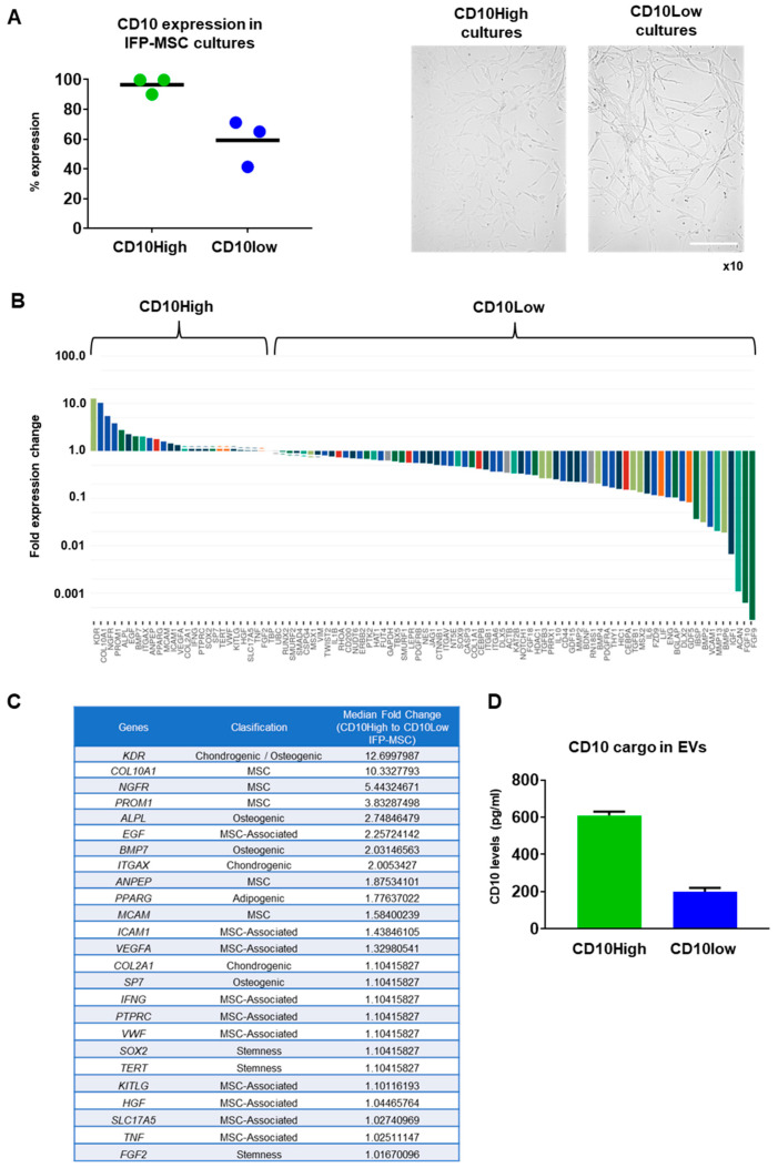
Figure 1 CD10High and CD10Low IFP-MSC cultures, and their molecular profiling. (A) IFP-MSC cultured in regulation-compliant media (Ch-R and hPL) media showed similar fibroblast-like morphology but had 96.5 ± 5.6% for Ch-R and 59.2 ± 15.7% for hPL CD10 expression levels. Magnification ×10. (B,C) Molecular profiling of CD10High versus CD10Low IFP-MSC revealed that 25 out of 90 genes tested were higher expressed in CD10High IFP-MSC, with 8 genes being more than twofold higher (KDR, COL10A1, NGFR, PROM1, ALPL, EGF, BMP7, ITGAX). Interestingly, genes tested were grouped in phenotype-related cohorts with MSC-associated, chondrogenic/osteogenic, and MSC cohorts showing overall the most prominent fold expression change between CD10High and CD10Low IFP-MSC cultures. (D) CD10High EVs had 610 ± 20.6 pg/mL whereas CD10Low EVs had 200 ± 20 pg/mL of CD10 protein as a cargo. Scale bar: 50 μm.
Figure 1 CD10High and CD10Low IFP-MSC cultures, and their molecular profiling. (A) IFP-MSC cultured in regulation-compliant media (Ch-R and hPL) media showed similar fibroblast-like morphology but had 96.5 ± 5.6% for Ch-R and 59.2 ± 15.7% for hPL CD10 expression levels. Magnification ×10. (B,C) Molecular profiling of CD10High versus CD10Low IFP-MSC revealed that 25 out of 90 genes tested were higher expressed in CD10High IFP-MSC, with 8 genes being more than twofold higher (KDR, COL10A1, NGFR, PROM1, ALPL, EGF, BMP7, ITGAX). Interestingly, genes tested were grouped in phenotype-related cohorts with MSC-associated, chondrogenic/osteogenic, and MSC cohorts showing overall the most prominent fold expression change between CD10High and CD10Low IFP-MSC cultures. (D) CD10High EVs had 610 ± 20.6 pg/mL whereas CD10Low EVs had 200 ± 20 pg/mL of CD10 protein as a cargo. Scale bar: 50 μm.CC BY · PubMed Central
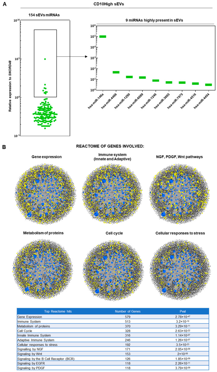
Figure 2 miRNA signature of CD10High sEVs. (A) From 166 MSC-related miRNAs analyzed, 154 miRNA cargos were present in CD10High sEVs. In CD10High sEVs, nine miRNA cargos were predominant (hsa-miR-146a, hsa-miR-4466, hsa-miR-1290, hsa-miR-6089, hsa-miR-1246, hsa-miR-3665, hsa-miR-7975, hsa-miR-4516, hsa-miR-4454). (B) Reactome analysis of miRNAs highly present in CD10High sEVs showed their involvement in the regulation of six gene groups related to gene expression, the immune system, NGF/PDGF/Wnt pathways, metabolism of proteins, the cell cycle, and cellular responses to stress (blue spots: miRNAs detected in sEVs, black spots: genes related to pathways, yellow spots: genes related to pathway that are affected by miRNAs detected in sEVs).
Figure 2 miRNA signature of CD10High sEVs. (A) From 166 MSC-related miRNAs analyzed, 154 miRNA cargos were present in CD10High sEVs. In CD10High sEVs, nine miRNA cargos were predominant (hsa-miR-146a, hsa-miR-4466, hsa-miR-1290, hsa-miR-6089, hsa-miR-1246, hsa-miR-3665, hsa-miR-7975, hsa-miR-4516, hsa-miR-4454). (B) Reactome analysis of miRNAs highly present in CD10High sEVs showed their involvement in the regulation of six gene groups related to gene expression, the immune system, NGF/PDGF/Wnt pathways, metabolism of proteins, the cell cycle, and cellular responses to stress (blue spots: miRNAs detected in sEVs, black spots: genes related to pathways, yellow spots: genes related to pathway that are affected by miRNAs detected in sEVs).CC BY · PubMed Central
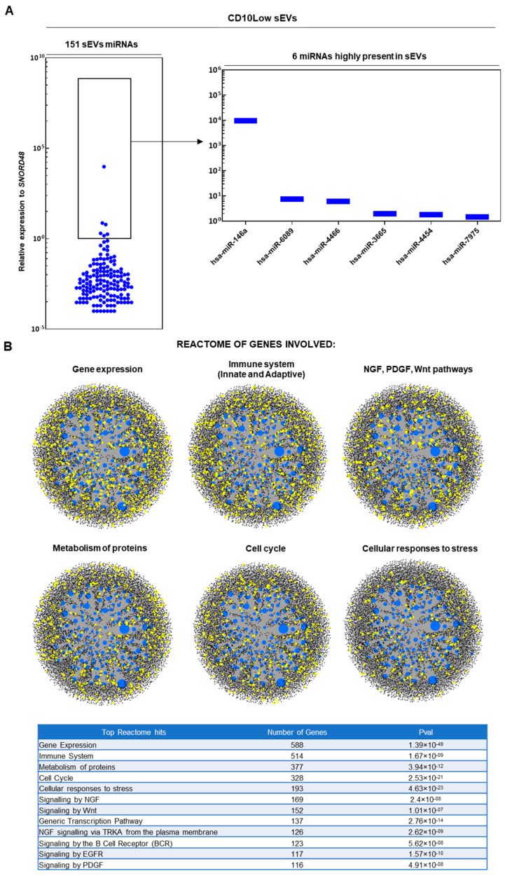
Figure 3 miRNA signature of CD10Low sEVs. (A) From 166 MSC-related miRNAs analyzed, 151 miRNA cargos were present in CD10Low sEVs. In CD10Low sEVs, six miRNAs cargo were highly present (hsa-miR-146a, hsa-miR-6089, hsa-miR-4466, hsa-miR-3665, hsa-miR-4454, hsa-miR-7975). (B) Reactome analysis of miRNAs highly present in CD10Low sEVs showed their involvement in the regulation of six gene groups related to gene expression, the immune system, NGF/PDGF/Wnt pathways, metabolism of proteins, the cell cycle, and cellular responses to stress (blue spots: miRNAs detected in sEVs, black spots: genes related to pathways, yellow spots: genes related to pathway that are affected by miRNAs detected in sEVs).
Figure 3 miRNA signature of CD10Low sEVs. (A) From 166 MSC-related miRNAs analyzed, 151 miRNA cargos were present in CD10Low sEVs. In CD10Low sEVs, six miRNAs cargo were highly present (hsa-miR-146a, hsa-miR-6089, hsa-miR-4466, hsa-miR-3665, hsa-miR-4454, hsa-miR-7975). (B) Reactome analysis of miRNAs highly present in CD10Low sEVs showed their involvement in the regulation of six gene groups related to gene expression, the immune system, NGF/PDGF/Wnt pathways, metabolism of proteins, the cell cycle, and cellular responses to stress (blue spots: miRNAs detected in sEVs, black spots: genes related to pathways, yellow spots: genes related to pathway that are affected by miRNAs detected in sEVs).CC BY · PubMed Central
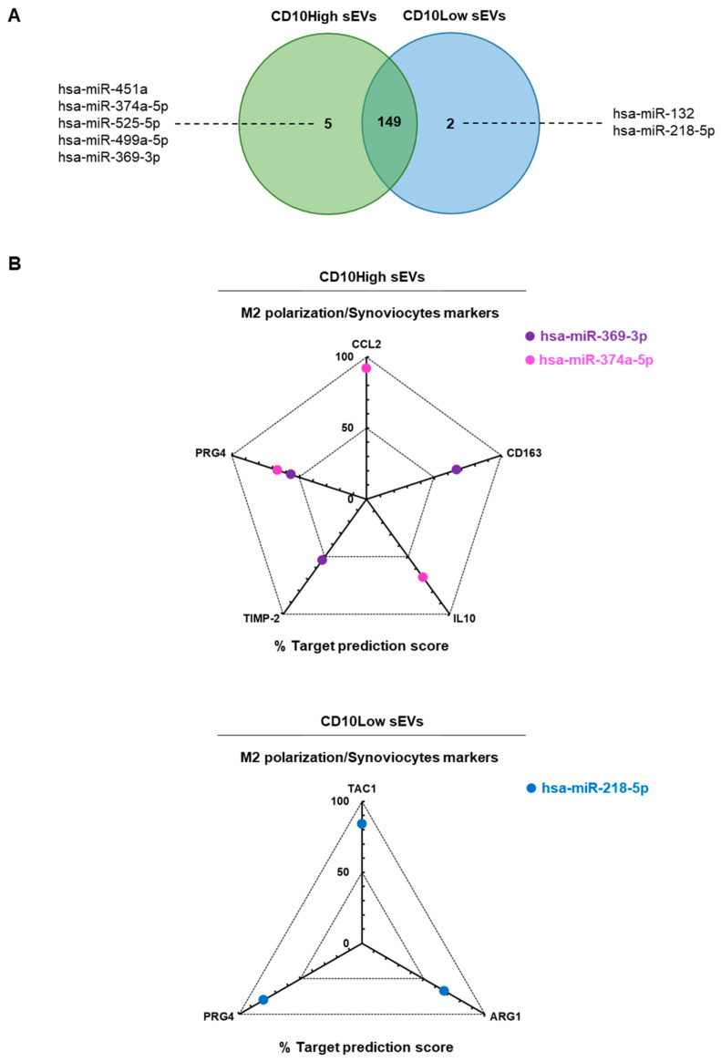
Figure 4 In silico analysis for functional correlation of identified miRNAs in IFP-MSC sEVs with known highly expressed target genes in macrophages and synoviocytes. (A) Overall, most detected miRNAs were commonly present in IFP-MSC sEVs, however, five miRNAs were specific for CD10High sEVs (hsa-miR-451a, hsa-miR-374a-5p, hsa-miR-525-5p, hsa-miR-499a-5p, hsa-miR-369-3p) and two miRNAs were specific for CD10Low sEVs (hsa-miR-132, hsa-miR-218-5p). (B) For CD10High sEVs, the prediction scoring system revealed CCL2 as a target for hsa-miR-374a-5p (92% probability), CD163 as target for hsa-miR-369-3p (67% probability), IL-10 as a target for hsa-miR-374a-5p (68% probability), TIMP-2 as a target for hsa-miR-369-3p (53% probability), and PRG4 as a target for hsa-miR-369-3p and hsa-miR-374a-5p (56% and 66%, respectively). For CD10Low sEVs, the prediction scoring system revealed TAC1, ARG1, and PRG4 as targets for hsa-miR-218-5p (84%, 67%, and 80%, respectively).
Figure 4 In silico analysis for functional correlation of identified miRNAs in IFP-MSC sEVs with known highly expressed target genes in macrophages and synoviocytes. (A) Overall, most detected miRNAs were commonly present in IFP-MSC sEVs, however, five miRNAs were specific for CD10High sEVs (hsa-miR-451a, hsa-miR-374a-5p, hsa-miR-525-5p, hsa-miR-499a-5p, hsa-miR-369-3p) and two miRNAs were specific for CD10Low sEVs (hsa-miR-132, hsa-miR-218-5p). (B) For CD10High sEVs, the prediction scoring system revealed CCL2 as a target for hsa-miR-374a-5p (92% probability), CD163 as target for hsa-miR-369-3p (67% probability), IL-10 as a target for hsa-miR-374a-5p (68% probability), TIMP-2 as a target for hsa-miR-369-3p (53% probability), and PRG4 as a target for hsa-miR-369-3p and hsa-miR-374a-5p (56% and 66%, respectively). For CD10Low sEVs, the prediction scoring system revealed TAC1, ARG1, and PRG4 as targets for hsa-miR-218-5p (84%, 67%, and 80%, respectively).CC BY · PubMed Central
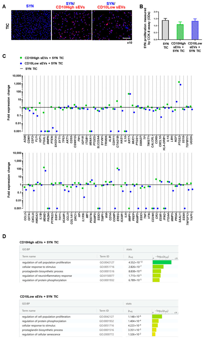
Figure 5 CD10High and CD10Low sEVs’ anti-inflammatory effects on synoviocytes. (A,B) Both CD10High and CD10Low sEVs were internalized by TIC-primed SYN (blue, nucleus; red, sEVs) whereas they tended to attenuate SYN TIC proliferation. (C) At the molecular level, SYN TIC cultures gene expression was strongly affected by both CD10High and CD10Low sEVs. Specifically, CD10High sEVs increased the expression levels of 45 genes in SYN TIC, with 13 genes more than twofold higher. Moreover, CD10Low sEVs increased the expression levels of 19 genes in SYN TIC with 9 genes more than twofold higher. (D) Reactome analysis of highly expressed genes in CD10High sEVs/SYN TICs and CD10Low sEVs/SYN TICs showed their involvement in the regulation of five molecular pathways. Scale bar: 50 μm.
Figure 5 CD10High and CD10Low sEVs’ anti-inflammatory effects on synoviocytes. (A,B) Both CD10High and CD10Low sEVs were internalized by TIC-primed SYN (blue, nucleus; red, sEVs) whereas they tended to attenuate SYN TIC proliferation. (C) At the molecular level, SYN TIC cultures gene expression was strongly affected by both CD10High and CD10Low sEVs. Specifically, CD10High sEVs increased the expression levels of 45 genes in SYN TIC, with 13 genes more than twofold higher. Moreover, CD10Low sEVs increased the expression levels of 19 genes in SYN TIC with 9 genes more than twofold higher. (D) Reactome analysis of highly expressed genes in CD10High sEVs/SYN TICs and CD10Low sEVs/SYN TICs showed their involvement in the regulation of five molecular pathways. Scale bar: 50 μm.CC BY · PubMed Central
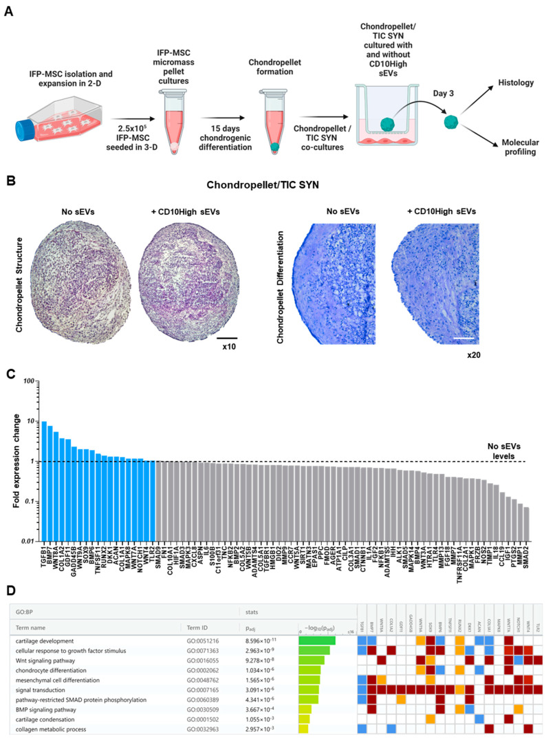
Figure 6 Effects of CD10High sEVs on chondrocytes in vitro. (A) Experimental strategy to simulate a pro-inflammatory microenvironment in vitro by co-culturing MSC-derived chondropellets with inflamed synoviocytes. (B) Chondropellet histological analysis revealed that although CD10High sEVs, treated and non-treated, have similar structure, CD10High sEV treatment resulted in enhanced chondrocyte differentiation and sulfated glycosaminoglycans production in vitro. (C) At the molecular level, CD10High sEV-treated chondropellets showed distinct transcriptional signature with higher expression of 19 genes. (D) Reactome analysis of highly expressed genes in CD10High sEV-treated chondropellets showed their involvement in the regulation of 10 chondroprotection- and cartilage homeostasis-related molecular pathways. Black scale bar: 50 μm, white scale bar: 25 μm.
Figure 6 Effects of CD10High sEVs on chondrocytes in vitro. (A) Experimental strategy to simulate a pro-inflammatory microenvironment in vitro by co-culturing MSC-derived chondropellets with inflamed synoviocytes. (B) Chondropellet histological analysis revealed that although CD10High sEVs, treated and non-treated, have similar structure, CD10High sEV treatment resulted in enhanced chondrocyte differentiation and sulfated glycosaminoglycans production in vitro. (C) At the molecular level, CD10High sEV-treated chondropellets showed distinct transcriptional signature with higher expression of 19 genes. (D) Reactome analysis of highly expressed genes in CD10High sEV-treated chondropellets showed their involvement in the regulation of 10 chondroprotection- and cartilage homeostasis-related molecular pathways. Black scale bar: 50 μm, white scale bar: 25 μm.CC BY · PubMed Central
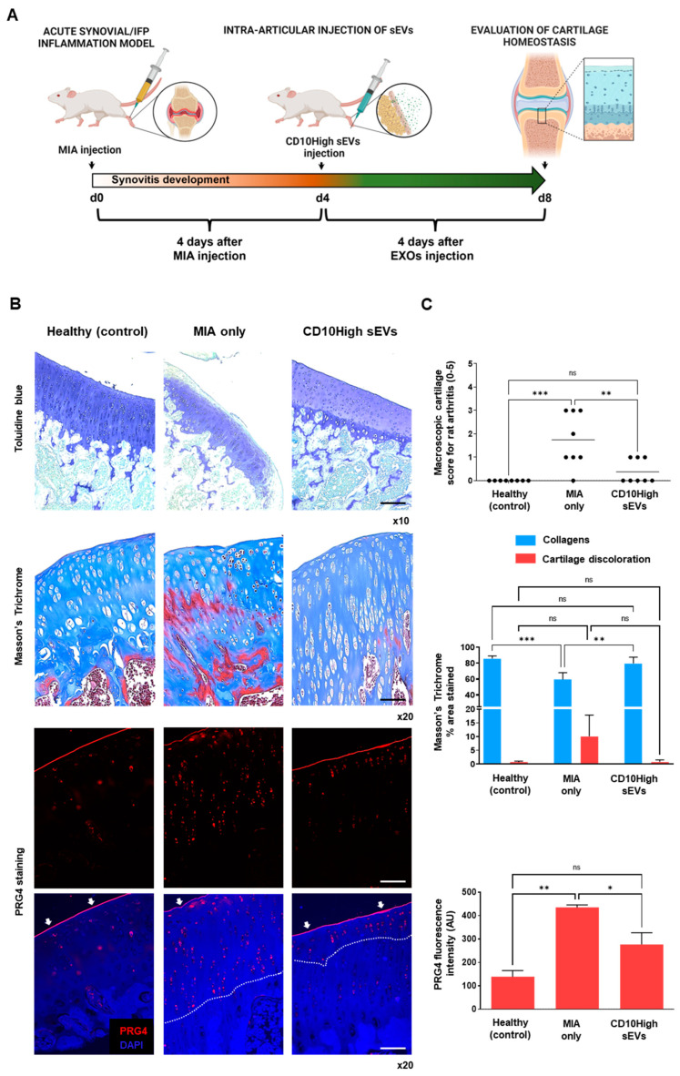
Figure 7 Effects of CD10High sEVs on articular cartilage homeostasis in vivo. (A) Schematic indicating the generation of acute synovitis/IFP fibrosis rat model, IFP-MSC sEVs’ therapeutic intervention and chronological evaluation. (B,C) Toluidine blue staining (top panel), Masson’s trichrome staining (middle panel), and PRG4 immunolocalization (lower panels) in sagittal-sectioned knees of representative rats for healthy, diseased (MIA only), or CD10High sEV treated groups. The diseased group demonstrated strong cartilage degeneration findings exemplified by reduced staining for sulfated proteoglycans. In contrast, CD10High sEV intra-articular infusion resulted in significantly reduced cartilage degeneration with only minor cartilage depressions and significantly increased collagen composition. Compared to the diseased group, the CD10High sEV group showed preservation of PRG4 expression on the upper cartilage surface and only minor expression from the intermediate zone chondrocytes (indicated by white arrows and dotted lines, ns: non-significant, * p < 0.05, ** p < 0.01, *** p < 0.001). Toluidine blue scale bar: 200 μm, Masson’s trichrome and PRG4 staining scale bars: 100 μm.
Figure 7 Effects of CD10High sEVs on articular cartilage homeostasis in vivo. (A) Schematic indicating the generation of acute synovitis/IFP fibrosis rat model, IFP-MSC sEVs’ therapeutic intervention and chronological evaluation. (B,C) Toluidine blue staining (top panel), Masson’s trichrome staining (middle panel), and PRG4 immunolocalization (lower panels) in sagittal-sectioned knees of representative rats for healthy, diseased (MIA only), or CD10High sEV treated groups. The diseased group demonstrated strong cartilage degeneration findings exemplified by reduced staining for sulfated proteoglycans. In contrast, CD10High sEV intra-articular infusion resulted in significantly reduced cartilage degeneration with only minor cartilage depressions and significantly increased collagen composition. Compared to the diseased group, the CD10High sEV group showed preservation of PRG4 expression on the upper cartilage surface and only minor expression from the intermediate zone chondrocytes (indicated by white arrows and dotted lines, ns: non-significant, * p < 0.05, ** p < 0.01, *** p < 0.001). Toluidine blue scale bar: 200 μm, Masson’s trichrome and PRG4 staining scale bars: 100 μm.CC BY · PubMed Central

Verification

Claims the paper makes

We couldn’t retrieve this paper’s full text, so its claims and self-check weren’t run. The registry and citation checks still apply.

Claims other papers make about this one

Each is checked against this paper’s own text: does it say what the citing paper says it does?

Citations

Cited by

Sorted by citations

Metadata

Authors
Kouroupis, D., Kaplan, L. D., Huard, J., & Best, T. M.
Affiliations
University of Miami (US)
Corresponding author
Kouroupis, Dimitrios
Venue
Cells
Publisher
MDPI AG
Published
2023-05-22
Volume
12
Issue
14
Pages
1824-1824
ISSN
2073-4409 (ISSN-L)
OpenAlex
W4384067747
Cited by
36
References
0
Citations by year
2026: 12 · 2025: 11 · 2024: 9 · 2023: 4
FWCI
3.21
Type
journal article
Language
en
Open access
Locations
  • Cells (publishedVersion, cc-by)
  • Preprints.org (acceptedVersion, cc-by)
  • PubMed Central (publishedVersion, cc-by)
  • PubMed (publishedVersion)
  • DOAJ (DOAJ: Directory of Open Access Journals) (submittedVersion, cc-by-sa)
Funders
  • Soffer Family Foundation
  • National Institutes of Health (5r21ar080388-02)
  • National Institute of Arthritis and Musculoskeletal and Skin Diseases (R21 AR080388)
Topics
  • Life Sciences › Biochemistry, Genetics and Molecular Biology › Molecular Biology › Extracellular vesicles in disease
  • Life Sciences › Biochemistry, Genetics and Molecular Biology › Cell Biology › Mesenchymal stem cell research
  • Health Sciences › Medicine › Rheumatology › Osteoarthritis Treatment and Mechanisms
Keywords
mesenchymal stem cells, small extracellular vesicles, CD10, neprilysin, synoviocytes, infrapatellar fat pad, synovitis, articular cartilage homeostasis, chondroprotection, microRNA cargo, immunomodulation, lubricin, cartilage degeneration
MeSH terms
Extracellular Vesicles, Animals, Cartilage, Articular, Fibrosis, Homeostasis, Humans, Knee Joint, Osteoarthritis, Synovitis, Neprilysin, Stromal Cells, MicroRNAs

About these checks

Registry, retraction, Cites and statistics results are computed, not judged, with one exception: a printed reference that no index lists is read once by a model before it counts as a citation error. Claims are extracted by a model and kept only when their quote appears verbatim in the text; each cited source is then checked, with a model’s help, for whether it says what the claim relies on. Those verdicts can be wrong — read the claim’s quote and the cited source before relying on one.

Statistics and arithmetic are recomputed, not judged.

Confidence bands are shown only for fields calibrated on our annotated test set. None is calibrated yet, so no band is shown.

What counts as a claim ›How we measure confidence ›How we check this ›

Are you an author, or do you see a problem with this page? Request a review