PreprintCited by 1Metalloenzymes and iron-sulfur proteinsEnzyme Structure and FunctionRNA and protein synthesis mechanisms

The DOI matches its Crossref record; 1 of the 1 references it cites were found in a registry or an index.

Functional Integrity of Radical SAM Enzyme Dph1•Dph2 Requires Non-canonical Cofactor Motifs with Tandem Cysteines

Open access

Preprint — published as 10.3390/biom14040470 · Paper page

Abstract

The Dph1•Dph2 heterodimer from yeast is a radical SAM (RS) enzyme that generates the 3-amino-3-carboxy-propyl (ACP) precursor for diphthamide, a clinically relevant modification on eukaryotic elongation factor 2 (eEF2). ACP formation requires SAM cleavage and atypical Cys-bound Fe-S clusters in each Dph1 and Dph2 subunit. Intriguingly, the first Cys residue in each motif locates next to another, ill-defined cysteine that we show in-here is conserved across eukaryotes. As judged from structural modeling, the orientation of these tandem cysteine motifs (TCM) suggests a candidate Fe-S cluster ligand role. Hence, we generated by site-directed DPH1 and DPH2 mutagenesis, Dph1•Dph2 variants with cysteines from each TCM replaced individually or in combination by serines. Assays diagnostic for diphthamide formation in vivo reveal that while single substitutions in the TCM of Dph2 cause mild defects, double mutations almost entirely inactivate the RS enzyme. Based on enhanced Dph1 and Dph2 subunit instability in response to cycloheximide chases, the variants with Cys substitutions in their cofactor motifs are particularly prone to protein degradation. In sum, we identify a fourth functionally cooperative Cys residue within the Fe-S motif of Dph2 and show that the Cys-based cofactor binding motifs in Dph1 and Dph2 are critical for the structural integrity of the dimeric RS enzyme in vivo.

Overview

The claims and statistics of this paper haven’t been checked yet.

Run the check — free, no account needed. It reads the open-access text and checks the sources it cites.

A few free checks a day · papers already verified open instantly · results are public

Integrity signals

Registry

Crossref · 10.20944/preprints202403.1547.v1

Cites

1 of 1 references verified0 citation errors · 0 not checkable

Statistics

Figures

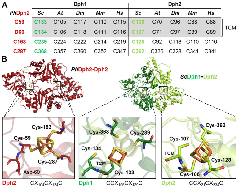
Figure 1 TCM conservation in atypical radical SAM and Fe-S motifs from Dph1•Dph2 dimers. (A) Dph1 and Dph2 alignment from indicated eukaryotic species to archaeal Dph2 (for full details, see Figure S1). (B) Structural comparisons. Left: structure of the PhDph2•Dph2 homodimer (firebrick red; PDB:3LZD) with the Cys-based (Cys-59; Cys-163; Cys-287) Fe-S binding motif previously established [7,9,10,12]. Note Cys-59 is next to Asp-60. Right: an AlphaFold model of the ScDph1•Dph2 heterodimer (forest and lemon green) confirming that cysteines in Dph1 (Cys-133; Cys-239; Cys-368) and Dph2 (Cys-C107; Cys-128; Cys-362) are proximal to respective Fe-S clusters. As part of TCMs, a fourth cysteine, Cys-134 next to Cys-133 in Dph1 and Cys-106 next to Cys-107 in Dph2, orients towards each Fe-S cluster. Fe-S motif close-ups are 66% transparent for emphasis on stick structures. Dph1•Dph2 was modelled and illustrated as previously described [10,21].
Figure 1 TCM conservation in atypical radical SAM and Fe-S motifs from Dph1•Dph2 dimers. (A) Dph1 and Dph2 alignment from indicated eukaryotic species to archaeal Dph2 (for full details, see Figure S1). (B) Structural comparisons. Left: structure of the PhDph2•Dph2 homodimer (firebrick red; PDB:3LZD) with the Cys-based (Cys-59; Cys-163; Cys-287) Fe-S binding motif previously established [7,9,10,12]. Note Cys-59 is next to Asp-60. Right: an AlphaFold model of the ScDph1•Dph2 heterodimer (forest and lemon green) confirming that cysteines in Dph1 (Cys-133; Cys-239; Cys-368) and Dph2 (Cys-C107; Cys-128; Cys-362) are proximal to respective Fe-S clusters. As part of TCMs, a fourth cysteine, Cys-134 next to Cys-133 in Dph1 and Cys-106 next to Cys-107 in Dph2, orients towards each Fe-S cluster. Fe-S motif close-ups are 66% transparent for emphasis on stick structures. Dph1•Dph2 was modelled and illustrated as previously described [10,21].CC BY · PubMed Central
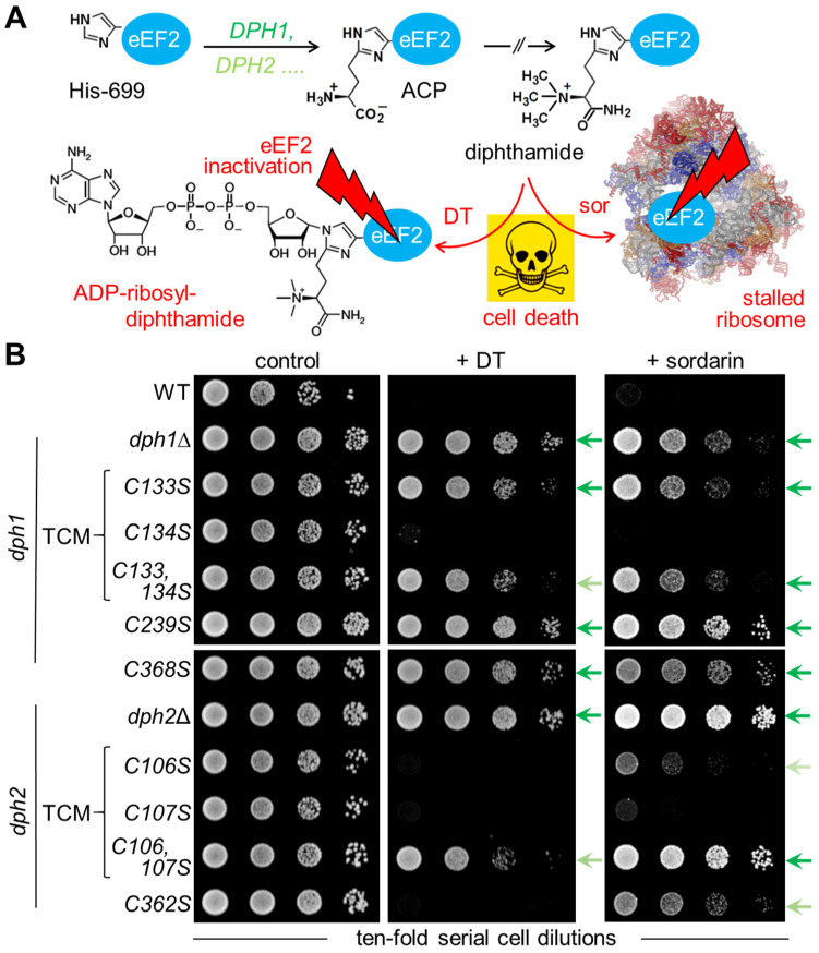
Figure 2 DPH1 and DPH2 mutagenesis to study TCM relevance for Dph1•Dph2 activity in vivo. (A) Simplified diphthamide pathway with DPH1 and DPH2 genes required to initiate ACP formation [21]; subsequent DPH products and steps for the completion of diphthamide synthesis [14,15,16,17] are not detailed. Diphthamide can be hijacked to induce cell death (skull–crossbones) either by DT for lethal ADP-ribosylation of eEF2 or sordarin (sor), which in a complex with the décor on eEF2 stalls ribosomes [40]. (B) Growth assays in response to DT and sordarin for the diagnosis of diphthamide defects in vivo. Yeast tester strains comprised wild-type (WT: DPH1 DPH2) and null mutants (dph1∆; dph2∆) together with single/double DPH1 or DPH2 gene substitutions, as indicated. Cells were grown w/o DT and sordarin (control: left panel), under DT-inducing conditions (+ DT: middle panel) [24], or with sordarin (+ sordarin: right panel) doses sufficient to inhibit WT cells. Arrows shaded in green denote various degrees of DT and sordarin resistance.
Figure 2 DPH1 and DPH2 mutagenesis to study TCM relevance for Dph1•Dph2 activity in vivo. (A) Simplified diphthamide pathway with DPH1 and DPH2 genes required to initiate ACP formation [21]; subsequent DPH products and steps for the completion of diphthamide synthesis [14,15,16,17] are not detailed. Diphthamide can be hijacked to induce cell death (skull–crossbones) either by DT for lethal ADP-ribosylation of eEF2 or sordarin (sor), which in a complex with the décor on eEF2 stalls ribosomes [40]. (B) Growth assays in response to DT and sordarin for the diagnosis of diphthamide defects in vivo. Yeast tester strains comprised wild-type (WT: DPH1 DPH2) and null mutants (dph1∆; dph2∆) together with single/double DPH1 or DPH2 gene substitutions, as indicated. Cells were grown w/o DT and sordarin (control: left panel), under DT-inducing conditions (+ DT: middle panel) [24], or with sordarin (+ sordarin: right panel) doses sufficient to inhibit WT cells. Arrows shaded in green denote various degrees of DT and sordarin resistance.CC BY · PubMed Central
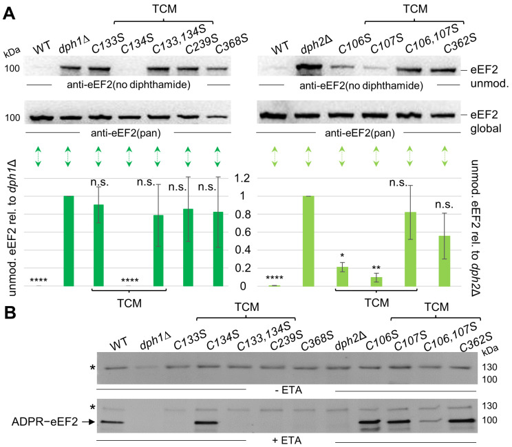
Figure 3 Accumulation of unmodified eEF2 in Cys-to-Ser Dph1•Dph2 mutants reveals functional roles of TCMs in diphthamide synthesis. (A) Western blots diagnostic for diphthamide synthesis on eEF2 from cell extracts of indicated yeast strains; anti-eEF2(no diphthamide) antibodies specific for unmodified eEF2 (upper panels) produce immune signals that associate with dysfunctional Dph1•Dph2 rather than WT enzymes, and anti-eEF2(pan) antibodies (middle panels) recognize eEF2 irrespective of modification state [31,32,33]. Technical repetitions (n = 3) were quantified (lower panels) using t-test statistics (* = p < 0.05; ** = p < 0.01; **** = p < 0.0001; n.s. = not significant). (B) ADP-ribosylation (ADPR) assay. Cell extracts incubated w/o (upper panel) or with exotoxin A (ETA) (500 ng, lower panel) with biotin-NAD+ allowed for diphthamide-dependent biotin-ADP-ribose transfer (ADPR-eEF2). Detection was performed via Western blot using a streptavidin-HPR-conjugate. Asterisks mark an unspecific ADPR band previously described [24]. Original images can be found in the Supplementary Materials (Figures S2–S4).
Figure 3 Accumulation of unmodified eEF2 in Cys-to-Ser Dph1•Dph2 mutants reveals functional roles of TCMs in diphthamide synthesis. (A) Western blots diagnostic for diphthamide synthesis on eEF2 from cell extracts of indicated yeast strains; anti-eEF2(no diphthamide) antibodies specific for unmodified eEF2 (upper panels) produce immune signals that associate with dysfunctional Dph1•Dph2 rather than WT enzymes, and anti-eEF2(pan) antibodies (middle panels) recognize eEF2 irrespective of modification state [31,32,33]. Technical repetitions (n = 3) were quantified (lower panels) using t-test statistics (* = p < 0.05; ** = p < 0.01; **** = p < 0.0001; n.s. = not significant). (B) ADP-ribosylation (ADPR) assay. Cell extracts incubated w/o (upper panel) or with exotoxin A (ETA) (500 ng, lower panel) with biotin-NAD+ allowed for diphthamide-dependent biotin-ADP-ribose transfer (ADPR-eEF2). Detection was performed via Western blot using a streptavidin-HPR-conjugate. Asterisks mark an unspecific ADPR band previously described [24]. Original images can be found in the Supplementary Materials (Figures S2–S4).CC BY · PubMed Central
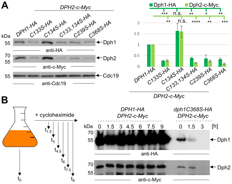
Figure 4 Malfunctional Cys-to-Ser variants of the Dph1•Dph2 dimer have significantly decreased amounts of Dph1 and Dph2 subunits. (A) Western blot detection of Dph1-HA (anti-HA) and Dph2-c-Myc (anti-c-Myc) from total extracts of indicated DPH1-HA substitution mutants co-expressing DPH2-c-Myc (left panel). Detection of Cdc19 (anti-Cdc19) served as internal control. Technical repetitions (n = 3) were followed by densitometric quantification of signal intensities (right panel) and t-test statistics (* = p < 0.05; ** = p < 0.01; *** = p < 0.001; **** = p < 0.0001; n.s. = not significant). (B) Cycloheximide chase of dph1C368S reveals accelerated decay of Dph1•Dph2. Schematic workflow of the cycloheximide chase (left panel). Yeast cells coding for HA and c-Myc-tagged Dph1 and Dph2 were grown to the exponential phase (t0) before the addition of cycloheximide. Samples were taken at indicated time points before (t0) and after cycloheximide addition for protein extraction and Western blot analysis (right panel). Original images can be found in the Supplementary Materials (Figures S5 and S8).
Figure 4 Malfunctional Cys-to-Ser variants of the Dph1•Dph2 dimer have significantly decreased amounts of Dph1 and Dph2 subunits. (A) Western blot detection of Dph1-HA (anti-HA) and Dph2-c-Myc (anti-c-Myc) from total extracts of indicated DPH1-HA substitution mutants co-expressing DPH2-c-Myc (left panel). Detection of Cdc19 (anti-Cdc19) served as internal control. Technical repetitions (n = 3) were followed by densitometric quantification of signal intensities (right panel) and t-test statistics (* = p < 0.05; ** = p < 0.01; *** = p < 0.001; **** = p < 0.0001; n.s. = not significant). (B) Cycloheximide chase of dph1C368S reveals accelerated decay of Dph1•Dph2. Schematic workflow of the cycloheximide chase (left panel). Yeast cells coding for HA and c-Myc-tagged Dph1 and Dph2 were grown to the exponential phase (t0) before the addition of cycloheximide. Samples were taken at indicated time points before (t0) and after cycloheximide addition for protein extraction and Western blot analysis (right panel). Original images can be found in the Supplementary Materials (Figures S5 and S8).CC BY · PubMed Central

Verification

Claims the paper makes

The claims and statistics of this paper haven’t been checked yet.

Run the check — free, no account needed. It reads the open-access text and checks the sources it cites.

A few free checks a day · papers already verified open instantly · results are public

Self-check

Percentages, sums and reported differences re-computed from the paper’s own numbers.

The self-check needs the paper’s full text.

Citations

Cites

Metadata

Authors
Ütkür, K., Mayer, K., Liu, S., Brinkmann, U., & Schaffrath, R.
Affiliations
  • University of Kassel (DE)
  • Roche Pharma AG (Germany) (DE)
  • University of Pittsburgh (US)
Corresponding author
Schaffrath, Raffael
Venue
Biomolecules
Publisher
MDPI AG
Published
2024-03-26
Volume
14
Issue
4
Pages
470-470
ISSN
2218-273X (ISSN-L)
OpenAlex
W4394764816
Cited by
1
References
1
Citations by year
2026: 1
FWCI
0.07
Type
journal article
Language
en
Open access
Locations
  • Biomolecules (publishedVersion, cc-by)
  • Preprints.org (acceptedVersion, cc-by)
  • PubMed Central (publishedVersion, cc-by)
  • PubMed (publishedVersion)
  • DOAJ (DOAJ: Directory of Open Access Journals) (submittedVersion, cc-by-sa)
Funders
  • Deutsche Forschungsgemeinschaft (Priority Program 1927 'Iron-Sulfur for Life' SCHA750/21-1)
  • Universität Kassel
Topics
  • Life Sciences › Biochemistry, Genetics and Molecular Biology › Biochemistry › Metalloenzymes and iron-sulfur proteins
  • Life Sciences › Biochemistry, Genetics and Molecular Biology › Structural Biology › Enzyme Structure and Function
  • Life Sciences › Biochemistry, Genetics and Molecular Biology › Molecular Biology › RNA and protein synthesis mechanisms
Keywords
radical SAM enzymes, diphthamide biosynthesis, iron-sulfur clusters, protein stability, protein degradation, site-directed mutagenesis, Saccharomyces cerevisiae
MeSH terms
Cysteine, Histidine, Repressor Proteins, Saccharomyces cerevisiae, Carbon-Sulfur Lyases, Mutagenesis, Site-Directed, Amino Acid Motifs, Saccharomyces cerevisiae Proteins, Protein Multimerization

About these checks

Registry, retraction, Cites and statistics results are computed, not judged, with one exception: a printed reference that no index lists is read once by a model before it counts as a citation error. Claims are extracted by a model and kept only when their quote appears verbatim in the text; each cited source is then checked, with a model’s help, for whether it says what the claim relies on. Those verdicts can be wrong — read the claim’s quote and the cited source before relying on one.

Statistics and arithmetic are recomputed, not judged.

Confidence bands are shown only for fields calibrated on our annotated test set. None is calibrated yet, so no band is shown.

What counts as a claim ›How we measure confidence ›How we check this ›

Are you an author, or do you see a problem with this page? Request a review