Cited by 5Pelvic floor disorders treatmentsGynecological conditions and treatmentsPregnancy-related medical research

The DOI matches its Crossref record; 9 of the 9 references it cites were found in a registry or an index.

A practical ligament-based diagnostic system for cure of pelvic symptoms and prolapse

Open access
Petros, Peter, Liedl, Bernhard, Palma, Paolo, Riccetto, Cassio, Ding, Shuqing·2024·Annals of Translational Medicine

Abstract

The Integral Theory Paradigm (ITP) has a 25-year track record of successfully treating bladder/bowel/pain symptoms caused by laxity in specific ligaments, even when the prolapse is minimal. The ITP-based treatment involves ligament support and can be nonsurgical or daycare surgical. An accurate diagnostic protocol is required. The Integral Theory Diagnostic system is performed in an outpatient setting. It a step-by-step "how to" resource for clinicians who wish to learn a practical anatomical diagnostic method which can quickly and accurately identify a ligament cause for bladder/bowel/pain symptoms, and therefore, potentially cure them. The structured ITP diagnosis flow chart uses symptoms to diagnose anatomical defects. It comprises 4 related steps. The ITP is holistic, and bladder, bowel, pain symptoms co-occur. The first step, therefore, is to establish all possible symptoms for transfer to the Diagnostic Algorithm which is the second step. Because patients complain of one main symptom, other symptoms must be located by direct questioning, using the Diagnostic Algorithm as an aide memoire, or a questionnaire to locate bladder, bowel, pain symptoms. Second step: symptoms are placed into 3 anatomical zones: anterior zone, pubourethral ligament (PUL) [stress urinary incontinence (SUI)]; middle zone, cardinal ligament (CL) (transverse defect cystocele); posterior zone, uterosacral ligament (USL) (uterine prolapse and enterocele). The third step is a vaginal examination to confirm the ligament damage (prolapses) in the three zones predicted by the algorithm. The fourth step is "simulated operations" (mechanical support of specific ligaments per vaginam) to validate the particular ligament indicated by the diagnostic algorithm, is indeed causing that symptom. For SUI, a hemostat test at midurethra supports PUL vaginally to stop urine loss on coughing; the lower blade of a bivalve speculum gently inserted into the vagina can relieve urge and pain.

Overview

The claims and statistics of this paper haven’t been checked yet.

Run the check — free, no account needed. It reads the open-access text and checks the sources it cites.

A few free checks a day · papers already verified open instantly · results are public

Integrity signals

Registry

Crossref · 10.21037/atm-23-1759

Cites

9 of 9 references verified0 citation errors · 0 not checkable

Statistics

Figures
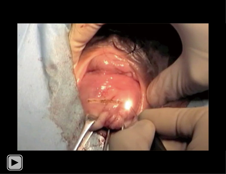
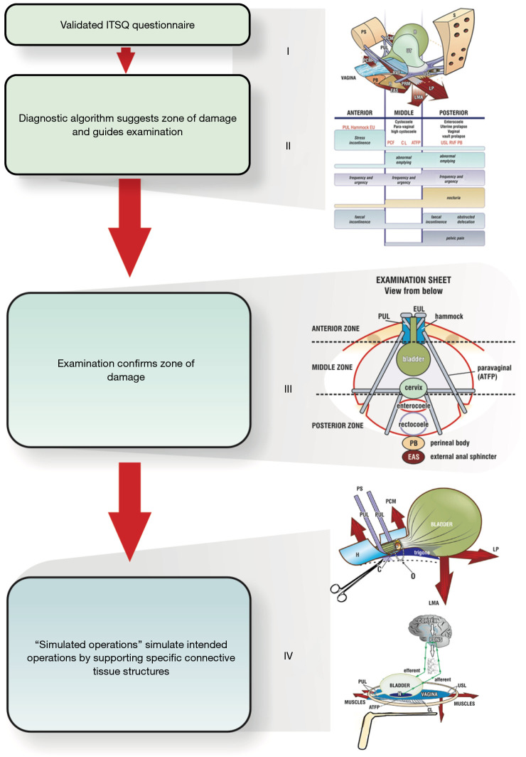
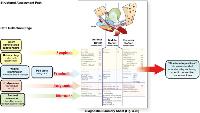
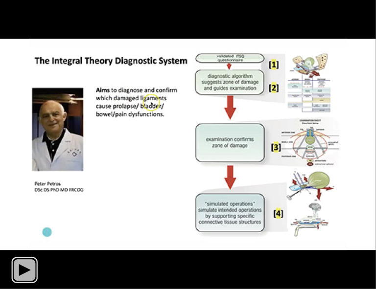
Figure 1 The diagnostic system flow chart has 4 parts: (I) the validated patient questionnaire ITSQ; (II) the pictorial diagnostic algorithm; (III) vaginal examination; (IV) simulated operations. Reused from (1). With permission from Peter Petros; retains ownership of the copyright.ITSQ, Integral Theory Symptom Questionnaire; S, sacrum; B, bladder; PS, pubic symphysis; PCM, pubococcygeus muscle; PUL, pubourethral ligament; UT, uterus; R, rectum; USL, uterosacral ligament; RVF, rectovaginal fascia; PB, perineal body; PRM, puborectalis muscle; EAS, external anal sphincter; LMA, conjoint longitudinal muscle of the anus; LP, levator plate; EUL, external urethral ligament; PCF, pubocervical fascia; CL, cardinal ligament; ATFP, arcus tendineus fascia pelvis; H, suburethral vaginal hammock; C, closed; O, open.
Figure 1 The diagnostic system flow chart has 4 parts: (I) the validated patient questionnaire ITSQ; (II) the pictorial diagnostic algorithm; (III) vaginal examination; (IV) simulated operations. Reused from (1). With permission from Peter Petros; retains ownership of the copyright.ITSQ, Integral Theory Symptom Questionnaire; S, sacrum; B, bladder; PS, pubic symphysis; PCM, pubococcygeus muscle; PUL, pubourethral ligament; UT, uterus; R, rectum; USL, uterosacral ligament; RVF, rectovaginal fascia; PB, perineal body; PRM, puborectalis muscle; EAS, external anal sphincter; LMA, conjoint longitudinal muscle of the anus; LP, levator plate; EUL, external urethral ligament; PCF, pubocervical fascia; CL, cardinal ligament; ATFP, arcus tendineus fascia pelvis; H, suburethral vaginal hammock; C, closed; O, open.CC BY-NC-ND · PubMed Central
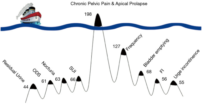
Figure 2 The Pescatori Iceberg diagram applied to CPP. Reused from (3). Copyright 2017, with permission from the Pelviperineology. The presenting symptom was CPP. Symptom prevalence is graphically indicated in the iceberg diagram. Latent symptoms are below the waterline, are numbered according to their prevalence and were derived from the ITSQ (4). CPP, chronic pelvic pain; ITSQ, Integral Theory Symptom Questionnaire; ODS, obstructive defecation syndrome; SUI, stress urinary incontinence; FI, fecal incontinence.
Figure 2 The Pescatori Iceberg diagram applied to CPP. Reused from (3). Copyright 2017, with permission from the Pelviperineology. The presenting symptom was CPP. Symptom prevalence is graphically indicated in the iceberg diagram. Latent symptoms are below the waterline, are numbered according to their prevalence and were derived from the ITSQ (4). CPP, chronic pelvic pain; ITSQ, Integral Theory Symptom Questionnaire; ODS, obstructive defecation syndrome; SUI, stress urinary incontinence; FI, fecal incontinence.CC BY-NC-ND · PubMed Central
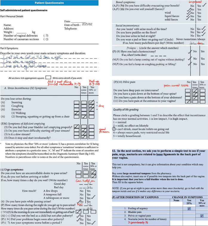
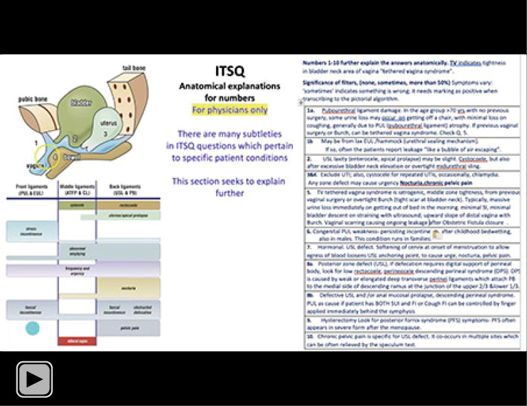
Figure 3 Self-administered patient questionnaire. A,M,P (Anterior, Middle, Posterior), on the left side of the questionnaire, indicate zones of connective tissue (ligament) looseness. The numbers 1–11 in the left column of the questionnaire explain the anatomical basis of the questions. This part is for the physician only and is located at the end of each questionnaire (see Appendix 1). The red writing shows the post-operative symptom results from midurethral and uterosacral sling operations. Reused from (1). With permission from Peter Petros; retains ownership of the copyright.
Figure 3 Self-administered patient questionnaire. A,M,P (Anterior, Middle, Posterior), on the left side of the questionnaire, indicate zones of connective tissue (ligament) looseness. The numbers 1–11 in the left column of the questionnaire explain the anatomical basis of the questions. This part is for the physician only and is located at the end of each questionnaire (see Appendix 1). The red writing shows the post-operative symptom results from midurethral and uterosacral sling operations. Reused from (1). With permission from Peter Petros; retains ownership of the copyright.CC BY-NC-ND · PubMed Central
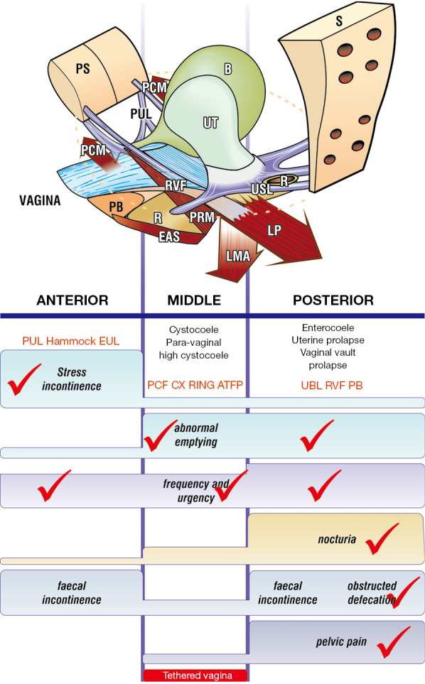
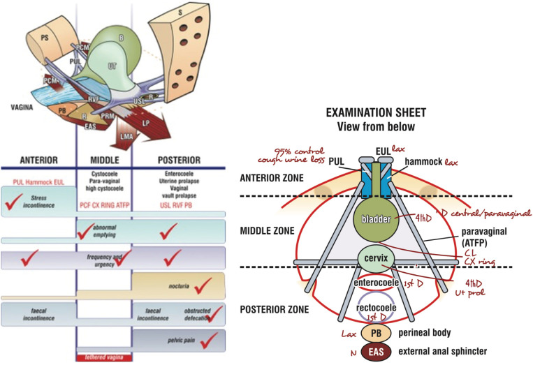
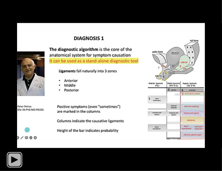
Figure 4 Patient algorithm has three zones of causation (ligament damage). It is marked with ITSQ answers. The prolapses in the algorithm correlate with ligament damage in the columns. Symptom groupings in the columns help deduce which ligaments cause which symptoms. Anterior zone runs from the external meatus to the bladder neck, and comprises EUL, PUL, suburethral vaginal hammock, and dislocated collagenous insertion of PCM also known as “levator avulsion”. Middle zone runs from the bladder neck to the anterior cervical ring and comprises PCF, CL, and ATFP. Posterior zone runs from the cervix to the PB. It comprises USL, RVF, and PB. Reused from (1). With permission from Peter Petros; retains ownership of the copyright. S, sacrum; B, bladder; PS, pubic symphysis; PCM, pubococcygeus muscle; PUL, pubourethral ligament; UT, uterus; R, rectum; USL, uterosacral ligament; RVF, rectovaginal fascia; PB, perineal body; PRM, puborectalis muscle; EAS, external anal sphincter; LMA, conjoint longitudinal muscle of the anus; LP, levator plate; EUL, external urethral ligament; PCF, pubocervical fascia; CX RING, cervical ring; USL, uterosacral ligament; ITSQ, Integral Theory Symptom Questionnaire; CL, cardinal ligament; ATFP, arcus tendineus fascia pelvis.
Figure 4 Patient algorithm has three zones of causation (ligament damage). It is marked with ITSQ answers. The prolapses in the algorithm correlate with ligament damage in the columns. Symptom groupings in the columns help deduce which ligaments cause which symptoms. Anterior zone runs from the external meatus to the bladder neck, and comprises EUL, PUL, suburethral vaginal hammock, and dislocated collagenous insertion of PCM also known as “levator avulsion”. Middle zone runs from the bladder neck to the anterior cervical ring and comprises PCF, CL, and ATFP. Posterior zone runs from the cervix to the PB. It comprises USL, RVF, and PB. Reused from (1). With permission from Peter Petros; retains ownership of the copyright. S, sacrum; B, bladder; PS, pubic symphysis; PCM, pubococcygeus muscle; PUL, pubourethral ligament; UT, uterus; R, rectum; USL, uterosacral ligament; RVF, rectovaginal fascia; PB, perineal body; PRM, puborectalis muscle; EAS, external anal sphincter; LMA, conjoint longitudinal muscle of the anus; LP, levator plate; EUL, external urethral ligament; PCF, pubocervical fascia; CX RING, cervical ring; USL, uterosacral ligament; ITSQ, Integral Theory Symptom Questionnaire; CL, cardinal ligament; ATFP, arcus tendineus fascia pelvis.CC BY-NC-ND · PubMed Central
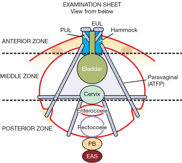
Figure 5 Completed examination sheet record. Completed examination sheet record for anatomical defects found in each of the three zones (for the completed ITSQ patient questionnaire). Reused from (1). With permission from Peter Petros; retains ownership of the copyright. ITSQ, Integral Theory Symptom Questionnaire; EUL, external urethral ligament; PUL, pubourethral ligament; PB, perineal body; EAS, external anal sphincter; ATFP, arcus tendineus fascia pelvis.
Figure 5 Completed examination sheet record. Completed examination sheet record for anatomical defects found in each of the three zones (for the completed ITSQ patient questionnaire). Reused from (1). With permission from Peter Petros; retains ownership of the copyright. ITSQ, Integral Theory Symptom Questionnaire; EUL, external urethral ligament; PUL, pubourethral ligament; PB, perineal body; EAS, external anal sphincter; ATFP, arcus tendineus fascia pelvis.CC BY-NC-ND · PubMed Central
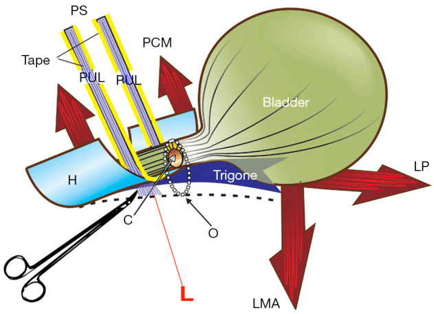
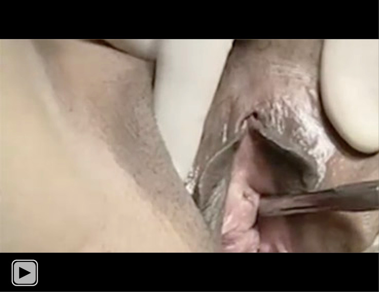
Figure 6 Anterior zone: Hemostat test. The hemostat test mechanically prevents elongation of the PUL in the same way as a midurethral sling. This is a 3-dimensional sagittal view of the bladder base and trigone supported by the vagina below (broken lines); PUL laxity “L”, allows PUL to stretch downwards; in the absence of a firm PUL anchoring point, the normal LP/LMA contraction which closes the bladder neck now forcibly opens the posterior wall of the urethra, from “C” (closed) to “O” (open), so urine is lost on effort. The hemostat mechanically prevents extension of the PUL, exactly as does a midurethral tape, so the bladder neck and distal urethra are closed as per normal. With permission from Peter Petros (1); retains ownership of the copyright. PUL, pubourethral ligament; PCM, pubococcygeus muscle; LP, levator plate; LMA, conjoint longitudinal muscle of the anus; H, suburethral vaginal hammock; C, closed; O, open; PS, pubic symphysis.
Figure 6 Anterior zone: Hemostat test. The hemostat test mechanically prevents elongation of the PUL in the same way as a midurethral sling. This is a 3-dimensional sagittal view of the bladder base and trigone supported by the vagina below (broken lines); PUL laxity “L”, allows PUL to stretch downwards; in the absence of a firm PUL anchoring point, the normal LP/LMA contraction which closes the bladder neck now forcibly opens the posterior wall of the urethra, from “C” (closed) to “O” (open), so urine is lost on effort. The hemostat mechanically prevents extension of the PUL, exactly as does a midurethral tape, so the bladder neck and distal urethra are closed as per normal. With permission from Peter Petros (1); retains ownership of the copyright. PUL, pubourethral ligament; PCM, pubococcygeus muscle; LP, levator plate; LMA, conjoint longitudinal muscle of the anus; H, suburethral vaginal hammock; C, closed; O, open; PS, pubic symphysis.CC BY-NC-ND · PubMed Central
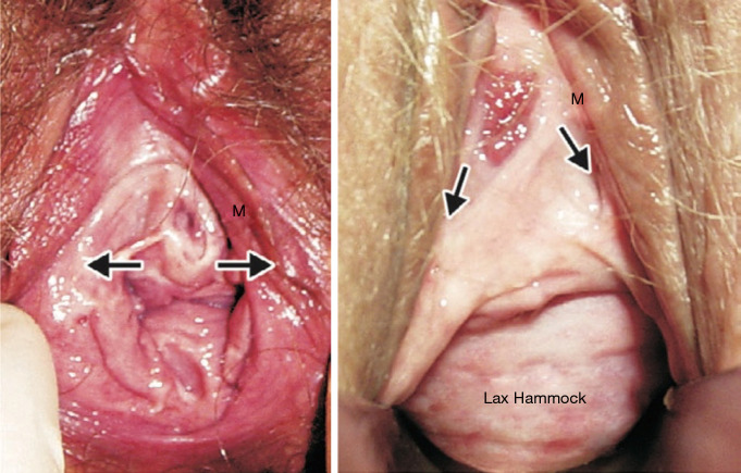
Figure 7 Anterior zone: EUL defect. Left: normal EUL. The urethral meatus (M) is well supported horizontally by the EUL (arrows) which attach it to the anterior surface of the symphysis. The hammock is in the normal plane. Note: the external urethral ligament is also called, “anterior pubourethral ligament”. Right: lax EUL and hammock. The urethral meatus (M) is lax and has caused the urethral mucosa to become everted. The lateral EUL supports are seen “drooping” downwards (arrows). The hammock is lax and angulated downwards. “Simulated operation”. Gentle mechanical support of EULs (“simulated operation”) will usually cause the everted mucosa to retract. With permission from Peter Petros (1); retains ownership of the copyright. EUL, external urethral ligament.
Figure 7 Anterior zone: EUL defect. Left: normal EUL. The urethral meatus (M) is well supported horizontally by the EUL (arrows) which attach it to the anterior surface of the symphysis. The hammock is in the normal plane. Note: the external urethral ligament is also called, “anterior pubourethral ligament”. Right: lax EUL and hammock. The urethral meatus (M) is lax and has caused the urethral mucosa to become everted. The lateral EUL supports are seen “drooping” downwards (arrows). The hammock is lax and angulated downwards. “Simulated operation”. Gentle mechanical support of EULs (“simulated operation”) will usually cause the everted mucosa to retract. With permission from Peter Petros (1); retains ownership of the copyright. EUL, external urethral ligament.CC BY-NC-ND · PubMed Central
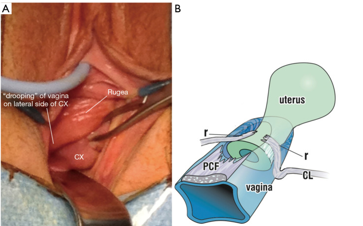
Figure 8 Middle zone: transverse defect. Findings of prolapsed vagina lateral to the CX, confirms CL dislocation or tearing. With permission from Peter Petros (1); retains ownership of the copyright. (A) Vaginal prolapse lateral to the cervix contains prolapsed “CL” as per (B); (B) pathogenesis: the vagina’s PCF, is torn from the “CL” and, now unsupported, prolapses down like a trapdoor as a “lateral defect” or “high” cystocele. r, rupture of the “CL”; PCF, pubocervical fascial layer; CL, cardinal ligament; CX, cervix.
Figure 8 Middle zone: transverse defect. Findings of prolapsed vagina lateral to the CX, confirms CL dislocation or tearing. With permission from Peter Petros (1); retains ownership of the copyright. (A) Vaginal prolapse lateral to the cervix contains prolapsed “CL” as per (B); (B) pathogenesis: the vagina’s PCF, is torn from the “CL” and, now unsupported, prolapses down like a trapdoor as a “lateral defect” or “high” cystocele. r, rupture of the “CL”; PCF, pubocervical fascial layer; CL, cardinal ligament; CX, cervix.CC BY-NC-ND · PubMed Central
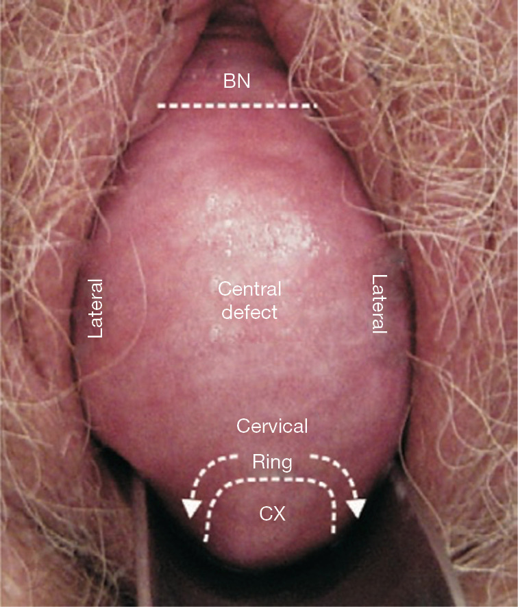
Figure 9 Middle zone: central defect cystocele and uterine prolapse. A large central defect (4th degree) extends laterally. Prolapse of the dislocated CL is seen around the CX (curved arrows), characteristic of CL/cervical ring defect. The central cystocele invariably has shiny vaginal epithelium. It is most likely caused by dislocation of the PCM from the symphysis. The central defect remained after the Allis forceps test to approximate displaced CLs and restore transverse defect. Posterior zone: uterine prolapse and cystocele, both 4th degree. The uterus is totally externalized. A dislocated CL is seen around the CX (curved arrows), characteristic of CL/cervical ring. With permission from Peter Petros (1); retains ownership of the copyright. BN, bladder neck; CX, cervix; CL, cardinal ligament; PCM, pubococcygeus muscle.
Figure 9 Middle zone: central defect cystocele and uterine prolapse. A large central defect (4th degree) extends laterally. Prolapse of the dislocated CL is seen around the CX (curved arrows), characteristic of CL/cervical ring defect. The central cystocele invariably has shiny vaginal epithelium. It is most likely caused by dislocation of the PCM from the symphysis. The central defect remained after the Allis forceps test to approximate displaced CLs and restore transverse defect. Posterior zone: uterine prolapse and cystocele, both 4th degree. The uterus is totally externalized. A dislocated CL is seen around the CX (curved arrows), characteristic of CL/cervical ring. With permission from Peter Petros (1); retains ownership of the copyright. BN, bladder neck; CX, cervix; CL, cardinal ligament; PCM, pubococcygeus muscle.CC BY-NC-ND · PubMed Central
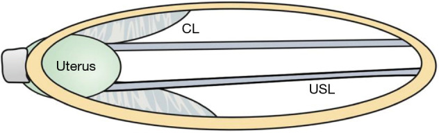
Figure 10 Posterior zone: 3rd degree uterine prolapse. Damage or weakening of the USL and CL at the entry to the birth canal contribute to uterine prolapse. Elongation of USLs also causes them to spread laterally, to cause an enterocele. It is important to remember that USLs attach to the lateral wall of the rectum with fine ligaments. As the uterus prolapses down, the anterior rectal wall may descend also, often as an intussusception. Surgical shortening and reinforcement of the USLs will also restore both the uterine prolapse and also, the intussusception (6). With permission from Peter Petros (1); retains ownership of the copyright. USL, uterosacral ligaments; CL, cardinal ligaments.
Figure 10 Posterior zone: 3rd degree uterine prolapse. Damage or weakening of the USL and CL at the entry to the birth canal contribute to uterine prolapse. Elongation of USLs also causes them to spread laterally, to cause an enterocele. It is important to remember that USLs attach to the lateral wall of the rectum with fine ligaments. As the uterus prolapses down, the anterior rectal wall may descend also, often as an intussusception. Surgical shortening and reinforcement of the USLs will also restore both the uterine prolapse and also, the intussusception (6). With permission from Peter Petros (1); retains ownership of the copyright. USL, uterosacral ligaments; CL, cardinal ligaments.CC BY-NC-ND · PubMed Central
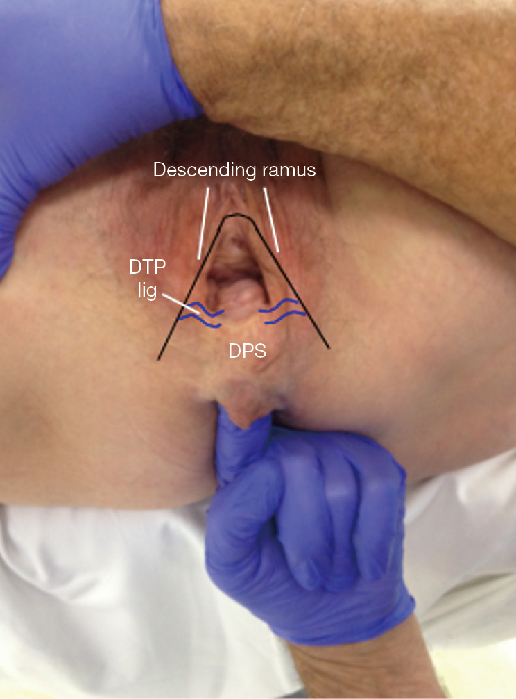
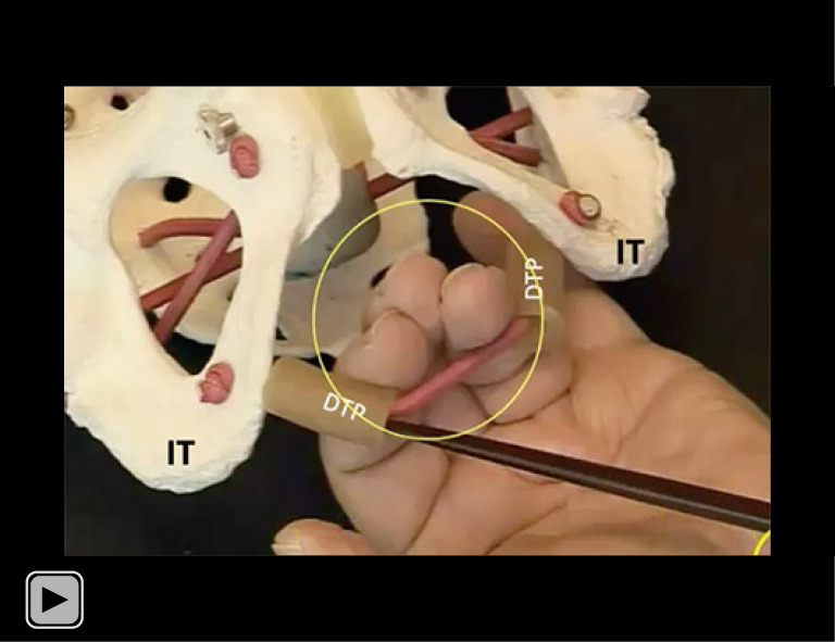
Figure 11 Posterior zone: rectal examination. DTP ligaments insert behind the junction of the upper 2/3 and lower 1/3 of the descending rami to suspend the perineal body. Reused from (1). With permission from Peter Petros; retains ownership of the copyright. DPS, descending perineal syndrome; DTP, deep transversus perinei.
Figure 11 Posterior zone: rectal examination. DTP ligaments insert behind the junction of the upper 2/3 and lower 1/3 of the descending rami to suspend the perineal body. Reused from (1). With permission from Peter Petros; retains ownership of the copyright. DPS, descending perineal syndrome; DTP, deep transversus perinei.CC BY-NC-ND · PubMed Central
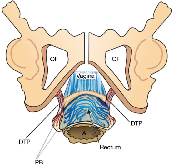
Figure 12 Posterior zone: pathogenesis of perineocele and descending perineal syndrome. Overstretching of the perineum at delivery may cause elongation of the DTP ligaments, with consequent displacement of the perineal bodies to below the ischial tuberosity. The rectum protrudes in the space of the displaced “PBs” and if the rectum’s muscular and serosal layers are ruptured, the rectal mucosa, often 1-mm thick, may adhere closely to the vagina, “PB” and pubic rami. Reused from (1). With permission from Peter Petros; retains ownership of the copyright. DTP, deep transversus perinei; OF, obturator fossa; PB, perineal body; A, anus.
Figure 12 Posterior zone: pathogenesis of perineocele and descending perineal syndrome. Overstretching of the perineum at delivery may cause elongation of the DTP ligaments, with consequent displacement of the perineal bodies to below the ischial tuberosity. The rectum protrudes in the space of the displaced “PBs” and if the rectum’s muscular and serosal layers are ruptured, the rectal mucosa, often 1-mm thick, may adhere closely to the vagina, “PB” and pubic rami. Reused from (1). With permission from Peter Petros; retains ownership of the copyright. DTP, deep transversus perinei; OF, obturator fossa; PB, perineal body; A, anus.CC BY-NC-ND · PubMed Central
Figure 13 Testing symptom predictions of ligament damage. Comparing algorithm, Figure 4 (left) with vaginal examination, Figure 5 (right). Reused from (1). With permission from Peter Petros; retains ownership of the copyright. USL, uterosacral ligament; PUL, pubourethral ligament; EUL, external urethral ligament; ATFP, arcus tendineus fascia pelvis; CL, cardinal ligament; LMA, conjoint longitudinal muscle of the anus; LP, levator plate; S, sacrum; B, bladder; PS, pubic symphysis; PCM, pubococcygeus muscle; PRM, puborectalis muscle; RVF, rectovaginal fascia; UT, uterus; PCF, pubocervical fascia; EAS, external anal sphincter; PB, perineal body; R, rectum; CX, cervix.
Figure 13 Testing symptom predictions of ligament damage. Comparing algorithm, Figure 4 (left) with vaginal examination, Figure 5 (right). Reused from (1). With permission from Peter Petros; retains ownership of the copyright. USL, uterosacral ligament; PUL, pubourethral ligament; EUL, external urethral ligament; ATFP, arcus tendineus fascia pelvis; CL, cardinal ligament; LMA, conjoint longitudinal muscle of the anus; LP, levator plate; S, sacrum; B, bladder; PS, pubic symphysis; PCM, pubococcygeus muscle; PRM, puborectalis muscle; RVF, rectovaginal fascia; UT, uterus; PCF, pubocervical fascia; EAS, external anal sphincter; PB, perineal body; R, rectum; CX, cervix.CC BY-NC-ND · PubMed Central
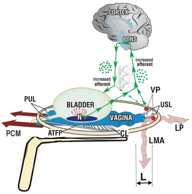
Figure 14 Speculum test (simulated operation). Gentle insertion of the speculum mechanically supports stretch receptors “N” to control urge. The speculum also supports lax USL and the VP to control pain. This test relieves pain and urge in 70–80% of women who have these symptoms. The pink wavy lines indicate weakened muscle forces, a consequence of the “LP” and “LMA” contracting against weakened USLs (L). Reused from (1). With permission from Peter Petros; retains ownership of the copyright. USL, uterosacral ligament; VP, visceral plexuses; PUL, pubourethral ligament; PCM, pubococcygeus muscle; ATFP, arcus tendineus fascia pelvis; CL, cardinal ligament; LMA, conjoint longitudinal muscle of the anus; LP, levator plate.
Figure 14 Speculum test (simulated operation). Gentle insertion of the speculum mechanically supports stretch receptors “N” to control urge. The speculum also supports lax USL and the VP to control pain. This test relieves pain and urge in 70–80% of women who have these symptoms. The pink wavy lines indicate weakened muscle forces, a consequence of the “LP” and “LMA” contracting against weakened USLs (L). Reused from (1). With permission from Peter Petros; retains ownership of the copyright. USL, uterosacral ligament; VP, visceral plexuses; PUL, pubourethral ligament; PCM, pubococcygeus muscle; ATFP, arcus tendineus fascia pelvis; CL, cardinal ligament; LMA, conjoint longitudinal muscle of the anus; LP, levator plate.CC BY-NC-ND · PubMed Central
Figure 15 Tertiary referral centre investigative assessment tool. Reused from (1). With permission from Peter Petros; retains ownership of the copyright.
Figure 15 Tertiary referral centre investigative assessment tool. Reused from (1). With permission from Peter Petros; retains ownership of the copyright.CC BY-NC-ND · PubMed Central
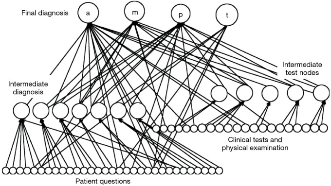
Figure 16 Schema for Bayesian Network output nodes. Top row: predicting the likelihood of defects in the anterior (a), middle (m), or posterior (p) zones, or a diagnosis of Tethered Vagina Syndrome (t). Intermediate diagnosis and test, and patient questions and clinical test nodes are as shown. Reused from (1). With permission from Peter Petros; retains ownership of the copyright.
Figure 16 Schema for Bayesian Network output nodes. Top row: predicting the likelihood of defects in the anterior (a), middle (m), or posterior (p) zones, or a diagnosis of Tethered Vagina Syndrome (t). Intermediate diagnosis and test, and patient questions and clinical test nodes are as shown. Reused from (1). With permission from Peter Petros; retains ownership of the copyright.CC BY-NC-ND · PubMed Central
Video S1 Overview.
Video S1 Overview.CC BY-NC-ND · PubMed Central
Video S2 Diagnostic algorithm.
Video S2 Diagnostic algorithm.CC BY-NC-ND · PubMed Central
Video S3 Integral Theory System Questionnaire.
Video S3 Integral Theory System Questionnaire.CC BY-NC-ND · PubMed Central
Video S4 Hemostat support of PUL for diagnosis of stress incontinence by permission of Professor Paolo Palma.
Video S4 Hemostat support of PUL for diagnosis of stress incontinence by permission of Professor Paolo Palma.CC BY-NC-ND · PubMed Central
Video S5 Role of both cardinal and uterosacral ligaments for uterine support by permission of Professor Yuki Sekiguchi.
Video S5 Role of both cardinal and uterosacral ligaments for uterine support by permission of Professor Yuki Sekiguchi.CC BY-NC-ND · PubMed Central
Video S6 Structural support of perineal body.
Video S6 Structural support of perineal body.CC BY-NC-ND · PubMed Central

Verification

Claims the paper makes

The claims and statistics of this paper haven’t been checked yet.

Run the check — free, no account needed. It reads the open-access text and checks the sources it cites.

A few free checks a day · papers already verified open instantly · results are public

Self-check

Percentages, sums and reported differences re-computed from the paper’s own numbers.

The self-check needs the paper’s full text.

Citations

Cites

Show all 9 references

Research works printed but not linked: 5

  1. [1]Petros PE. Chapter 3, Diagnosis of Connective tissue damage. In: The female pelvic floor: function, dysfunction and management according to the Integral Theory. Heidelberg: Springer Verlag; 2010:81-103.
  2. [3]Goeschen K Gold DM. Surgical cure of chronic pelvic pain, associated bladder & bowel symptoms by posterior sling in 198 patients validates the Pescatori Iceberg principle of pelvic symptom co-occurrence. Pelviperineology 2017; 36: 84- 8.
  3. [5]Scheffler KU Petros PE Oliver W A hypothesis for urinary stream divergence in the female: unilateral dislocation of the pubovisceral muscle. Pelviperineology 2014; 33: 10- 3.
  4. [6]Abendstein B Brugger BA Furtschegger A Role of the uterosacral ligaments in the causation of rectal intussusception, abnormal bowel emptying, and fecal incontinence-a prospective study. Pelviperineology 2008; 27: 118- 21.
  5. [14]Hunt M, von Konsky B, Venkatesh S, et al. Bayesian networks and decision trees in the diagnosis of female urinary incontinence. Engineering in Medicine and Biology Society 2000. Proceedings of the 22nd Annual International Conference of the IEEE 2000;1:551-4.

Cited by

Sorted by citations

See all 5

Metadata

Authors
Petros, P., Liedl, B., Palma, P., Riccetto, C., & Ding, S.
Affiliations
  • Urologische Klinik München (DE)
  • Northwestern Health Sciences University (US)
Corresponding author
Petros, Peter
Venue
Annals of Translational Medicine
Published
2024-04
Volume
12
Issue
2
Pages
27-27
ISSN
  • 2305-5839 (ISSN-L)
  • 2305-5847
OpenAlex
W4394957284
Cited by
4
References
9
Citations by year
2026: 1 · 2025: 1 · 2024: 2
FWCI
1.30
Type
journal article
Language
en
Open access
Licence
cc-by-nc-nd
Locations
  • Annals of Translational Medicine (publishedVersion, cc-by-nc-nd)
  • PubMed Central (publishedVersion, cc-by-nc-nd)
  • PubMed (publishedVersion)
Topics
  • Health Sciences › Medicine › Urology › Pelvic floor disorders treatments
  • Health Sciences › Medicine › Obstetrics and Gynecology › Gynecological conditions and treatments
  • Health Sciences › Medicine › Obstetrics and Gynecology › Pregnancy-related medical research
Keywords
pelvic organ prolapse, stress urinary incontinence, cystocele, enterocele, pubourethral ligament, cardinal ligament, uterosacral ligament, ligamentous laxity, diagnostic algorithm, vaginal examination, bladder symptoms, bowel symptoms, pelvic pain

About these checks

Registry, retraction, Cites and statistics results are computed, not judged, with one exception: a printed reference that no index lists is read once by a model before it counts as a citation error. Claims are extracted by a model and kept only when their quote appears verbatim in the text; each cited source is then checked, with a model’s help, for whether it says what the claim relies on. Those verdicts can be wrong — read the claim’s quote and the cited source before relying on one.

Statistics and arithmetic are recomputed, not judged.

Confidence bands are shown only for fields calibrated on our annotated test set. None is calibrated yet, so no band is shown.

What counts as a claim ›How we measure confidence ›How we check this ›

Are you an author, or do you see a problem with this page? Request a review

How to cite

Cite CoChat for the checks on this page, and the paper for its own findings.

This page
CoChat. (2026). Citation check: A practical ligament-based diagnostic system for cure of pelvic symptoms and prolapse. https://app.cochat.ai/papers/10.21037/atm-23-1759
The paper
Petros, P., Liedl, B., Palma, P., Riccetto, C., & Ding, S. (2024). A practical ligament-based diagnostic system for cure of pelvic symptoms and prolapse. Annals of Translational Medicine. https://doi.org/10.21037/atm-23-1759