PreprintCited by 46Receptor Mechanisms and SignalingPhagocytosis and Immune RegulationSphingolipid Metabolism and Signaling

The DOI matches its Crossref record; 1 of the 1 references it cites were found in a registry or an index.

Pro-phagocytic function and structural basis of GPR84 signaling

Open access

Preprint — published as 10.1038/s41467-023-41201-0 · Paper page

Abstract

GPR84 is a unique orphan G protein-coupled receptor (GPCR) that can be activated by endogenous medium-chain fatty acids (MCFAs). The signaling of GPR84 is largely pro-inflammatory, which can augment inflammatory response, and GPR84 also functions as a pro-phagocytic receptor to enhance the phagocytic activities of macrophages. In this study, we first showed that the activation of GPR84 by the synthetic agonist 6-OAU could synergize with the blockade of CD47 on cancer cells to induce phagocytosis of cancer cells by macrophages. Then, we determined a high-resolution structure of the GPR84-Gi signaling complex with 6-OAU. This structure revealed a completely occluded binding pocket for 6-OAU, the molecular basis of receptor activation involving non-conserved structural motifs of GPR84, and an unusual Gi-coupling interface. Together with computational docking and simulations studies, our structure also suggested the mechanism for the high selectivity of GPR84 for MCFAs and the potential routes of ligand binding and dissociation. Our results provide a framework for understanding GPR84 signaling and developing new drugs targeting GPR84.

Overview

The claims and statistics of this paper haven’t been checked yet.

Run the check — free, no account needed. It reads the open-access text and checks the sources it cites.

A few free checks a day · papers already verified open instantly · results are public

Integrity signals

Registry

Crossref · 10.21203/rs.3.rs-2535247/v1

Cites

1 of 1 references verified0 citation errors · 0 not checkable

Statistics

Figures

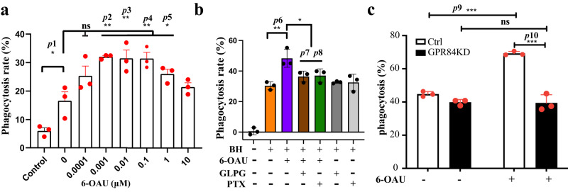
Fig. 1 GPR84-Gi signaling facilitates cancer cell phagocytosis. a Dose-dependent pro-phagocytic effect of 6-OAU. b GLPG1205 and pertussis toxin (PTX) abolished the pro-phagocytic effect of 6-OAU. BH means B6H12, the CD47-blocking antibody. PTX means pertussis toxin. c Knockdown of GPR84 expression in macrophages abolished the pro-phagocytic effect of 6-OAU. Each column represents means ± S.D. (n = 3) Data are representative of at least two independent experiments. Dunnett’s one-way ANOVA test was performed to compare the means of two data groups. *p < 0.1, **p < 0.01, ***p < 0.001 (p1 = 0.0262, p2 = 0.0012, p3 = 0.0017, p4 = 0.0017, p5 = 0.0278, p6 = 0.0011, p7 = 0.0108, p8 = 0.0158, p9 = 0.000025, p10 = 0.000006). ns means no significant difference between the groups.
Fig. 1 GPR84-Gi signaling facilitates cancer cell phagocytosis. a Dose-dependent pro-phagocytic effect of 6-OAU. b GLPG1205 and pertussis toxin (PTX) abolished the pro-phagocytic effect of 6-OAU. BH means B6H12, the CD47-blocking antibody. PTX means pertussis toxin. c Knockdown of GPR84 expression in macrophages abolished the pro-phagocytic effect of 6-OAU. Each column represents means ± S.D. (n = 3) Data are representative of at least two independent experiments. Dunnett’s one-way ANOVA test was performed to compare the means of two data groups. *p < 0.1, **p < 0.01, ***p < 0.001 (p1 = 0.0262, p2 = 0.0012, p3 = 0.0017, p4 = 0.0017, p5 = 0.0278, p6 = 0.0011, p7 = 0.0108, p8 = 0.0158, p9 = 0.000025, p10 = 0.000006). ns means no significant difference between the groups.CC BY · PubMed Central
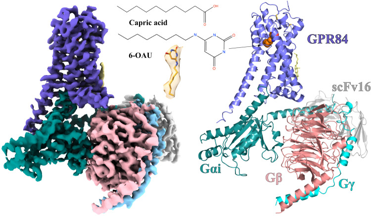
Fig. 2 Overall structure of the 6-OAU-GPR84-Gi complex. The left and right panels show the cryo-EM density map and the overall structure, respectively. The chemical structures of capric acid and 6-OAU and the cryo-EM density of 6-OAU contoured at xx are shown in the middle. GPR84 is colored in blue. Gαi, Gβ, and Gγ subunits are colored in cyan, pink, and light blue, respectively. ScFv16 is colored in gray.
Fig. 2 Overall structure of the 6-OAU-GPR84-Gi complex. The left and right panels show the cryo-EM density map and the overall structure, respectively. The chemical structures of capric acid and 6-OAU and the cryo-EM density of 6-OAU contoured at xx are shown in the middle. GPR84 is colored in blue. Gαi, Gβ, and Gγ subunits are colored in cyan, pink, and light blue, respectively. ScFv16 is colored in gray.CC BY · PubMed Central
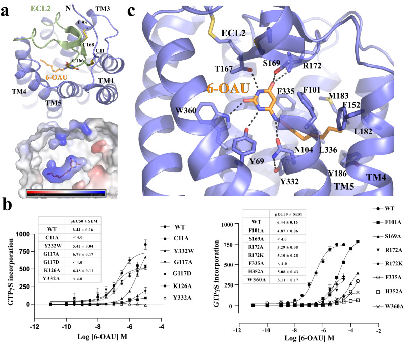
Fig. 3 6-OAU binding in GPR84. a Occluded binding pocket for 6-OAU covered by ECL2. The lower panel indicates the charge potential of the 6-OAU binding pocket. The bar shows the levels of negative (red) and positive (blue) charge potential. b Mutagenesis data using GTPγS incorporation assays. Data represent mean ± S.E.M. from at least three independent experiments. c Interactions between 6-OAU and GPR84. The polar interactions are shown as dashed lines. 6-OAU is shown as orange sticks in all figures.
Fig. 3 6-OAU binding in GPR84. a Occluded binding pocket for 6-OAU covered by ECL2. The lower panel indicates the charge potential of the 6-OAU binding pocket. The bar shows the levels of negative (red) and positive (blue) charge potential. b Mutagenesis data using GTPγS incorporation assays. Data represent mean ± S.E.M. from at least three independent experiments. c Interactions between 6-OAU and GPR84. The polar interactions are shown as dashed lines. 6-OAU is shown as orange sticks in all figures.CC BY · PubMed Central
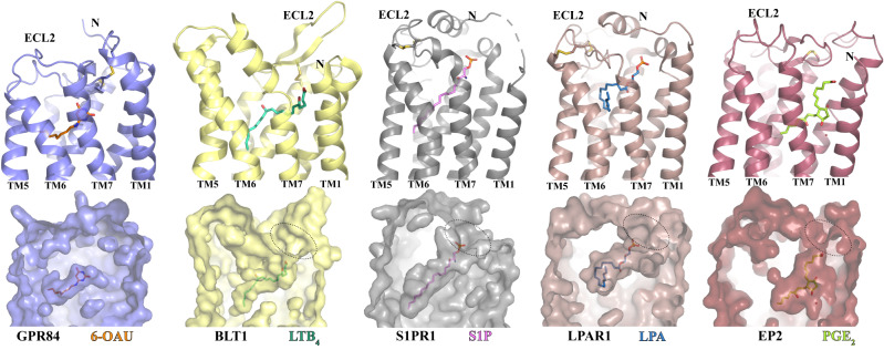
Fig. 4 Comparison of the ligand-binding pockets in GPR84 and four other lipid GPCRs. BLT1, S1PR1, LPAR1, and EP2 are receptors of LTB4, S1P, LPA, and PGE2, respectively. The structures of GPR84, BLT1 (PDB ID 7VKT), S1PR1 (PDB ID 7TD3), LPAR1 (PDB ID 7TD0), and EP2 (PDB ID 7CX2) are colored slate, light yellow, gray, brown, and dark red, respectively. All ligands are shown in sticks. In each column, the cartoon model and surface representation are used for the same receptor. The structures of the five receptors are placed side by side in each column after structural alignment, providing a consistent viewpoint from the same angle.
Fig. 4 Comparison of the ligand-binding pockets in GPR84 and four other lipid GPCRs. BLT1, S1PR1, LPAR1, and EP2 are receptors of LTB4, S1P, LPA, and PGE2, respectively. The structures of GPR84, BLT1 (PDB ID 7VKT), S1PR1 (PDB ID 7TD3), LPAR1 (PDB ID 7TD0), and EP2 (PDB ID 7CX2) are colored slate, light yellow, gray, brown, and dark red, respectively. All ligands are shown in sticks. In each column, the cartoon model and surface representation are used for the same receptor. The structures of the five receptors are placed side by side in each column after structural alignment, providing a consistent viewpoint from the same angle.CC BY · PubMed Central
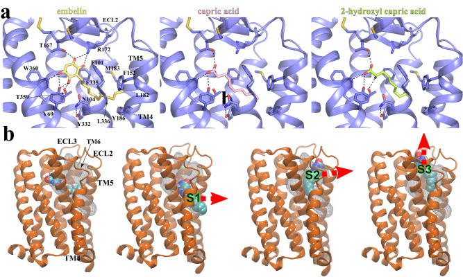
Fig. 5 Docking of GPR84 agonists and MD simulations of 6-OAU-bound GPR84. a Interactions of docked embelin (light yellow), capric acid (pink), and 2-hydroxyl capric acid (lime) with GPR84 (slate). Hydrogen bonds are shown as black dashed lines. b Putative exit routes of 6-OAU in MD simulations. The 6-OAU movement during the simulations is shown as density in white grid. Red arrows indicate possible ligand exit routes via metastable sites S1, S2 or S3. 6-OAU is shown as cyan spheres and GPR84 is shown in orange.
Fig. 5 Docking of GPR84 agonists and MD simulations of 6-OAU-bound GPR84. a Interactions of docked embelin (light yellow), capric acid (pink), and 2-hydroxyl capric acid (lime) with GPR84 (slate). Hydrogen bonds are shown as black dashed lines. b Putative exit routes of 6-OAU in MD simulations. The 6-OAU movement during the simulations is shown as density in white grid. Red arrows indicate possible ligand exit routes via metastable sites S1, S2 or S3. 6-OAU is shown as cyan spheres and GPR84 is shown in orange.CC BY · PubMed Central
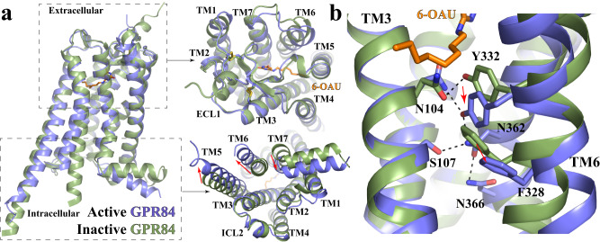
Fig. 6 Active conformation of GPR84. a Superimposition of the active GPR84 structure (blue) to the Alphafold predicted inactive GPR84 structure (green). The extracellular and intracellular regions are shown in the left upper and lower panels, respectively. The red arrows indicate conformational changes of TMs. b Residues involved in the receptor activation at the core region of GPR84.
Fig. 6 Active conformation of GPR84. a Superimposition of the active GPR84 structure (blue) to the Alphafold predicted inactive GPR84 structure (green). The extracellular and intracellular regions are shown in the left upper and lower panels, respectively. The red arrows indicate conformational changes of TMs. b Residues involved in the receptor activation at the core region of GPR84.CC BY · PubMed Central
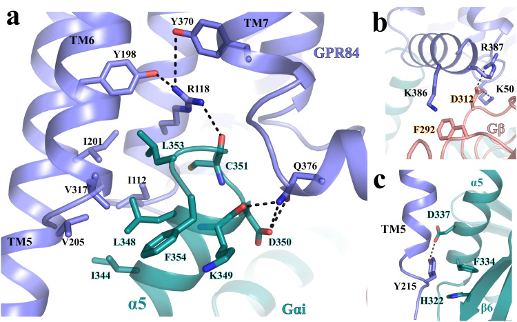
Fig. 7 Gi-coupling to GPR84. a Interactions between GPR84 (blue) and the α5 of Gαi (cyan). b Interactions between GPR84 (blue) and Gβ (salmon). c Interactions between the C-terminal end of TM5 of GPR84 (blue) and Gαi (cyan). All polar interactions are shown as dashed lines.
Fig. 7 Gi-coupling to GPR84. a Interactions between GPR84 (blue) and the α5 of Gαi (cyan). b Interactions between GPR84 (blue) and Gβ (salmon). c Interactions between the C-terminal end of TM5 of GPR84 (blue) and Gαi (cyan). All polar interactions are shown as dashed lines.CC BY · PubMed Central

Verification

Claims the paper makes

The claims and statistics of this paper haven’t been checked yet.

Run the check — free, no account needed. It reads the open-access text and checks the sources it cites.

A few free checks a day · papers already verified open instantly · results are public

Self-check

Percentages, sums and reported differences re-computed from the paper’s own numbers.

The self-check needs the paper’s full text.

Citations

Cites

Metadata

Authors
Zhang, X., Wang, Y., Supekar, S., Cao, X., Zhou, J., Dang, J., Chen, S., Jenkins, L., Marsango, S., Li, X., Liu, G., Milligan, G., Feng, M., Fan, H., Gong, W., & Zhang, C.
Affiliations
  • University of Science and Technology of China (CN)
  • Agency for Science, Technology and Research (SG)
  • City Of Hope National Medical Center (US)
  • University of Glasgow (GB)
  • University of Pittsburgh (US)
Corresponding author
  • Milligan, Graeme
  • Feng, Mingye
  • Fan, Hao
  • Gong, Weimin
  • Zhang, Cheng
Venue
Nature Communications
Published
2023-02-15
Volume
14
Issue
1
Pages
5706-5706
ISSN
2041-1723 (ISSN-L)
OpenAlex
W4386732685
Cited by
46
References
1
Citations by year
2026: 16 · 2025: 17 · 2024: 9 · 2023: 4
FWCI
4.09
Type
journal article
Language
en
Open access
Locations
  • Nature Communications (publishedVersion, cc-by)
  • Research Square (acceptedVersion, cc-by)
  • PubMed Central (publishedVersion, cc-by)
  • PubMed (publishedVersion)
  • ENLIGHTEN (Jurnal Bimbingan dan Konseling Islam) (acceptedVersion, cc-by)
  • PubMed Central (submittedVersion, cc-by)
  • RePEc: Research Papers in Economics (submittedVersion)
  • DOAJ (DOAJ: Directory of Open Access Journals) (submittedVersion, cc-by-sa)
Funders
  • U.S. Department of Health and Human Services (R35GM128641)
  • UK Research and Innovation
  • Research Councils UK (BB/T000562/1)
  • National Natural Science Foundation of China (T2221005)
  • University of Science and Technology of China
  • National Supercomputing Centre Singapore
  • National Institutes of Health
  • Biotechnology and Biological Sciences Research Council (BB/T000562/1)
  • Biomedical Research Council
  • National Institute of General Medical Sciences (R35GM128641)
Topics
  • Life Sciences › Biochemistry, Genetics and Molecular Biology › Molecular Biology › Receptor Mechanisms and Signaling
  • Life Sciences › Immunology and Microbiology › Immunology › Phagocytosis and Immune Regulation
  • Life Sciences › Biochemistry, Genetics and Molecular Biology › Molecular Biology › Sphingolipid Metabolism and Signaling
Keywords
GPR84, orphan G protein-coupled receptor, medium-chain fatty acids, CD47 blockade, phagocytosis, macrophages, binding pocket, fatty acid selectivity, pro-inflammatory signaling

About these checks

Registry, retraction, Cites and statistics results are computed, not judged, with one exception: a printed reference that no index lists is read once by a model before it counts as a citation error. Claims are extracted by a model and kept only when their quote appears verbatim in the text; each cited source is then checked, with a model’s help, for whether it says what the claim relies on. Those verdicts can be wrong — read the claim’s quote and the cited source before relying on one.

Statistics and arithmetic are recomputed, not judged.

Confidence bands are shown only for fields calibrated on our annotated test set. None is calibrated yet, so no band is shown.

What counts as a claim ›How we measure confidence ›How we check this ›

Are you an author, or do you see a problem with this page? Request a review