PreprintCited by 13Tuberculosis Research and EpidemiologyMycobacterium research and diagnosisInfectious Diseases and Tuberculosis

The DOI matches its Crossref record; 1 of the 1 references it cites were found in a registry or an index.

Rv2231c, a unique histidinol phosphate aminotransferase from Mycobacterium tuberculosis, supports virulence by inhibiting host-directed defense

Open access

Preprint — published as 10.1007/s00018-024-05200-8 · Paper page

Abstract

Nitrogen metabolism of M. tuberculosis is critical for its survival in infected host cells. M. tuberculosis has evolved sophisticated strategies to switch between de novo synthesis and uptake of various amino acids from host cells for metabolic demands. Pyridoxal phosphate-dependent histidinol phosphate aminotransferase-HspAT enzyme is critically required for histidine biosynthesis. HspAT is involved in metabolic synthesis of histidine, phenylalanine, tyrosine, tryptophan, and novobiocin. We showed that M. tuberculosis Rv2231c is a conserved enzyme with HspAT activity. Rv2231c is a monomeric globular protein that contains α-helices and β-sheets. It is a secretory and cell wall-localized protein that regulates critical pathogenic attributes. Rv2231c enhances the survival and virulence of recombinant M. smegmatis in infected RAW264.7 macrophage cells. Rv2231c interacts with TLR4 immune receptor and modulates host immune response suppressing the secretion of antibacterial pro-inflammatory IL-12 and IL-6 cytokines. It also inhibits the expression of co-stimulatory molecules CD80 and CD86 along with antigen presenting molecule MHC-I on macrophage and suppresses reactive nitrogen species formation, thereby promoting M2 macrophage polarization. Rv2231c inhibits intrinsic apoptosis while activating necroptosis and pyroptosis, thereby promoting efficient bacterial survival and proliferation to increase virulence. Our results indicate that Rv2231c is a moonlighting protein that regulates multiple functions of M. tuberculosis pathophysiology to increase its virulence. These mechanistic insights can be used to better understand the pathogenesis of M. tuberculosis and to design strategies for tuberculosis mitigation.

Overview

The claims and statistics of this paper haven’t been checked yet.

Run the check — free, no account needed. It reads the open-access text and checks the sources it cites.

A few free checks a day · papers already verified open instantly · results are public

Integrity signals

Registry

Crossref · 10.21203/rs.3.rs-3073689/v1

Cites

1 of 1 references verified0 citation errors · 0 not checkable

Statistics

Figures

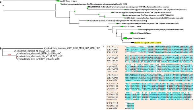
Fig. 1 Rv2231c is a unique pathogenic Mycobacterial protein. a A BLAST tree view, constructed using BLAST pairwise alignment and the fast minimum evolution tree approach, showed homology between M. tb and M. bovis HspAT. b A phylogenetic tree of M. tb-Rv2231c showing closely related proteins from different mycobacterial species. The number of substitutions per site was directly correlated with branch length in the tree. (c) Sequence alignment of M. tb-Rv2231c with related Mycobacterial species revealed 100% identity with M. bovis. The most conserved residues are highlighted in similar colors using BLOSUM62.M.a.ATCC_1: M. abscessus, M.tubercul: M. tuberculosis, M.b.AF2122: M. bovis, IDMT18b_29: M. tuberculosis 18b
Fig. 1 Rv2231c is a unique pathogenic Mycobacterial protein. a A BLAST tree view, constructed using BLAST pairwise alignment and the fast minimum evolution tree approach, showed homology between M. tb and M. bovis HspAT. b A phylogenetic tree of M. tb-Rv2231c showing closely related proteins from different mycobacterial species. The number of substitutions per site was directly correlated with branch length in the tree. (c) Sequence alignment of M. tb-Rv2231c with related Mycobacterial species revealed 100% identity with M. bovis. The most conserved residues are highlighted in similar colors using BLOSUM62.M.a.ATCC_1: M. abscessus, M.tubercul: M. tuberculosis, M.b.AF2122: M. bovis, IDMT18b_29: M. tuberculosis 18bCC BY · PubMed Central
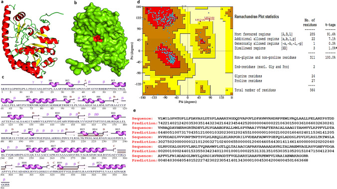
Fig. 2 Structural analysis of the Rv2231c protein using AlphaFold. a and b Cartoons predicting the tertiary structure of M. tuberculosis Rv2231c from the alpha-fold. c The secondary structure of Rv2231c contains 20 α-helices, 13 β-strands, three β-sheets, three β-hairpins, two β-α-β units, 18 helix-helix interactions, 26 β-turns, and four γ-turns. d Ramachandran plot of Rv2231c showing the allowed and disallowed amino acid regions. e Predicted solvent accessibility of amino acid residues of Rv2231c. Scores ranged from 0 (buried residues) to 8 (heavily exposed residues)
Fig. 2 Structural analysis of the Rv2231c protein using AlphaFold. a and b Cartoons predicting the tertiary structure of M. tuberculosis Rv2231c from the alpha-fold. c The secondary structure of Rv2231c contains 20 α-helices, 13 β-strands, three β-sheets, three β-hairpins, two β-α-β units, 18 helix-helix interactions, 26 β-turns, and four γ-turns. d Ramachandran plot of Rv2231c showing the allowed and disallowed amino acid regions. e Predicted solvent accessibility of amino acid residues of Rv2231c. Scores ranged from 0 (buried residues) to 8 (heavily exposed residues)CC BY · PubMed Central
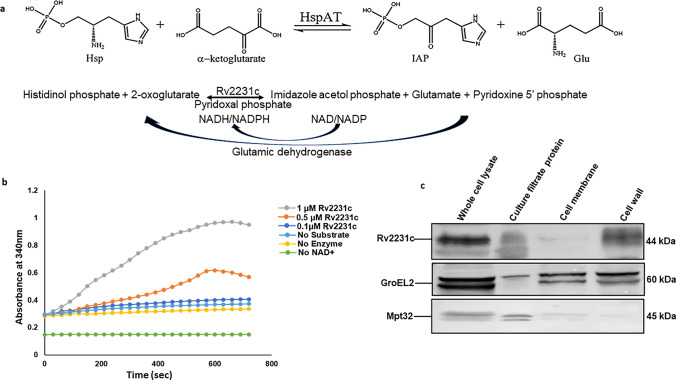
Fig. 3 M. tb Rv2231c is a functional HspAT. a Schematic representation of the GDH-coupled assay system showing the two steps used to measure the Rv2231c aminotransferase activity. b Activity of purified recombinant M. tb-Rv2231c was assessed at 340 nm and an increase in absorbance was recorded over time. This increase in absorbance was attributed to the increased conversion of NAD+ to NADH. An enzyme-free reaction was used as a negative control (c) Western blot analysis of different purified fractions of M. tb showed that Rv2231c is localized to multiple sites in M. tb. GroEL2 and Mpt32 localization was considered as control
Fig. 3 M. tb Rv2231c is a functional HspAT. a Schematic representation of the GDH-coupled assay system showing the two steps used to measure the Rv2231c aminotransferase activity. b Activity of purified recombinant M. tb-Rv2231c was assessed at 340 nm and an increase in absorbance was recorded over time. This increase in absorbance was attributed to the increased conversion of NAD+ to NADH. An enzyme-free reaction was used as a negative control (c) Western blot analysis of different purified fractions of M. tb showed that Rv2231c is localized to multiple sites in M. tb. GroEL2 and Mpt32 localization was considered as controlCC BY · PubMed Central
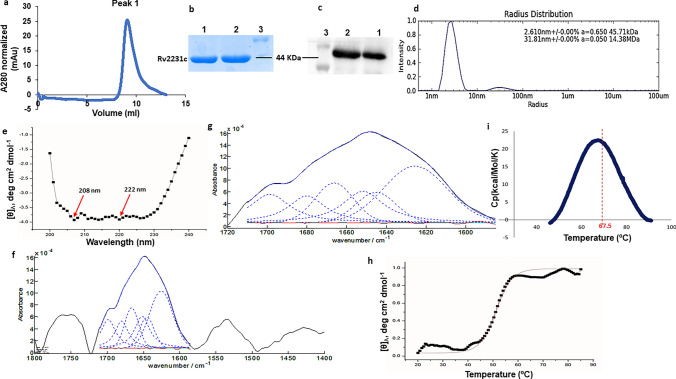
Fig. 4 M. tb Rv2231c is a thermostable protein present as a folded globular monomer in solution. a Analytical gel filtration chromatography (GFC) chromatogram of Rv2231c; blue curve represents the elution profile of Rv2231c. b and c SDS-PAGE and western blot analysis of Rv2231c after Ni–NTA and gel filtration chromatography. Lane 1: Rv2231c obtained after GFC; lane 2: affinity-purified Rv2231c; Lane 3: protein size markers (BLUeye prestained protein ladder). d Size distribution analysis of purified Rv2231c. Hydrodynamic radii were determined using Dynamic Light Scattering. e Far-UV CD spectrum of Rv2231c. Purified Rv2231c was loaded into a quartz cuvette, and spectra were measured at 200–250 nm. The secondary structure content was analyzed using K2D2 software. f and g FTIR spectrum of Rv2231c. Each protein sample (1 mg/ml) was scanned using a Pro MID/FAR IR instrument (Agilent Technologies) in the spectral range of 800 to 3800 cm−1. h and i The thermal transition curve of Rv2231c was estimated by CD and DSC spectroscopy
Fig. 4 M. tb Rv2231c is a thermostable protein present as a folded globular monomer in solution. a Analytical gel filtration chromatography (GFC) chromatogram of Rv2231c; blue curve represents the elution profile of Rv2231c. b and c SDS-PAGE and western blot analysis of Rv2231c after Ni–NTA and gel filtration chromatography. Lane 1: Rv2231c obtained after GFC; lane 2: affinity-purified Rv2231c; Lane 3: protein size markers (BLUeye prestained protein ladder). d Size distribution analysis of purified Rv2231c. Hydrodynamic radii were determined using Dynamic Light Scattering. e Far-UV CD spectrum of Rv2231c. Purified Rv2231c was loaded into a quartz cuvette, and spectra were measured at 200–250 nm. The secondary structure content was analyzed using K2D2 software. f and g FTIR spectrum of Rv2231c. Each protein sample (1 mg/ml) was scanned using a Pro MID/FAR IR instrument (Agilent Technologies) in the spectral range of 800 to 3800 cm−1. h and i The thermal transition curve of Rv2231c was estimated by CD and DSC spectroscopyCC BY · PubMed Central
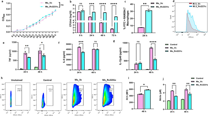
Fig. 5 M. smegmatis expressing M. tb-Rv2231c protein showed higher intracellular survival in RAW264.7 macrophage cells and inhibits secretion of inflammatory cytokines. Macrophage cells were infected with FITC-labelled Ms_Vc and Ms_Rv2231c at MOI of 1:10 and incubated for 4 h. The cells were washed to remove extracellular bacteria and replenished with DMEM. a Growth kinetics of M. smegmatis expressing Rv2231c or vector-only cells. The 7H9 Middlebrook broth was inoculated with primary cultures (OD 0.01) of Ms_Vc or Ms_Rv2231c and incubated at 37 °C at 200 rpm. OD600 nm was measured at intervals of 3 h to 40 h. b Enumeration of phagocytosed and surviving bacteria at 0, 24, 48, and 72 h post infection was carried out using CFU assay. c and d Flow cytometry analysis of bacterial survival in macrophages 24 h after infection. e–g Estimation of pro-inflammatory cytokines upon infection. RAW264.7 cells (0.25 × 10⁶ cells/well) were infected with Ms_Vc and Ms_Rv2231c at MOI of 1:10. ELISA was done to measure the cytokines TNF-α, IL-6, and IL-12p40 in cell culture supernatants collected 24 and 48 h after infection. h FACS plot showing the ROS production after 48 h. i Bar graph showing ROS production levels. j The NO content was estimated spectrophotometrically at 545 nm using Griess reagent. Standard deviation (SD) was calculated using the means of two independent experiments. Graphs were created with the help of GraphPad software. *p ≤ 0.05, **p ≤ 0.01, ***p ≤ 0.001, ****p ≤ 0.0001, ns; not significant
Fig. 5 M. smegmatis expressing M. tb-Rv2231c protein showed higher intracellular survival in RAW264.7 macrophage cells and inhibits secretion of inflammatory cytokines. Macrophage cells were infected with FITC-labelled Ms_Vc and Ms_Rv2231c at MOI of 1:10 and incubated for 4 h. The cells were washed to remove extracellular bacteria and replenished with DMEM. a Growth kinetics of M. smegmatis expressing Rv2231c or vector-only cells. The 7H9 Middlebrook broth was inoculated with primary cultures (OD 0.01) of Ms_Vc or Ms_Rv2231c and incubated at 37 °C at 200 rpm. OD600 nm was measured at intervals of 3 h to 40 h. b Enumeration of phagocytosed and surviving bacteria at 0, 24, 48, and 72 h post infection was carried out using CFU assay. c and d Flow cytometry analysis of bacterial survival in macrophages 24 h after infection. e–g Estimation of pro-inflammatory cytokines upon infection. RAW264.7 cells (0.25 × 10⁶ cells/well) were infected with Ms_Vc and Ms_Rv2231c at MOI of 1:10. ELISA was done to measure the cytokines TNF-α, IL-6, and IL-12p40 in cell culture supernatants collected 24 and 48 h after infection. h FACS plot showing the ROS production after 48 h. i Bar graph showing ROS production levels. j The NO content was estimated spectrophotometrically at 545 nm using Griess reagent. Standard deviation (SD) was calculated using the means of two independent experiments. Graphs were created with the help of GraphPad software. *p ≤ 0.05, **p ≤ 0.01, ***p ≤ 0.001, ****p ≤ 0.0001, ns; not significantCC BY · PubMed Central
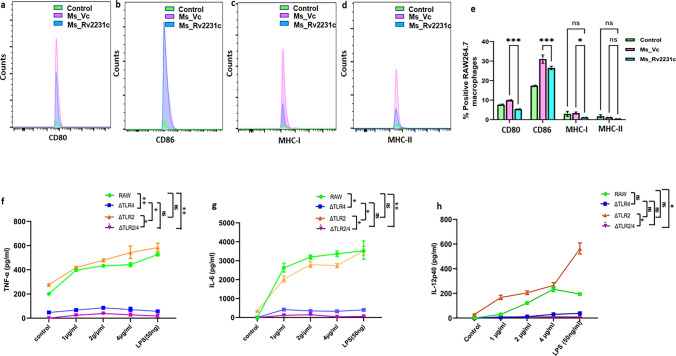
Fig. 6 Rv2231c inhibits M2 macrophage activation. RAW264.7 macrophage cells were infected with Ms_Vc and Ms_Rv2231c at MOI of 1:10. Infected cells were harvested after 48 h, stained with fluorescent tagged antibodies CD80, CD86, MHC-I, and MHC-II, and analyzed using flow cytometry. The culture supernatant from 48 h was collected to estimate the NO concentrations. Recombinant M. smegmatis expressing Rv2231c suppressed antigen presentation. FACS data show percent of RAW264.7 cells infected with Ms_Vc and Ms_2231c expressing a CD80, b CD86, c MHC-I, and d MHC-II. e Flow cytometry histogram showing the expression levels of CD80, CD86, MHC I, and MHC II. f–h RAW264.7, RAW-ΔTLR2, and RAW-ΔTLR4 knockout cells were stimulated with Rv2231c (1, 2, and 4 µg/ml) and LPS (50 ng/ml) for 48 h. TNF, IL-6, and IL-12p40 levels were estimated by ELISA. Standard deviation was calculated using the means of two independent experiments. All graphs were created using GraphPad software. *p ≤ 0.05, ***p ≤ 0.001, ns; not significant
Fig. 6 Rv2231c inhibits M2 macrophage activation. RAW264.7 macrophage cells were infected with Ms_Vc and Ms_Rv2231c at MOI of 1:10. Infected cells were harvested after 48 h, stained with fluorescent tagged antibodies CD80, CD86, MHC-I, and MHC-II, and analyzed using flow cytometry. The culture supernatant from 48 h was collected to estimate the NO concentrations. Recombinant M. smegmatis expressing Rv2231c suppressed antigen presentation. FACS data show percent of RAW264.7 cells infected with Ms_Vc and Ms_2231c expressing a CD80, b CD86, c MHC-I, and d MHC-II. e Flow cytometry histogram showing the expression levels of CD80, CD86, MHC I, and MHC II. f–h RAW264.7, RAW-ΔTLR2, and RAW-ΔTLR4 knockout cells were stimulated with Rv2231c (1, 2, and 4 µg/ml) and LPS (50 ng/ml) for 48 h. TNF, IL-6, and IL-12p40 levels were estimated by ELISA. Standard deviation was calculated using the means of two independent experiments. All graphs were created using GraphPad software. *p ≤ 0.05, ***p ≤ 0.001, ns; not significantCC BY · PubMed Central
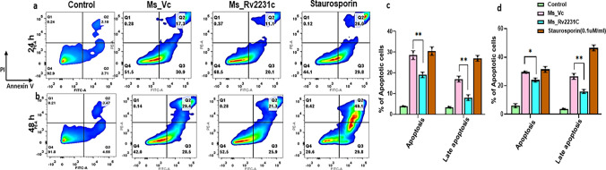
Fig. 7 Recombinant M. smegmatis containing Rv2231c inhibits apoptosis of RAW264.7 cells. RAW264.7 cells were infected with Ms_Vc and Ms_Rv2231c at MOI of 1:10. At 24 and 48 h post-infection, the cells were stained with the Annexin V/PI kit, and apoptosis was analyzed. FACS analysis of different stages, that is, early and late apoptosis in Uninfected Control group, Ms_Vc-and Ms_Rv2231c infected murine macrophages a 24 h and b 48 h of infection along with staurosporine stimulation as a positive control. The bar graph shows the early and late apoptotic cells in different samples after 24 h and 48 h, as shown in Figure (c and d)
Fig. 7 Recombinant M. smegmatis containing Rv2231c inhibits apoptosis of RAW264.7 cells. RAW264.7 cells were infected with Ms_Vc and Ms_Rv2231c at MOI of 1:10. At 24 and 48 h post-infection, the cells were stained with the Annexin V/PI kit, and apoptosis was analyzed. FACS analysis of different stages, that is, early and late apoptosis in Uninfected Control group, Ms_Vc-and Ms_Rv2231c infected murine macrophages a 24 h and b 48 h of infection along with staurosporine stimulation as a positive control. The bar graph shows the early and late apoptotic cells in different samples after 24 h and 48 h, as shown in Figure (c and d)CC BY · PubMed Central
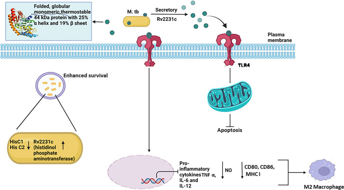
Fig. 8 M. tb Rv2231c is recognized by TLR4 receptor to regulate downstream signaling cascades. This figure illustrates a summary of the possible mechanism of action M. tuberculosis Rv2231c protein
Fig. 8 M. tb Rv2231c is recognized by TLR4 receptor to regulate downstream signaling cascades. This figure illustrates a summary of the possible mechanism of action M. tuberculosis Rv2231c proteinCC BY · PubMed Central

Verification

Claims the paper makes

The claims and statistics of this paper haven’t been checked yet.

Run the check — free, no account needed. It reads the open-access text and checks the sources it cites.

A few free checks a day · papers already verified open instantly · results are public

Self-check

Percentages, sums and reported differences re-computed from the paper’s own numbers.

The self-check needs the paper’s full text.

Citations

Cites

Metadata

Authors
Zarin, S., Shariq, M., Rastogi, N., Ahuja, Y., Manjunath, P., Alam, A., Hasnain, S. E., & Ehtesham, N. Z.
Affiliations
  • Jamia Hamdard (IN)
  • Indian Council of Medical Research (IN)
  • Sharda University (IN)
  • Indian Institute of Technology Delhi (IN)
Corresponding author
  • Hasnain, Seyed Ehtesham
  • Ehtesham, Nasreen Zafar
Venue
Cellular and Molecular Life Sciences
Published
2023-07-25
Volume
81
Issue
1
Pages
203-203
ISSN
1420-682X (ISSN-L)
OpenAlex
W4396574859
Cited by
13
References
1
Citations by year
2026: 5 · 2025: 7 · 2024: 1
FWCI
4.47
Type
journal article
Language
en
Open access
Locations
  • Cellular and Molecular Life Sciences (publishedVersion, cc-by)
  • Research Square (acceptedVersion, cc-by)
  • PubMed Central (publishedVersion, cc-by)
  • PubMed (publishedVersion)
Funders
  • Department of Science and Technology, Ministry of Science and Technology, India
  • Indian Council of Medical Research
  • Council of Scientific and Industrial Research, India
  • Science and Engineering Research Board
Topics
  • Health Sciences › Medicine › Infectious Diseases › Tuberculosis Research and Epidemiology
  • Health Sciences › Medicine › Epidemiology › Mycobacterium research and diagnosis
  • Health Sciences › Medicine › Infectious Diseases › Infectious Diseases and Tuberculosis
Keywords
Mycobacterium tuberculosis, histidine biosynthesis, moonlighting proteins, bacterial virulence, TLR4, macrophage polarization, M2 macrophage polarization, cytokine inhibition, CD80/CD86, MHC class I, reactive nitrogen species, apoptosis, Mycobacterium smegmatis, tuberculosis pathogenesis
MeSH terms
RAW 264.7 Cells, Transaminases, Animals, Bacterial Proteins, Humans, Immunity, Innate, Macrophages, Mycobacterium tuberculosis, Tuberculosis, Virulence, Cytokines, Mycobacterium smegmatis, Toll-Like Receptor 4, Mice, Host-Pathogen Interactions

About these checks

Registry, retraction, Cites and statistics results are computed, not judged, with one exception: a printed reference that no index lists is read once by a model before it counts as a citation error. Claims are extracted by a model and kept only when their quote appears verbatim in the text; each cited source is then checked, with a model’s help, for whether it says what the claim relies on. Those verdicts can be wrong — read the claim’s quote and the cited source before relying on one.

Statistics and arithmetic are recomputed, not judged.

Confidence bands are shown only for fields calibrated on our annotated test set. None is calibrated yet, so no band is shown.

What counts as a claim ›How we measure confidence ›How we check this ›

Are you an author, or do you see a problem with this page? Request a review