PreprintCited by 27Bacterial Genetics and BiotechnologyBiochemical and Molecular ResearchBacterial biofilms and quorum sensing

The DOI matches its Crossref record; 1 of the 1 references it cites were found in a registry or an index.

Ubiquitous purine sensor modulates diverse signal transduction pathways in bacteria

Open access
Krell, Tino, Monteagudo-Cascales, Elizabet, Gumerov, Vadim, Fernández, Matilde, Matilla, Miguel, Gavira, Jose, Zhulin, Igor·2023·Nature Communications

Preprint — published as 10.1038/s41467-024-50275-3 · Paper page

Abstract

Purines and their derivatives are key molecules for controlling intracellular energy homeostasis and nucleotide synthesis. In eukaryotes, including humans, purines also act as signaling molecules that mediate extracellular communication and control key cellular processes, such as proliferation, migration, differentiation, and apoptosis. However, the signaling role of purines in bacteria is largely unknown. Here, by combining structural and sequence information, we define a purine-binding motif, which is present in sensor domains of thousands of bacterial receptors that modulate motility, gene expression, metabolism and second messenger turnover. The screening of compound libraries and microcalorimetric titrations of selected sensor domains validated their ability to specifically bind purine derivatives. The physiological relevance of purine sensing was demonstrated in a second messenger signaling system that modulates c-di-GMP levels.

Overview

The claims and statistics of this paper haven’t been checked yet.

Run the check — free, no account needed. It reads the open-access text and checks the sources it cites.

A few free checks a day · papers already verified open instantly · results are public

Integrity signals

Registry

Crossref · 10.21203/rs.3.rs-3494331/v1

Cites

1 of 1 references verified0 citation errors · 0 not checkable

Statistics

Figures

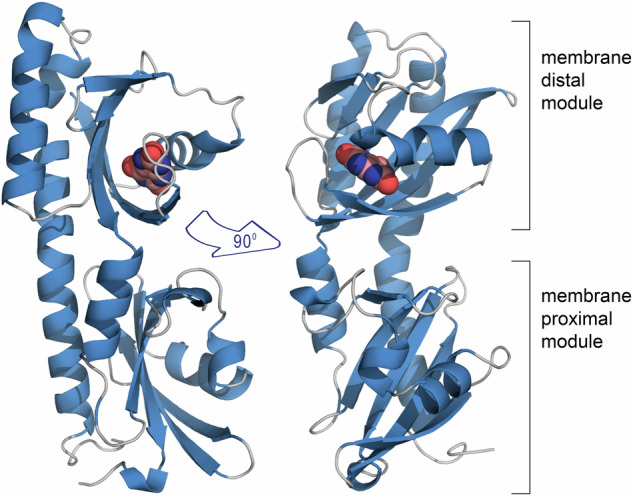
Fig. 1 The overall structure of McpH-LBD in complex with uric acid. Ribbon diagram with bound uric acid shown in spacefill mode.
Fig. 1 The overall structure of McpH-LBD in complex with uric acid. Ribbon diagram with bound uric acid shown in spacefill mode.CC BY · PubMed Central
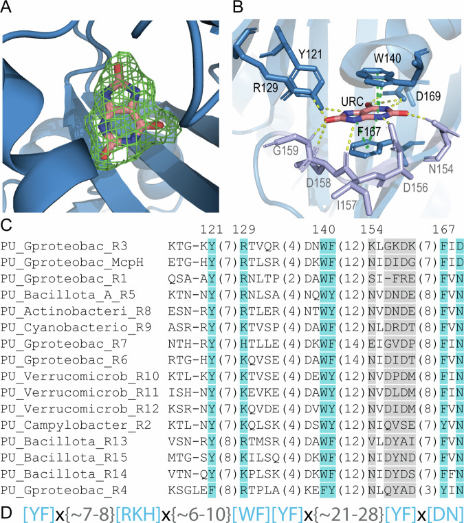
Fig. 2 Computationally derived purine binding motif. A, B The molecular detail of uric acid recognition at McpH-LBD. A The mesh corresponds to the initial |Fo-Fc| electron density map containing bound uric acid contoured at 1.5 σ level. The uric acid model placed into this density is shown in stick mode. B Ligand binding pocket of the McpH dCache_1 domain. Residues constituting the purine binding motif are shown in blue, variable residues involved in ligand binding are shown in light purple. Contacts in yellow—hydrogen bonds; contacts in green—pi-stacking; URC—uric acid. Molecular interactions were identified by Mol* Viewer using a cut-off of 3.5 Å72. C Multiple sequence alignment showing the predicted purine binding motif (positions in blue as well as an aromatic position corresponding to F141 in McpH). Positions shown in light gray form contacts with the ligand but are not conserved across the full alignment. Phylum/class names are provided. Endings R1, R2, etc. in protein names refer to the proteins analyzed in this study (Table 2). A full alignment can be found in Supplementary Fig. 3. D The purine binding motif. Residues in brackets indicate that any of the specified residue can be at the corresponding position. “X” denotes any residue, numbers in curly brackets indicate the distance in amino acids between the specified positions.
Fig. 2 Computationally derived purine binding motif. A, B The molecular detail of uric acid recognition at McpH-LBD. A The mesh corresponds to the initial |Fo-Fc| electron density map containing bound uric acid contoured at 1.5 σ level. The uric acid model placed into this density is shown in stick mode. B Ligand binding pocket of the McpH dCache_1 domain. Residues constituting the purine binding motif are shown in blue, variable residues involved in ligand binding are shown in light purple. Contacts in yellow—hydrogen bonds; contacts in green—pi-stacking; URC—uric acid. Molecular interactions were identified by Mol* Viewer using a cut-off of 3.5 Å72. C Multiple sequence alignment showing the predicted purine binding motif (positions in blue as well as an aromatic position corresponding to F141 in McpH). Positions shown in light gray form contacts with the ligand but are not conserved across the full alignment. Phylum/class names are provided. Endings R1, R2, etc. in protein names refer to the proteins analyzed in this study (Table 2). A full alignment can be found in Supplementary Fig. 3. D The purine binding motif. Residues in brackets indicate that any of the specified residue can be at the corresponding position. “X” denotes any residue, numbers in curly brackets indicate the distance in amino acids between the specified positions.CC BY · PubMed Central
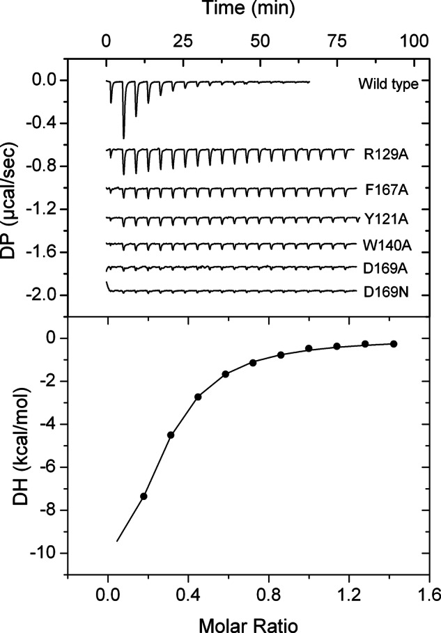
Fig. 3 Assessment of the contribution of motif amino acids to binding. Microcalorimetric titrations of McpH-LBD and site-directed mutants with adenine. Upper panel: heat changes for the titration of 9–18 µM protein with 12.8 μl aliquots of 300 μM adenine. Lower panel: integrated, dilution heat corrected and concentration normalized titration data of the wild-type protein fitted with the “One binding site model” of the MicroCal version of ORIGIN. Titrations of mutant proteins were repeated with the maximal possible adenine concentration (Supplementary Fig. 5) to derive, if possible, the binding parameters.
Fig. 3 Assessment of the contribution of motif amino acids to binding. Microcalorimetric titrations of McpH-LBD and site-directed mutants with adenine. Upper panel: heat changes for the titration of 9–18 µM protein with 12.8 μl aliquots of 300 μM adenine. Lower panel: integrated, dilution heat corrected and concentration normalized titration data of the wild-type protein fitted with the “One binding site model” of the MicroCal version of ORIGIN. Titrations of mutant proteins were repeated with the maximal possible adenine concentration (Supplementary Fig. 5) to derive, if possible, the binding parameters.CC BY · PubMed Central
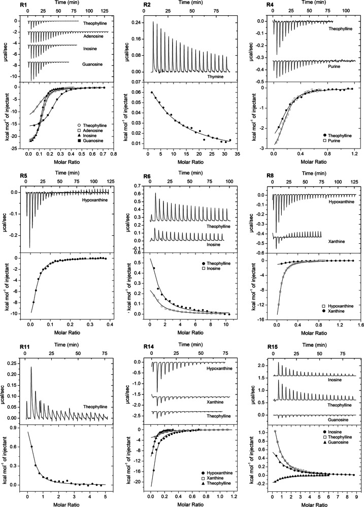
Fig. 4 Binding studies of purine derivatives to dCache_1PU domains from various receptor families. Microcalorimetric titration of 45–138 μM protein with 0.2–20 mM solutions of purine and pyrimidine derivatives. Upper panels: raw titration data. Lower panels: integrated, dilution heat corrected and concentration normalized raw data. The lines are the best fits using the “One binding site” model of the MicroCal version of ORIGIN. The derived dissociation constants are provided in Table 3.
Fig. 4 Binding studies of purine derivatives to dCache_1PU domains from various receptor families. Microcalorimetric titration of 45–138 μM protein with 0.2–20 mM solutions of purine and pyrimidine derivatives. Upper panels: raw titration data. Lower panels: integrated, dilution heat corrected and concentration normalized raw data. The lines are the best fits using the “One binding site” model of the MicroCal version of ORIGIN. The derived dissociation constants are provided in Table 3.CC BY · PubMed Central
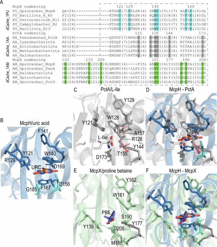
Fig. 5 Comparison of dCache_1PU, dCache_1AA, and dCache_1AM sensors. A Multiple protein sequence alignment of purine, amino acid, and amine binding dCache_1 domains. The corresponding motifs are shown in blue (purine motif), gray (amino acid motif), and green (amine motif). A dashed rectangle denotes a sequence region corresponding to the ligand binding site. A full alignment can be found in Supplementary Fig. 3. B Ligand binding pocket of the purine receptor McpH from Pseudomonas putida. The two residues in turquoise are in positions corresponding to the amino acid as well as amine motif residues but are not part of the purine motif. URC—uric acid. C Ligand binding pocket of the amino acid receptor PctA from Pseudomonas aeruginosa with bound L-isoleucine. The two residues in red are in positions corresponding to the purine motif residues but they are not part of the amino acid motif. D Structural superimposition of the ligand binding pockets of McpH and PctA. E Ligand binding pocket of the amine receptor McpX from Sinorhizobium meliloti in complex with proline betaine (PBE). F Structural superimposition of the ligand binding pockets of McpH and McpX.
Fig. 5 Comparison of dCache_1PU, dCache_1AA, and dCache_1AM sensors. A Multiple protein sequence alignment of purine, amino acid, and amine binding dCache_1 domains. The corresponding motifs are shown in blue (purine motif), gray (amino acid motif), and green (amine motif). A dashed rectangle denotes a sequence region corresponding to the ligand binding site. A full alignment can be found in Supplementary Fig. 3. B Ligand binding pocket of the purine receptor McpH from Pseudomonas putida. The two residues in turquoise are in positions corresponding to the amino acid as well as amine motif residues but are not part of the purine motif. URC—uric acid. C Ligand binding pocket of the amino acid receptor PctA from Pseudomonas aeruginosa with bound L-isoleucine. The two residues in red are in positions corresponding to the purine motif residues but they are not part of the amino acid motif. D Structural superimposition of the ligand binding pockets of McpH and PctA. E Ligand binding pocket of the amine receptor McpX from Sinorhizobium meliloti in complex with proline betaine (PBE). F Structural superimposition of the ligand binding pockets of McpH and McpX.CC BY · PubMed Central
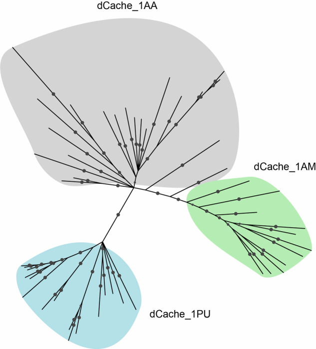
Fig. 6 Purine sensors share an evolutionary ancestor with amino acid sensors. The phylogenetic tree was constructed using the Bayesian inference method implemented in MrBayes71. Probabilities with the value greater than 70 are shown as filled gray circles. dCache_1 amino acid sequences from major phyla of each shown family were used. The tree with information about the proteins used and phyla in which they occur can be found in Supplementary Fig. 13, the tree in NEXUS format can be found at https://github.com/ToshkaDev/Purine-pyrimidine_motif. Bayesian analyses settings are provided in Supplementary Data 3.
Fig. 6 Purine sensors share an evolutionary ancestor with amino acid sensors. The phylogenetic tree was constructed using the Bayesian inference method implemented in MrBayes71. Probabilities with the value greater than 70 are shown as filled gray circles. dCache_1 amino acid sequences from major phyla of each shown family were used. The tree with information about the proteins used and phyla in which they occur can be found in Supplementary Fig. 13, the tree in NEXUS format can be found at https://github.com/ToshkaDev/Purine-pyrimidine_motif. Bayesian analyses settings are provided in Supplementary Data 3.CC BY · PubMed Central
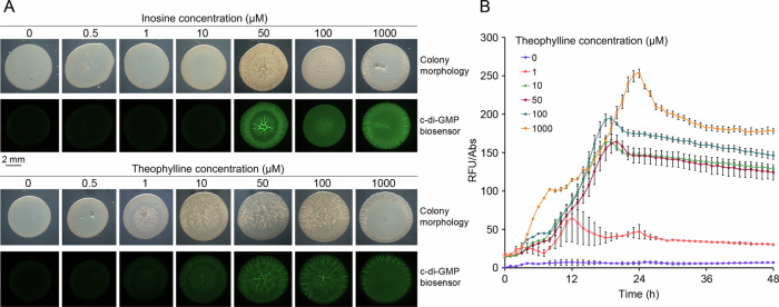
Fig. 7 Inosine and theophylline modulate the activity of a diguanylate cyclase (R6) of Vibrio cholerae. A Colony morphology and c-di-GMP levels (green fluorescence) of P. putida harboring plasmid pCdrA::gfpC (c-di-GMP biosensor) and pBBR1MCS-2_START_R6 (R6 expression plasmid) in the absence and presence of increasing inosine and theophylline concentrations. The brightness of the colony morphology images has been adjusted to optimize visibility. Pictures were taken after 24 h of incubation at 30 °C. B c-di-GMP levels during growth in liquid cultures of P. putida harboring plasmid pCdrA::gfpC in the absence and presence of different theophylline concentrations. Shown are means and standard deviations derived from three biological replicates each conducted in triplicate. The control experiments containing the P. putida harboring the empty plasmid pBBR1MCS-2_START instead of pBBR1MCS-2_START_R6 are shown in Supplementary Fig. 15. RFU relative fluorescence units, Abs absorbance at 600 nm.
Fig. 7 Inosine and theophylline modulate the activity of a diguanylate cyclase (R6) of Vibrio cholerae. A Colony morphology and c-di-GMP levels (green fluorescence) of P. putida harboring plasmid pCdrA::gfpC (c-di-GMP biosensor) and pBBR1MCS-2_START_R6 (R6 expression plasmid) in the absence and presence of increasing inosine and theophylline concentrations. The brightness of the colony morphology images has been adjusted to optimize visibility. Pictures were taken after 24 h of incubation at 30 °C. B c-di-GMP levels during growth in liquid cultures of P. putida harboring plasmid pCdrA::gfpC in the absence and presence of different theophylline concentrations. Shown are means and standard deviations derived from three biological replicates each conducted in triplicate. The control experiments containing the P. putida harboring the empty plasmid pBBR1MCS-2_START instead of pBBR1MCS-2_START_R6 are shown in Supplementary Fig. 15. RFU relative fluorescence units, Abs absorbance at 600 nm.CC BY · PubMed Central

Verification

Claims the paper makes

The claims and statistics of this paper haven’t been checked yet.

Run the check — free, no account needed. It reads the open-access text and checks the sources it cites.

A few free checks a day · papers already verified open instantly · results are public

Self-check

Percentages, sums and reported differences re-computed from the paper’s own numbers.

The self-check needs the paper’s full text.

Citations

Cites

Metadata

Authors
Krell, T., Monteagudo-Cascales, E., Gumerov, V., Fernández, M., Matilla, M., Gavira, J., & Zhulin, I.
Affiliations
  • Consejo Superior de Investigaciones Científicas (ES)
  • Estación Experimental del Zaidín
  • The Ohio State University (US)
  • Universidad de Granada (ES)
  • IACT (CSIC-UGR)
Corresponding author
  • Krell, Tino
  • Zhulin, Igor
Venue
Nature Communications
Published
2023-11-16
Volume
15
Issue
1
Pages
5867-5867
ISSN
2041-1723 (ISSN-L)
OpenAlex
W4400573468
Cited by
27
References
1
Citations by year
2026: 6 · 2025: 15 · 2024: 6
FWCI
7.14
Type
journal article
Language
en
Open access
Locations
  • Nature Communications (publishedVersion, cc-by)
  • bioRxiv (Cold Spring Harbor Laboratory) (acceptedVersion)
  • Research Square (acceptedVersion, cc-by)
  • PubMed Central (publishedVersion, cc-by)
  • PubMed (publishedVersion)
  • DIGITAL.CSIC (Spanish National Research Council (CSIC)) (acceptedVersion, other-oa)
  • Institutional Repository of the University of Granada (University of Granada) (submittedVersion, cc-by)
  • PubMed Central (submittedVersion)
  • RePEc: Research Papers in Economics (submittedVersion)
  • Institutional Repository of the University of Granada (University of Granada) (submittedVersion, cc-by-nc-nd)
  • DIGITAL.CSIC (Spanish National Research Council (CSIC)) (submittedVersion, cc-by)
  • DOAJ (DOAJ: Directory of Open Access Journals) (submittedVersion, cc-by-sa)
Funders
  • Consejería de Economía, Innovación, Ciencia y Empleo, Junta de Andalucía (P18‐FR‐1621)
  • Junta de Andalucía (P18-FR-1621)
  • National Institutes of Health
  • Agencia Estatal de Investigación (501100011033, PID2020‐116261GB‐I00, 13039, .13039/501100011033, PID2019‐103972GA‐I00, PID2020‐112612GB‐I00, PID2019)
  • National Institute of General Medical Sciences (R35GM131760)
Topics
  • Life Sciences › Biochemistry, Genetics and Molecular Biology › Genetics › Bacterial Genetics and Biotechnology
  • Life Sciences › Biochemistry, Genetics and Molecular Biology › Molecular Biology › Biochemical and Molecular Research
  • Life Sciences › Biochemistry, Genetics and Molecular Biology › Molecular Biology › Bacterial biofilms and quorum sensing
Keywords
bacterial receptors, sensor domain, second messenger signaling, c-di-GMP, signal transduction, purine derivatives, amino acid receptors, evolutionary origin, bacterial motility, gene expression regulation

About these checks

Registry, retraction, Cites and statistics results are computed, not judged, with one exception: a printed reference that no index lists is read once by a model before it counts as a citation error. Claims are extracted by a model and kept only when their quote appears verbatim in the text; each cited source is then checked, with a model’s help, for whether it says what the claim relies on. Those verdicts can be wrong — read the claim’s quote and the cited source before relying on one.

Statistics and arithmetic are recomputed, not judged.

Confidence bands are shown only for fields calibrated on our annotated test set. None is calibrated yet, so no band is shown.

What counts as a claim ›How we measure confidence ›How we check this ›

Are you an author, or do you see a problem with this page? Request a review