

The DOI matches its Crossref record; 1 of the 1 references it cites were found in a registry or an index.
Profiling of pathogenic variants in Japanese patients with sarcoglycanopathy
Open accessPreprint — published as 10.1186/s13023-024-03521-2 · Paper page
Abstract
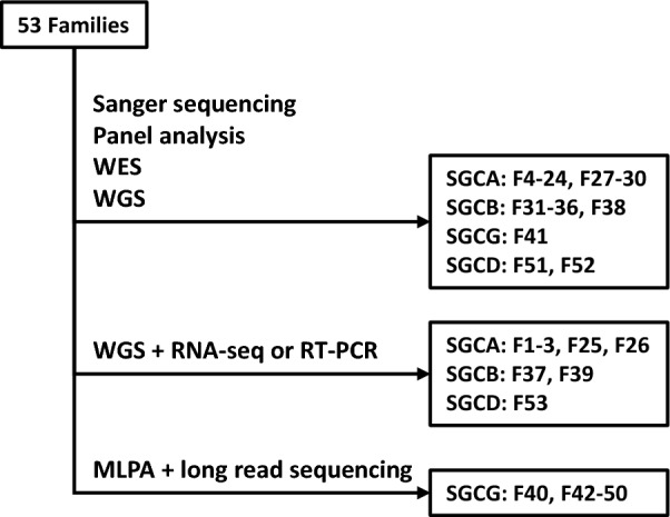
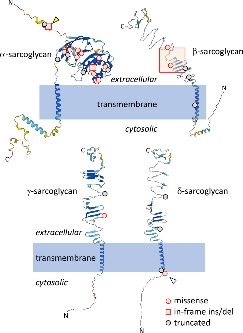
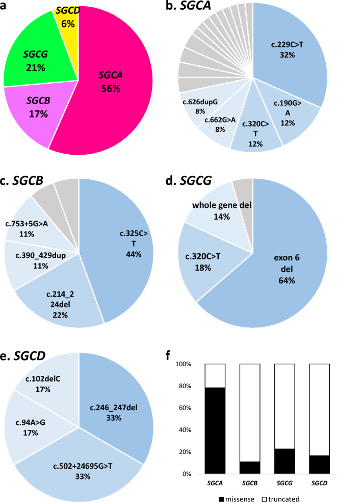
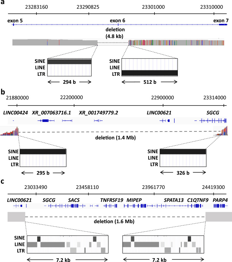
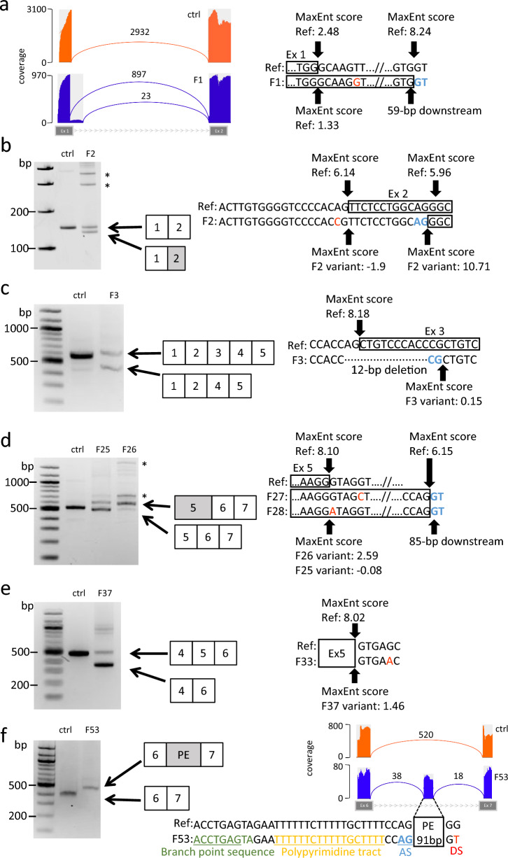
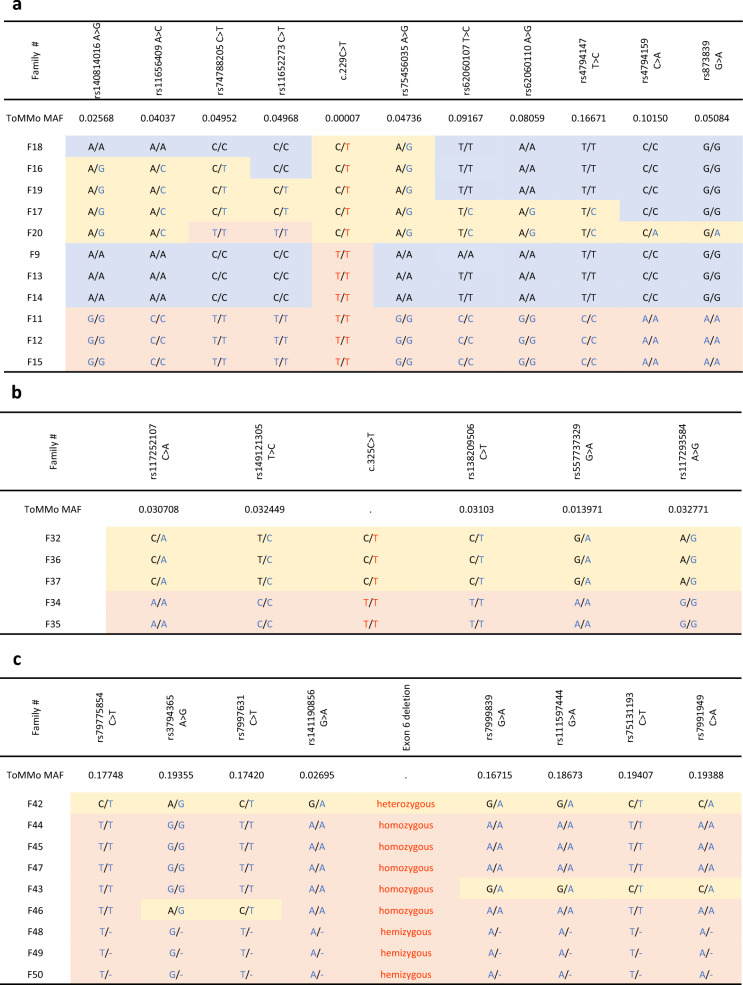
Background Sarcoglycanopathies (SGPs) are limb-girdle muscular dystrophies (LGMDs) that can be classified into four types, LGMDR3, LGMDR4, LGMDR5, and LGMDR6, caused by mutations in the genes, SGCA, SGCB, SGCG, and SGCD, respectively. SGPs are relatively rare in Japan. This study aims to profile the genetic variants that cause SGPs in Japanese patients. Methods Clinical course and pathological findings were retrospectively reviewed in Japanese patients with SGP. Genetic analyses were performed using a combination of targeted resequencing with a hereditary muscle disease panel, whole genome sequencing, multiplex ligation-dependent probe amplification, and long-read sequencing. The structures of transcripts with aberrant splicing were also determined by RT-PCR, RNA-seq, and in silico prediction. Results We identified biallelic variants in SGC genes in 53 families, including three families with LGMDR6, which had not been identified in Japan so far. SGCA was the most common causative gene, accounting for 56% of cases, followed by SGCG, SGCB, and SGCD, at 17%, 21%, and 6%, respectively. Missense variants in SGCA were very frequent at 78.3%, while they were relatively rare in SGCB, SGCG, and SGCD at 11.1%, 18.2%, and 16.6%, respectively. We also analyzed the haplotypes of alleles carrying three variants found in multiple cases: c.229C > T in SGCA, c.325C > T in SGCB, and exon 6 deletion in SGCG; two distinct haplotypes were found for c.229C > T in SGCA, while each of the latter two variants was on single haplotypes. Conclusions We present genetic profiles of Japanese patients with SGPs. Haplotype analysis indicated common ancestors of frequent variants. Our findings will support genetic diagnosis and gene therapy.
Overview
The claims and statistics of this paper haven’t been checked yet.
Run the check — free, no account needed. It reads the open-access text and checks the sources it cites.
Integrity signals
Registry
Cites
Statistics
Figures
Verification
Claims the paper makes
The claims and statistics of this paper haven’t been checked yet.
Run the check — free, no account needed. It reads the open-access text and checks the sources it cites.
Self-check
Percentages, sums and reported differences re-computed from the paper’s own numbers.
The self-check needs the paper’s full text.
Citations
Cites
Highly accurate protein structure prediction with AlphaFold
Jumper, J., Evans, R., Pritzel, A., Green, T., Figurnov, M., Ronneberger, O., Tunyasuvunakool, K., Bates, R., Žídek, A., Potapenko, A., et al. (2021) · Nature · doi:10.1038/s41586-021-03819-2 · Cited by 43,764
Research works printed but not linked: 53
- Straub V, Murphy A, Udd B, LGMD workshop study group. 229th ENMC international workshop: Limb girdle muscular dystrophies - Nomenclature and reformed classification Naarden, the Netherlands, 17–19 March 2017. Neuromuscul Disord. 2018;28:702–10.
- Xie Z, Hou Y, Yu M, Liu Y, Fan Y, Zhang W, et al. Clinical and genetic spectrum of sarcoglycanopathies in a large cohort of Chinese patients. Orphanet J Rare Dis. 2019;14:43. · DOI
- Alonso-Pérez J, González-Quereda L, Bello L, Guglieri M, Straub V, Gallano P, et al. New genotype-phenotype correlations in a large European cohort of patients with sarcoglycanopathy. Brain. 2020;143:2696–708. · DOI
- Dalichaouche I, Sifi Y, Roudaut C, Sifi K, Hamri A, Rouabah L, et al. γ-sarcoglycan and dystrophin mutation spectrum in an Algerian cohort. Muscle Nerve. 2017;56:129–35. · DOI
- Liang W-C, Chou P-C, Hung C-C, Su Y-N, Kan T-M, Chen W-Z, et al. Probable high prevalence of limb-girdle muscular dystrophy type 2D in Taiwan. J Neurol Sci. 2016;362:304–8. · DOI
- Alavi A, Esmaeili S, Nilipour Y, Nafissi S, Tonekaboni SH, Zamani G, et al. LGMD2E is the most common type of sarcoglycanopathies in the Iranian population. J Neurogenet. 2017;31:161–9. · DOI
- Mojbafan M, Bahmani R, Bagheri SD, Sharifi Z, Zeinali S. Mutational spectrum of autosomal recessive limb-girdle muscular dystrophies in a cohort of 112 Iranian patients and reporting of a possible founder effect. Orphanet J Rare Dis. 2020;15:14. · DOI
- Sanga S, Chakraborty S, Bardhan M, Polavarapu K, Kumar VP, Bhattacharya C, et al. Identification of a shared, common haplotype segregating with an SGCB c.544 T > G mutation in Indian patients affected with sarcoglycanopathy. Sci Rep. 2023;13:15095. · DOI
- Noguchi S, McNally EM, Ben Othmane K, Hagiwara Y, Mizuno Y, Yoshida M, et al. Mutations in the dystrophin-associated protein gamma-sarcoglycan in chromosome 13 muscular dystrophy. Science. 1995;270:819–22. · DOI
- Higuchi I, Kawai H, Umaki Y, Kawajiri M, Adachi K, Fukunaga H, et al. Different manners of sarcoglycan expression in genetically proven alpha-sarcoglycan deficiency and gamma-sarcoglycan deficiency. Acta Neuropathol. 1998;96:202–6. · DOI
- Lee T, Tokunaga S, Taniguchi N, Misaki M, Shimomura H, Nishino I, et al. Underlying diseases in sporadic presentation of high creatine kinase levels in girls. Clin Chim Acta. 2021;519:198–203. · DOI
- Okubo M, Noguchi S, Awaya T, Hosokawa M, Tsukui N, Ogawa M et al. RNA-seq analysis, targeted long-read sequencing and in silico prediction to unravel pathogenic intronic events and complicated splicing abnormalities in dystrophinopathy. Hum Genet [Internet]. 2022; http://dx.doi.org/10.1007/s00439-022-02485-2 · DOI
- Jaganathan K, Kyriazopoulou Panagiotopoulou S, McRae JF, Darbandi SF, Knowles D, Li YI, et al. Predicting Splicing from Primary Sequence with Deep Learning. Cell. 2019;176:535–e54824. · DOI · Paper page
- Malicdan MCV, Noguchi S, Nishino I. Chapter 19 Monitoring Autophagy in Muscle Diseases. Methods in Enzymology. Academic; 2009. pp. 379–96.
- Endo Y, Noguchi S, Hara Y, Hayashi YK, Motomura K, Miyatake S, et al. Dominant mutations in ORAI1 cause tubular aggregate myopathy with hypocalcemia via constitutive activation of store-operated Ca2+ channels. Hum Mol Genet. 2015;24:637–48. · DOI
- Okubo M, Minami N, Goto K, Goto Y, Noguchi S, Mitsuhashi S, et al. Genetic diagnosis of Duchenne/Becker muscular dystrophy using next-generation sequencing: validation analysis of DMD mutations. J Hum Genet. 2016;61:483–9. · DOI
- Shamsani J, Kazakoff SH, Armean IM, McLaren W, Parsons MT, Thompson BA, et al. A plugin for the Ensembl Variant Effect Predictor that uses MaxEntScan to predict variant spliceogenicity. Bioinformatics. 2019;35:2315–7. · DOI
- Kurosawa R, Iida K, Ajiro M, Awaya T, Yamada M, Kosaki K, et al. PDIVAS: Pathogenicity predictor for Deep-Intronic Variants causing Aberrant Splicing. BMC Genomics. 2023;24:601. · DOI
- Duncan DR, Kang PB, Rabbat JC, Briggs CE, Lidov HGW, Darras BT, et al. A novel mutation in two families with limb-girdle muscular dystrophy type 2C. Neurology. 2006;67:167–9. · DOI
- Tétreault M, Srour M, Allyson J, Thiffault I, Loisel L, Robitaille Y, et al. Founder mutation for α-sarcoglycan-LGMD2D in a Magdalen Islands Acadian cluster. Can J Neurol Sci. 2011;38:747–52. · DOI
- Reddy HM, Hamed SA, Lek M, Mitsuhashi S, Estrella E, Jones MD, et al. Homozygous nonsense mutation in SGCA is a common cause of limb-girdle muscular dystrophy in Assiut, Egypt. Muscle Nerve. 2016;54:690–5. · DOI
- Ferreira AFB, Carvalho MS, Resende MBD, Wakamatsu A, Reed UC, Marie SKN. Phenotypic and immunohistochemical characterization of sarcoglycanopathies. Clinics. 2011;66:1713–9.
- Nalini A, Gayathri N, Thaha F, Das S, Shylashree S. Sarcoglycanopathy: clinical and histochemical characteristics in 66 patients. Neurol India. 2010;58:691–6. · DOI
- Alonso-Pérez J, González-Quereda L, Bruno C, Panicucci C, Alavi A, Nafissi S, et al. Clinical and genetic spectrum of a large cohort of patients with δ-sarcoglycan muscular dystrophy. Brain. 2022;145:596–606. · DOI
- Moreira ES, Vainzof M, Suzuki OT, Pavanello RCM, Zatz M, Passos-Bueno MR. Genotype-phenotype correlations in 35 Brazilian families with sarcoglycanopathies including the description of three novel mutations. J Med Genet. 2003;40:E12. · DOI
- Hackman P, Juvonen V, Sarparanta J, Penttinen M, Aärimaa T, Uusitalo M, et al. Enrichment of the R77C alpha-sarcoglycan gene mutation in Finnish LGMD2D patients. Muscle Nerve. 2005;31:199–204. · DOI
- Tetreault M, Srour M, Allyson J, Thiffault I. Founder mutation for a-sarcoglycan-LGMD2D in a Magdalen Islands Acadian cluster. Canadian journal of [Internet]. 2011; https://www.cambridge.org/core/journals/canadian-journal-of-neurological-sciences/article/founder-mutation-for-asarcoglycanlgmd2d-in-a-magdalen-islands-acadian-cluster/11284447A55DE04D1C0F94A0F9FC58A3
- Parks MM, Lawrence CE, Raphael BJ. Detecting non-allelic homologous recombination from high-throughput sequencing data. Genome Biol. 2015;16:72. · DOI
- Engert JC, Bérubé P, Mercier J, Doré C, Lepage P, Ge B, et al. ARSACS, a spastic ataxia common in northeastern Québec, is caused by mutations in a new gene encoding an 11.5-kb ORF. Nat Genet. 2000;24:120–5. · DOI · Paper page
- Eldomery MK, Akdemir ZC, Vögtle F-N, Charng W-L, Mulica P, Rosenfeld JA, et al. MIPEP recessive variants cause a syndrome of left ventricular non-compaction, hypotonia, and infantile death. Genome Med. 2016;8:106. · DOI
- Komaki H, Takeshima Y, Matsumura T, Ozasa S, Funato M, Takeshita E, et al. Viltolarsen in Japanese Duchenne muscular dystrophy patients: A phase 1/2 study. Ann Clin Transl Neurol. 2020;7:2393–408. · DOI
- Ohara H, Hosokawa M, Awaya T, Hagiwara A, Kurosawa R, Sako Y, et al. Branchpoints as potential targets of exon-skipping therapies for genetic disorders. Mol Therapy - Nucleic Acids. 2023;33:404–12. · DOI
- Trabelsi M, Kavian N, Daoud F, Commere V, Deburgrave N, Beugnet C, et al. Revised spectrum of mutations in sarcoglycanopathies. Eur J Hum Genet. 2008;16:793–803. · DOI
- Varadi M, Anyango S, Deshpande M, Nair S, Natassia C, Yordanova G, et al. AlphaFold Protein Structure Database: massively expanding the structural coverage of protein-sequence space with high-accuracy models. Nucleic Acids Res. 2021;50:D439–44. · DOI · Paper page
- Betto R, Senter L, Ceoldo S, Tarricone E, Biral D, Salviati G. Ecto-ATPase activity of alpha-sarcoglycan (adhalin). J Biol Chem. 1999;274:7907–12. · DOI
- Noguchi S, Wakabayashi E, Imamura M, Yoshida M, Ozawa E. Formation of sarcoglycan complex with differentiation in cultured myocytes. Eur J Biochem. 2000;267:640–8. · DOI
- Chan YM, Bönnemann CG, Lidov HG, Kunkel LM. Molecular organization of sarcoglycan complex in mouse myotubes in culture. J Cell Biol. 1998;143:2033–44. · DOI
- Bannai H, Tamada Y, Maruyama O, Nakai K, Miyano S. Extensive feature detection of N-terminal protein sorting signals. Bioinformatics. 2002;18:298–305. · DOI
- Horton P, Park K-J, Obayashi T, Fujita N, Harada H, Adams-Collier CJ, et al. WoLF PSORT: protein localization predictor. Nucleic Acids Res. 2007;35:W585–7. · DOI
- Bardhan M, Anjanappa RM, Polavarapu K, Preethish-Kumar V, Vengalil S, Nashi S, et al. Clinical, genetic profile and disease progression of sarcoglycanopathies in a large cohort from India: high prevalence of SGCB c.544A > C. Neurogenetics. 2022;23:187–202. · DOI
- Piccolo F, Roberds SL, Jeanpierre M, Leturcq F, Azibi K, Beldjord C, et al. Primary adhalinopathy: a common cause of autosomal recessive muscular dystrophy of variable severity. Nat Genet. 1995;10:243–5. · DOI
- Tian X, Liang W-C, Feng Y, Wang J, Zhang VW, Chou C-H, et al. Expanding genotype/phenotype of neuromuscular diseases by comprehensive target capture/NGS. Neurol Genet. 2015;1:e14. · DOI
- Cantero D, Hernández-Laín A, Martínez JFG, Pérez MR, Ruano Y, Lleixà C, et al. Milder forms of α-sarcoglicanopathies diagnosed in adulthood by NGS analysis. J Neurol Sci. 2018;394:63–7. · DOI
- Higuchi I, Iwaki H, Kawai H, Endo T, Kunishige M, Fukunaga H, et al. New missense mutation in the alpha-sarcoglycan gene in a Japanese patient with severe childhood autosomal recessive muscular dystrophy with incomplete alpha-sarcoglycan deficiency. J Neurol Sci. 1997;153:100–5. · DOI
- Bueno MR, Moreira ES, Vainzof M, Chamberlain J, Marie SK, Pereira L, et al. A common missense mutation in the adhalin gene in three unrelated Brazilian families with a relatively mild form of autosomal recessive limb-girdle muscular dystrophy. Hum Mol Genet. 1995;4:1163–7. · DOI
- Dinçer P, Leturcq F, Richard I, Piccolo F, Yalnizoglu D, de Toma C, et al. A biochemical, genetic, and clinical survey of autosomal recessive limb girdle muscular dystrophies in Turkey. Ann Neurol. 1997;42:222–9. · DOI
- Carrié A, Piccolo F, Leturcq F, de Toma C, Azibi K, Beldjord C, et al. Mutational diversity and hot spots in the alpha-sarcoglycan gene in autosomal recessive muscular dystrophy (LGMD2D). J Med Genet. 1997;34:470–5. · DOI
- Boito C, Fanin M, Siciliano G, Angelini C, Pegoraro E. Novel sarcoglycan gene mutations in a large cohort of Italian patients. J Med Genet. 2003;40:e67. · DOI
- Xiong HY, Alipanahi B, Lee LJ, Bretschneider H, Merico D, Yuen RKC, et al. RNA splicing. The human splicing code reveals new insights into the genetic determinants of disease. Science. 2015;347:1254806. · DOI
- Guglieri M, Magri F, D’Angelo MG, Prelle A, Morandi L, Rodolico C, et al. Clinical, molecular, and protein correlations in a large sample of genetically diagnosed Italian limb girdle muscular dystrophy patients. Hum Mutat. 2008;29:258–66. · DOI
- Winckler PB, da Silva AMS, Coimbra-Neto AR, Carvalho E, Cavalcanti EBU, Sobreira CFR, et al. Clinicogenetic lessons from 370 patients with autosomal recessive limb-girdle muscular dystrophy. Clin Genet. 2019;96:341–53. · DOI
- Semplicini C, Vissing J, Dahlqvist JR, Stojkovic T, Bello L, Witting N, et al. Clinical and genetic spectrum in limb-girdle muscular dystrophy type 2E. Neurology. 2015;84:1772–81. · DOI
- Takano A, Bönnemann CG, Honda H, Sakai M, Feener CA, Kunkel LM, et al. Intrafamilial phenotypic variation in limb-girdle muscular dystrophy type 2C with compound heterozygous mutations. Muscle Nerve. 2000;23:807–10. · DOI
Metadata
- Authors
- Shimazaki, R., Saito, Y., Awaya, T., Minami, N., Kurosawa, R., Hosokawa, M., Ohara, H., Hayashi, S., Takeuchi, A., Hagiwara, M., Hayashi, Y. K., Noguchi, S., & Nishino, I.
- Affiliations
- National Center of Neurology and Psychiatry (JP)
- Kyoto University (JP)
- Ehime University (JP)
- Tokyo Medical University (JP)
- Corresponding author
- Noguchi, Satoru
- ORCID
- Venue
- Orphanet Journal of Rare Diseases
- Published
- 2024-12-18
- Volume
- 20
- Issue
- 1
- Pages
- 1-1
- ISSN
- 1750-1172 (ISSN-L)
- PMC
- PMC11699685
- OpenAlex
- W4406052185
- Cited by
- 4
- References
- 1
- Citations by year
- 2026: 2 · 2025: 2
- FWCI
- 1.20
- Type
- journal article
- Language
- en
- Open access
- gold
- doi.org
- Locations
- Orphanet Journal of Rare Diseases (publishedVersion, cc-by)
- Research Square (acceptedVersion, cc-by)
- PubMed Central (publishedVersion, cc-by)
- PubMed (publishedVersion)
- DOAJ (DOAJ: Directory of Open Access Journals) (submittedVersion, cc-by-sa)
- Funders
- Japan Agency for Medical Research and Development (JP23ak0101195s0101, JP23ek0109617s0402, JP20ek0109490h0001)
- National Center of Neurology and Psychiatry
- Japan Society for the Promotion of Science (22KJ2023, 23K24059, 23K16771)
- Topics
- Life Sciences › Biochemistry, Genetics and Molecular Biology › Molecular Biology › Muscle Physiology and Disorders
- Life Sciences › Biochemistry, Genetics and Molecular Biology › Genetics › Genomics and Rare Diseases
- Health Sciences › Medicine › Cardiology and Cardiovascular Medicine › Cardiomyopathy and Myosin Studies
- Keywords
- sarcoglycanopathies, limb-girdle muscular dystrophy, SGCA, SGCD, pathogenic variants, haplotype analysis, missense mutations, exon deletion, Japanese patients, genetic diagnosis, gene therapy
- MeSH terms
- East Asian People, Adolescent, Adult, Child, Child, Preschool, Female, Humans, Japan, Male, Middle Aged, Mutation, Retrospective Studies, Sarcoglycans, Young Adult, Sarcoglycanopathies
About these checks
Registry, retraction, Cites and statistics results are computed, not judged, with one exception: a printed reference that no index lists is read once by a model before it counts as a citation error. Claims are extracted by a model and kept only when their quote appears verbatim in the text; each cited source is then checked, with a model’s help, for whether it says what the claim relies on. Those verdicts can be wrong — read the claim’s quote and the cited source before relying on one.
Statistics and arithmetic are recomputed, not judged.
Confidence bands are shown only for fields calibrated on our annotated test set. None is calibrated yet, so no band is shown.
What counts as a claim ›How we measure confidence ›How we check this ›
Are you an author, or do you see a problem with this page? Request a review
