PreprintCited by 28Microbial Metabolic Engineering and BioproductionPlant biochemistry and biosynthesisSteroid Chemistry and Biochemistry

The DOI matches its Crossref record; 1 of the 1 references it cites were found in a registry or an index.

Electron transfer engineering of artificially designed cell factory for complete biosynthesis of natural products

Open access

Preprint — published as 10.1038/s41467-025-58926-9

Abstract

Biosynthesis of natural products (NPs) by artificially designed cell factories often involves numerous nicotinamide adenine dinucleotide phosphate (NADPH)-dependent enzymes that mediate electron transfer reactions. However, the mechanisms of electron transfer from regeneration to the final delivery to the active centers of various types of NADPH-dependent enzymes remain elusive, limiting our ability to systematically engineer electron transfer (ETE) to improve NPs production. Here, we elucidated the electron transfer mechanisms of NADPH-dependent enzymes, which were further contributed to systematically ETE of Saccharomyces cerevisiae, including step-by-step engineering the electron transfer residues of 7-Dehydrocholesterol reductase (DHCR7) and P450 sterol side chain cleaving enzyme (P450 scc), electron transfer components for directing carbon flux, and NADPH regeneration pathways, for high-level production of the cholesterol (1.78 g/L) and pregnenolone (0.83 g/L). Additionally, computational chemical analysis indicated that the ETE process make the electron transfer chains shorter and more stable which significantly accelerated proton coupled electron transfer process. This study underscores the significance of ETE strategies in NPs biosynthesis and expands synthetic biology approaches.

Overview

The claims and statistics of this paper haven’t been checked yet.

Run the check — free, no account needed. It reads the open-access text and checks the sources it cites.

A few free checks a day · papers already verified open instantly · results are public

Integrity signals

Registry

Crossref · 10.21203/rs.3.rs-4718218/v1

Cites

1 of 1 references verified0 citation errors · 0 not checkable

Statistics

Figures

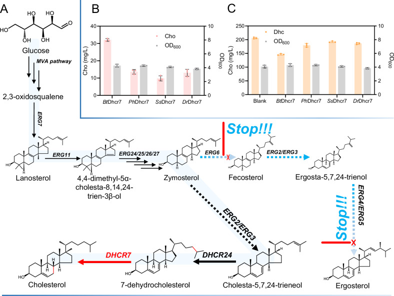
Fig. 1 Construction of a cholesterol biosynthetic pathway in Saccharomyces cerevisiae. A Schematic of the yeast mevalonate (MVA) pathway for cholesterol (Cho) biosynthesis. Endogenous synthesis of 2,3-epoxysqualene proceeds via the MVA pathway, followed by cyclization through ERG7 to form lanosterol. In the endoplasmic reticulum, lanosterol is sequentially converted by ERG2/3/7/11/24/25/26/27 into cholesta-5,7,24-trien-3β-ol and ergosterol. Exogenous 24-dehydrocholesterol reductase (DHCR24) and 7-dehydrocholesterol reductase (DHCR7) then convert cholesta-5,7,24-trien-3β-ol toward Cho. ERG5 and ERG6 are knocked out to block flux toward ergosterol. B Cho titers (pink bars) and optical density at 600 nm (OD600) (gray bars) in strain D7-1 expressing Bos taurus (BtDHCR7), Pangasianodon hypophthalmus (PhDHCR7), Sus scrofa (SsDHCR7), or Danio rerio (DrDHCR7) DHCR7. C 7-dehydrocholesterol (Dhc) titers (pink bars) and OD600 (gray bars) in D7-1 expressing BtDHCR7, PhDHCR7, SsDHCR7, or DrDHCR7. Data are presented as mean ± s.d. from three biologically independent replicates. Source data are provided as a Source Data file.
Fig. 1 Construction of a cholesterol biosynthetic pathway in Saccharomyces cerevisiae. A Schematic of the yeast mevalonate (MVA) pathway for cholesterol (Cho) biosynthesis. Endogenous synthesis of 2,3-epoxysqualene proceeds via the MVA pathway, followed by cyclization through ERG7 to form lanosterol. In the endoplasmic reticulum, lanosterol is sequentially converted by ERG2/3/7/11/24/25/26/27 into cholesta-5,7,24-trien-3β-ol and ergosterol. Exogenous 24-dehydrocholesterol reductase (DHCR24) and 7-dehydrocholesterol reductase (DHCR7) then convert cholesta-5,7,24-trien-3β-ol toward Cho. ERG5 and ERG6 are knocked out to block flux toward ergosterol. B Cho titers (pink bars) and optical density at 600 nm (OD600) (gray bars) in strain D7-1 expressing Bos taurus (BtDHCR7), Pangasianodon hypophthalmus (PhDHCR7), Sus scrofa (SsDHCR7), or Danio rerio (DrDHCR7) DHCR7. C 7-dehydrocholesterol (Dhc) titers (pink bars) and OD600 (gray bars) in D7-1 expressing BtDHCR7, PhDHCR7, SsDHCR7, or DrDHCR7. Data are presented as mean ± s.d. from three biologically independent replicates. Source data are provided as a Source Data file.CC BY-NC-ND · PubMed Central
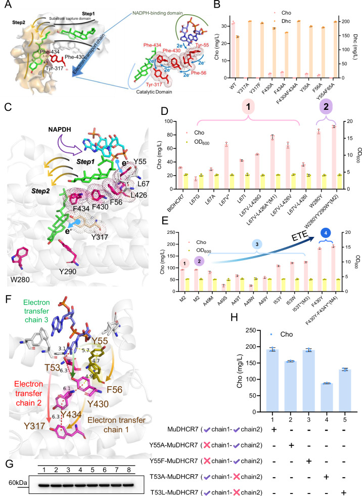
Fig. 2 Engineering the electron transfer system of BtDHCR7. A The catalysis of 7-dehydrocholesterol (Dhc) by 7-dehydrocholesterol reductase (DHCR7) involved two steps: substrate capture (white region) and substrate binding (yellow region). Recognition sites (white region) are involved in substrate binding; F430 and F434 serve as binding residues. In the catalytic pocket, Y317 donates electrons for catalysis. Y55, F56, F430, F434, and Y317 (marked in red) together form the electron transfer chain. B Residues in the electron transport chain were identified by alanine scanning or by substituting tyrosine with phenylalanine. Y317 of codon-optimized DHCR7 (BtDHCR7) was mutated to Phe and lost activity. C Rationally engineering BtDHCR7 was divided into four steps: 1. Engineering the L67 and L426 of DHCR7’s substrate-recognizing domain; 2. Engineering the W280 and Y290 of DHCR7’s substrate-binding domain; 3. Engineering the A49 and I53 of DHCR7’s NADPH-binding domain; 4. Engineering the F430 and F434 of DHCR7’s electron transfer chain. ETE of BtDHCR7 was step 3 and 4. The two blue arrows represent the capture and transfer of electrons from NADPH to C7 = C8, respectively. The yellow arrow represents DHC entering the catalytic pocket (step 2) after being captured by DHCR7 (step 2). D Rationally engineering BtDHCR7 by engineering substrate-recognizing domain and substrate-binding domain. W280 and Y290 were subjected to saturation mutagenesis, and the mutants with the highest Cho production were shown. L67V-L426A-BtDHCR7 was named M1. L67V-L426A-W280Y-Y290W-BtDHCR7 was named M2. The final OD600 value and Cho yield of fermentation are provided. (E) Rationally engineering BtDHCR7 by engineering NADPH-binding domain and electron transfer chain. I53T-M2 was named as M3. I53T-F430Y-F434Y-M2 was named M4. The final OD600 value and Cho yield of fermentation are provided. F Electron transfer chain 2 was another electronic transfer chain in M4. G Western blot analysis of BtDHCR7 and its variants. 1: WT-BtDHCR7, 2: L67V-BtDHCR7, 3: M1, 4: M2, 5: I53W-M2, 6: M3, 7: F430Y-M3, 8: M4. H Engineering the electron transfer chain of MuBtDHCR7 demonstrated the electron transfer chain 1 was the primary chain. Values are shown as mean ± s. d. from three biological replicates. Source data are provided as a Source Data file.
Fig. 2 Engineering the electron transfer system of BtDHCR7. A The catalysis of 7-dehydrocholesterol (Dhc) by 7-dehydrocholesterol reductase (DHCR7) involved two steps: substrate capture (white region) and substrate binding (yellow region). Recognition sites (white region) are involved in substrate binding; F430 and F434 serve as binding residues. In the catalytic pocket, Y317 donates electrons for catalysis. Y55, F56, F430, F434, and Y317 (marked in red) together form the electron transfer chain. B Residues in the electron transport chain were identified by alanine scanning or by substituting tyrosine with phenylalanine. Y317 of codon-optimized DHCR7 (BtDHCR7) was mutated to Phe and lost activity. C Rationally engineering BtDHCR7 was divided into four steps: 1. Engineering the L67 and L426 of DHCR7’s substrate-recognizing domain; 2. Engineering the W280 and Y290 of DHCR7’s substrate-binding domain; 3. Engineering the A49 and I53 of DHCR7’s NADPH-binding domain; 4. Engineering the F430 and F434 of DHCR7’s electron transfer chain. ETE of BtDHCR7 was step 3 and 4. The two blue arrows represent the capture and transfer of electrons from NADPH to C7 = C8, respectively. The yellow arrow represents DHC entering the catalytic pocket (step 2) after being captured by DHCR7 (step 2). D Rationally engineering BtDHCR7 by engineering substrate-recognizing domain and substrate-binding domain. W280 and Y290 were subjected to saturation mutagenesis, and the mutants with the highest Cho production were shown. L67V-L426A-BtDHCR7 was named M1. L67V-L426A-W280Y-Y290W-BtDHCR7 was named M2. The final OD600 value and Cho yield of fermentation are provided. (E) Rationally engineering BtDHCR7 by engineering NADPH-binding domain and electron transfer chain. I53T-M2 was named as M3. I53T-F430Y-F434Y-M2 was named M4. The final OD600 value and Cho yield of fermentation are provided. F Electron transfer chain 2 was another electronic transfer chain in M4. G Western blot analysis of BtDHCR7 and its variants. 1: WT-BtDHCR7, 2: L67V-BtDHCR7, 3: M1, 4: M2, 5: I53W-M2, 6: M3, 7: F430Y-M3, 8: M4. H Engineering the electron transfer chain of MuBtDHCR7 demonstrated the electron transfer chain 1 was the primary chain. Values are shown as mean ± s. d. from three biological replicates. Source data are provided as a Source Data file.CC BY-NC-ND · PubMed Central
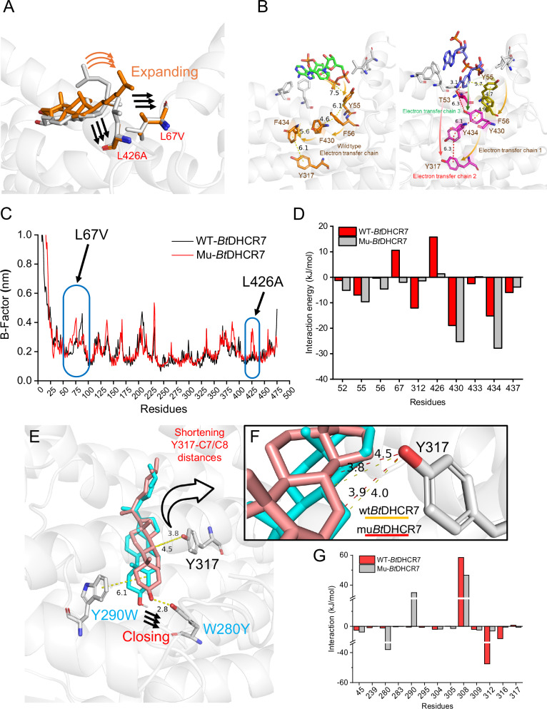
Fig. 3 Explaining the mechanism of DHCR7 activity enhancement. A L67 and L426 of DHCR7’s substrate-recognizing domain were engineered. L67V and L426A allow the steroid side chain to expand. The Dhc marked in the yellow and grey stick represented the conformation in the modified SRD and wild-type SRD, respectively. B Left: NADPH located in the wild-type DHCR7. Residues with electron transfer functions are rendered in yellow sticks. Right: NADPH located in the MuBtDHCR7. Residues with electron transfer chain 1 and 2 functions are rendered in red and yellow sticks, respectively. C B-Factor values of SRD of WTBtDHCR7 (black) and MuBtDHCR7 (red) with Dhc complexes in 100 ns. D Free energy calculation for residues within 4 Å of Dhc in the DHCR7’s SRD. The red bar represents wild-type BtDHCR7 (WT-BtDHCR7). The gray bar represents mutant BtDHCR7 (Mu-BtDHCR7). E Y290 and W280 were engineered for shortening Y317-C7/C8 distances. The DHC marked in the yellow and pink stick represented the conformation in the wild-type substrate-binding domain and mutant substrate-binding domain, respectively. F The distances between Y317 and C7/C8 of Dhc. G Free energy calculation for residues within 4 Å of Dhc in the DHCR7’s SBD. The red bar represents WT-BtDHCR7. The gray bar represents mutant Mu-BtDHCR7. Source data are provided as a Source Data file.
Fig. 3 Explaining the mechanism of DHCR7 activity enhancement. A L67 and L426 of DHCR7’s substrate-recognizing domain were engineered. L67V and L426A allow the steroid side chain to expand. The Dhc marked in the yellow and grey stick represented the conformation in the modified SRD and wild-type SRD, respectively. B Left: NADPH located in the wild-type DHCR7. Residues with electron transfer functions are rendered in yellow sticks. Right: NADPH located in the MuBtDHCR7. Residues with electron transfer chain 1 and 2 functions are rendered in red and yellow sticks, respectively. C B-Factor values of SRD of WTBtDHCR7 (black) and MuBtDHCR7 (red) with Dhc complexes in 100 ns. D Free energy calculation for residues within 4 Å of Dhc in the DHCR7’s SRD. The red bar represents wild-type BtDHCR7 (WT-BtDHCR7). The gray bar represents mutant BtDHCR7 (Mu-BtDHCR7). E Y290 and W280 were engineered for shortening Y317-C7/C8 distances. The DHC marked in the yellow and pink stick represented the conformation in the wild-type substrate-binding domain and mutant substrate-binding domain, respectively. F The distances between Y317 and C7/C8 of Dhc. G Free energy calculation for residues within 4 Å of Dhc in the DHCR7’s SBD. The red bar represents WT-BtDHCR7. The gray bar represents mutant Mu-BtDHCR7. Source data are provided as a Source Data file.CC BY-NC-ND · PubMed Central
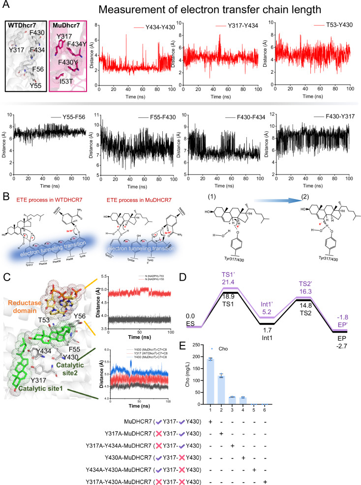
Fig. 4 Mechanism analysis of DHCR7 by MD simulation and QM/MM calculation. A Analyzing electron transport chain length of DHCR7 by replica exchange molecular dynamics (REMD) simulation in 100 ns. Red: analyzing the distances of MuDHCR7. Black: analyzing the distances of WTDHCR7. B 2D sketch of the electron transfer process in wild type DHCR7 (WTDhcr7) and mutant DHCR7 (MuDHCR7). The electron transfer chain was rendered in blue. (1) and (2) showed Y317 or Y430 provided two electrons to C7 = C8. C Analyzing the electron capture and catalytic residues of DHCR7. An another electron transfer chain was formed by T53, Y430, Y434, and Y317. Y317 and Y430 were both the catalytic centers of MuDHCR7. MD simulation was used to determine the distance changes from the active groups of NADPH to T53 (red) and Y55 (black) for giving electrons within 100 ns. The distance changes from C7 = C8 to Y317 (MuDHCR7, black), Y317 (WTDHCR7, red), and Y430 (MuDHCR7, blue) for receiving electrons within 100 ns. D QM/MM calculated energy profile (in kcal/mol) for the C7 = C8 addition reaction in engineered catalytic pocket 1 (black) and catalytic pocket 2 (purple). QM/MM calculation optimized structures of key residues involved in the reaction, and key distances are given in angstroms. TS1 and TS2 were shown the Y317 for catalyzing Dhc. TS1’ and TS2’ were shown the Y430 for catalyzing Dhc. E The catalytic residues of MuDHCR7 were determined by alanine scanning. Y317 and Y430 in mutant DHCR7 were determined as the catalytic residues. Values are shown as mean±s. d. from three biological replicates. Source data are provided as a Source Data file.
Fig. 4 Mechanism analysis of DHCR7 by MD simulation and QM/MM calculation. A Analyzing electron transport chain length of DHCR7 by replica exchange molecular dynamics (REMD) simulation in 100 ns. Red: analyzing the distances of MuDHCR7. Black: analyzing the distances of WTDHCR7. B 2D sketch of the electron transfer process in wild type DHCR7 (WTDhcr7) and mutant DHCR7 (MuDHCR7). The electron transfer chain was rendered in blue. (1) and (2) showed Y317 or Y430 provided two electrons to C7 = C8. C Analyzing the electron capture and catalytic residues of DHCR7. An another electron transfer chain was formed by T53, Y430, Y434, and Y317. Y317 and Y430 were both the catalytic centers of MuDHCR7. MD simulation was used to determine the distance changes from the active groups of NADPH to T53 (red) and Y55 (black) for giving electrons within 100 ns. The distance changes from C7 = C8 to Y317 (MuDHCR7, black), Y317 (WTDHCR7, red), and Y430 (MuDHCR7, blue) for receiving electrons within 100 ns. D QM/MM calculated energy profile (in kcal/mol) for the C7 = C8 addition reaction in engineered catalytic pocket 1 (black) and catalytic pocket 2 (purple). QM/MM calculation optimized structures of key residues involved in the reaction, and key distances are given in angstroms. TS1 and TS2 were shown the Y317 for catalyzing Dhc. TS1’ and TS2’ were shown the Y430 for catalyzing Dhc. E The catalytic residues of MuDHCR7 were determined by alanine scanning. Y317 and Y430 in mutant DHCR7 were determined as the catalytic residues. Values are shown as mean±s. d. from three biological replicates. Source data are provided as a Source Data file.CC BY-NC-ND · PubMed Central
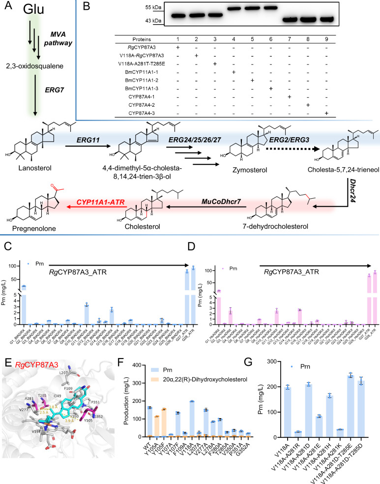
Fig. 5 Constructing Prn biosynthetic pathway in S. cerevisiae. A Overviewing the de novo biosynthetic pathway of Prn production in S. cerevisiae. B Western blot analysis of BmCYP11A1, CYP87A4, RgCYP87A3 and its variants. C Cho production strain D7-1 was engineered to de novo biosynthesize Prn by introducing different combinations of CYP11A1 and CPR. RgCYP11A1 from R. glutinosa with ATR from A. thaliana produced 93.5 mg/L Prn. G1-G26 of CYP11A1 were derived from animals and compared with Bos taurus ADR and Bos taurus ADX. G27-G28 of CYP87A were derived from plants and compared with Arabidopsis thaliana cytochrome P450 reductase 2 (ATR2). The accession numbers of G1-G6 were shown in Supplementary Data 6. D G1-G26 of CYP11A1 were derived from animals and compared with Homo sapiens ADR and Homo sapiens ADX. G27-G28 of CYP87A were derived from plants and compared with Arabidopsis thaliana cytochrome P450 reductase 2 (ATR2). E The residues within a range of 4 Å near Cho in the heme center in WTRgCYP87A3 are shown. F Alanine scanning the residues within a range of 4 Å near Cho in WTRgCYP87A3 for determining residues for side chain cleaving. Moreover, Y105 and Y107 were also mutated into phenylalanine for determining the side chain cleaving function. G Engineering V118A-RgCYP87A3 using ETE strategy. V118 and A281 were mutated into acidic residues for proton extraction, respectively. V118A-A281D-T285E produced 243 mg/L Prn. ADR/X means ADR and ADX. Values are shown as mean ± s. d. from three biological replicates. Source data are provided as a Source Data file.
Fig. 5 Constructing Prn biosynthetic pathway in S. cerevisiae. A Overviewing the de novo biosynthetic pathway of Prn production in S. cerevisiae. B Western blot analysis of BmCYP11A1, CYP87A4, RgCYP87A3 and its variants. C Cho production strain D7-1 was engineered to de novo biosynthesize Prn by introducing different combinations of CYP11A1 and CPR. RgCYP11A1 from R. glutinosa with ATR from A. thaliana produced 93.5 mg/L Prn. G1-G26 of CYP11A1 were derived from animals and compared with Bos taurus ADR and Bos taurus ADX. G27-G28 of CYP87A were derived from plants and compared with Arabidopsis thaliana cytochrome P450 reductase 2 (ATR2). The accession numbers of G1-G6 were shown in Supplementary Data 6. D G1-G26 of CYP11A1 were derived from animals and compared with Homo sapiens ADR and Homo sapiens ADX. G27-G28 of CYP87A were derived from plants and compared with Arabidopsis thaliana cytochrome P450 reductase 2 (ATR2). E The residues within a range of 4 Å near Cho in the heme center in WTRgCYP87A3 are shown. F Alanine scanning the residues within a range of 4 Å near Cho in WTRgCYP87A3 for determining residues for side chain cleaving. Moreover, Y105 and Y107 were also mutated into phenylalanine for determining the side chain cleaving function. G Engineering V118A-RgCYP87A3 using ETE strategy. V118 and A281 were mutated into acidic residues for proton extraction, respectively. V118A-A281D-T285E produced 243 mg/L Prn. ADR/X means ADR and ADX. Values are shown as mean ± s. d. from three biological replicates. Source data are provided as a Source Data file.CC BY-NC-ND · PubMed Central
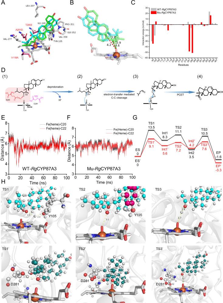
Fig. 6 ETE of RgCYP87A3 for Prn production and mechanism analysis. A The conformation of Cho in the catalytic pocket of wild-type (cyan stick) and Mutant (green stick) RgCYP87A3. V118A, A281D, and T285E were shown in red. B In the wild-type catalytic pocket, the distances of C20 and C22 of Cho from the FeO center are 3.9 Å each. In the mutant catalytic pocket, these distances are 4.2 Å for C20 and 2.6 Å for C22. C Free energy calculation for residues within 4 Å of Cho in wild type (WT-RgCYP87A3, red) and mutant (Mu-RgCYP87A3, gray) RgCYP87A3’s heme center. D Schematic drawings the three steps of side chain cleaving process by D281, E285, and heme center. E Distances between FeO (heme) of WTRgCYP87A3 and C20/22 of Cho were measured by MD simulation in 100 ns. F Distances between FeO (heme) of MuRgCYP87A3 and C20/22 of Cho were measured by MD simulation in 100 ns. G QM/MM calculation calculated energy profile (in kcal/mol) for RgCYP87A3 cleaving Cho side chain process in engineered catalytic pocket (red) and wild type pocket (black). (H) QM/MM-optimized the structures in the RgCYP87A3 and mutant RgCYP87A3 cleaving reaction. Key distances of TS1, TS2, TS3, TS1’, TS2’, and TS3’ are given in Å. Values are shown as mean±s. d. from three biological replicates. Source data are provided as a Source Data file.
Fig. 6 ETE of RgCYP87A3 for Prn production and mechanism analysis. A The conformation of Cho in the catalytic pocket of wild-type (cyan stick) and Mutant (green stick) RgCYP87A3. V118A, A281D, and T285E were shown in red. B In the wild-type catalytic pocket, the distances of C20 and C22 of Cho from the FeO center are 3.9 Å each. In the mutant catalytic pocket, these distances are 4.2 Å for C20 and 2.6 Å for C22. C Free energy calculation for residues within 4 Å of Cho in wild type (WT-RgCYP87A3, red) and mutant (Mu-RgCYP87A3, gray) RgCYP87A3’s heme center. D Schematic drawings the three steps of side chain cleaving process by D281, E285, and heme center. E Distances between FeO (heme) of WTRgCYP87A3 and C20/22 of Cho were measured by MD simulation in 100 ns. F Distances between FeO (heme) of MuRgCYP87A3 and C20/22 of Cho were measured by MD simulation in 100 ns. G QM/MM calculation calculated energy profile (in kcal/mol) for RgCYP87A3 cleaving Cho side chain process in engineered catalytic pocket (red) and wild type pocket (black). (H) QM/MM-optimized the structures in the RgCYP87A3 and mutant RgCYP87A3 cleaving reaction. Key distances of TS1, TS2, TS3, TS1’, TS2’, and TS3’ are given in Å. Values are shown as mean±s. d. from three biological replicates. Source data are provided as a Source Data file.CC BY-NC-ND · PubMed Central
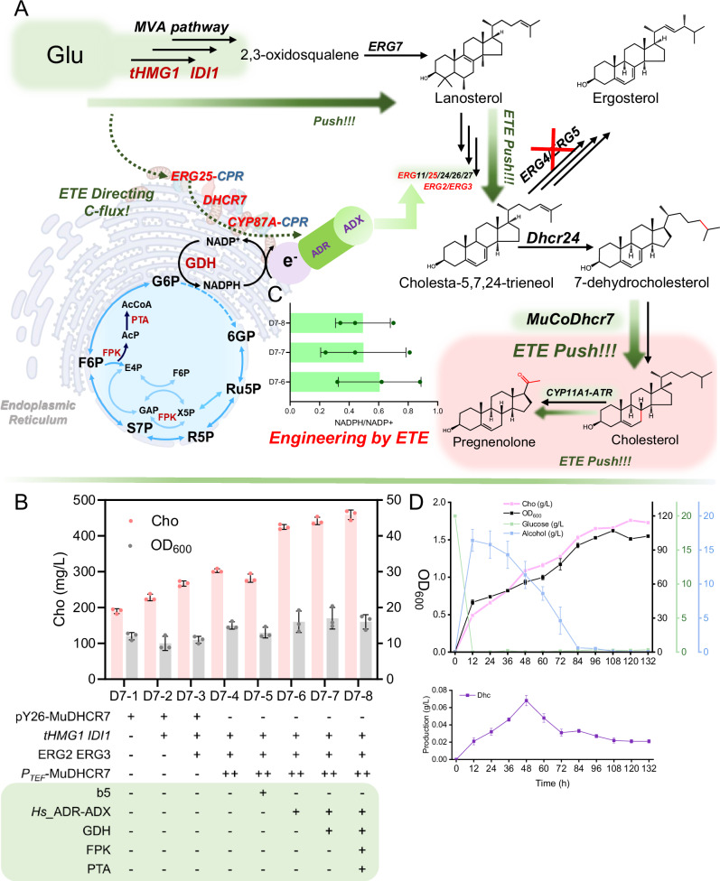
Fig. 7 Engineering the electron transfer system for Cho production. A Primary and secondary metabolic pathways for Cho and Prn synthesis in S. cerevisiae, along with the synthesis pathway for the electron donor (NADPH). Subcellular structure was created in BioRender. Chen, Q. (2025) https://BioRender.com/9smi93r. B Metabolic engineering and ETE of S. cerevisiae for Cho production. 3-hydroxy-3-methylglutaryl-CoA reductase 1 (tHMG1) encoded truncated 3-hydroxy-3-methylglutaryl-CoA reductase. IDI1 encoded isopentenyl-diphosphate delta isomerase 1. ERG2 encoded C-8 sterol isomerase. ERG3 encoded C-5 sterol desaturase. Hs_ADR encoded adriamycin from H. sapiens. Hs_ADX encoded adrenodoxin from H. sapiens. GDH encoded glutamate dehydrogenase from Bacillus subtilis. FPK encoded phosphoketolase from Bifidobacterium breve. PTA encoded phosphotransacetylase from Clostridium kluyveri. Gpp1 encoded glycerol-3-phosphate phosphatase 1. C Cellular NADPH/NADP+ ratios in the engineered D7−6, D7-7, and D7-8; samples for NADP(H) quantification were harvested after 48 h. The NADPH/NADP+ ratio was determined using the NADP+/NADPH quantitation kit. D High-density fermentation for Cho production in a medium supplemented with glucose for about 16 h and nitrogen for 72 h. Fermentation is divided into three stages, shown from light to dark. Glucose was added at 16 h, and YPD was added at 72 h. Values are shown as mean±s. d. from three biological replicates. Source data are provided as a Source Data file.
Fig. 7 Engineering the electron transfer system for Cho production. A Primary and secondary metabolic pathways for Cho and Prn synthesis in S. cerevisiae, along with the synthesis pathway for the electron donor (NADPH). Subcellular structure was created in BioRender. Chen, Q. (2025) https://BioRender.com/9smi93r. B Metabolic engineering and ETE of S. cerevisiae for Cho production. 3-hydroxy-3-methylglutaryl-CoA reductase 1 (tHMG1) encoded truncated 3-hydroxy-3-methylglutaryl-CoA reductase. IDI1 encoded isopentenyl-diphosphate delta isomerase 1. ERG2 encoded C-8 sterol isomerase. ERG3 encoded C-5 sterol desaturase. Hs_ADR encoded adriamycin from H. sapiens. Hs_ADX encoded adrenodoxin from H. sapiens. GDH encoded glutamate dehydrogenase from Bacillus subtilis. FPK encoded phosphoketolase from Bifidobacterium breve. PTA encoded phosphotransacetylase from Clostridium kluyveri. Gpp1 encoded glycerol-3-phosphate phosphatase 1. C Cellular NADPH/NADP+ ratios in the engineered D7−6, D7-7, and D7-8; samples for NADP(H) quantification were harvested after 48 h. The NADPH/NADP+ ratio was determined using the NADP+/NADPH quantitation kit. D High-density fermentation for Cho production in a medium supplemented with glucose for about 16 h and nitrogen for 72 h. Fermentation is divided into three stages, shown from light to dark. Glucose was added at 16 h, and YPD was added at 72 h. Values are shown as mean±s. d. from three biological replicates. Source data are provided as a Source Data file.CC BY-NC-ND · PubMed Central
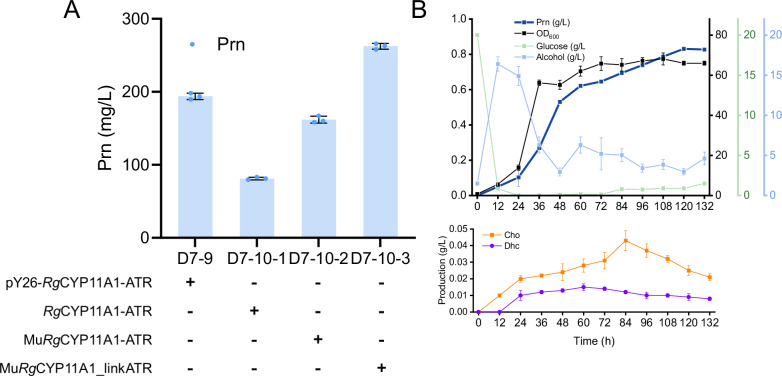
Fig. 8 Engineering the electron transfer system for Prn production. A Engineering S. cerevisiae for Prn production using ETE strategy. Promoting electron transfer efficiency using peptides to shorten the distance between RgCYP87A3 and ATR. B High-density fermentation for Prn production in a medium supplemented with glucose for about 16 h and nitrogen for 72 h. Values are shown as mean±s. d. from three biological replicates. Source data are provided as a Source Data file.
Fig. 8 Engineering the electron transfer system for Prn production. A Engineering S. cerevisiae for Prn production using ETE strategy. Promoting electron transfer efficiency using peptides to shorten the distance between RgCYP87A3 and ATR. B High-density fermentation for Prn production in a medium supplemented with glucose for about 16 h and nitrogen for 72 h. Values are shown as mean±s. d. from three biological replicates. Source data are provided as a Source Data file.CC BY-NC-ND · PubMed Central

Verification

Claims the paper makes

The claims and statistics of this paper haven’t been checked yet.

Run the check — free, no account needed. It reads the open-access text and checks the sources it cites.

A few free checks a day · papers already verified open instantly · results are public

Self-check

Percentages, sums and reported differences re-computed from the paper’s own numbers.

The self-check needs the paper’s full text.

Citations

Cites

Show all 1 references

Research works printed but not linked: 28

  1. Park SY, Eun H, Lee MH, Lee SY (2022) Metabolic engineering of Escherichia coli with electron channelling for the production of natural products. Nat Catal 5(8):726–737 · DOI
  2. Li X, Roberti R, Blobel G (2015) Structure of an integral membrane sterol reductase from Methylomicrobium alcaliphilum. Nature 517(7532):104–107 · DOI
  3. Chen R, Gao J, Yu W, Chen X, Zhai X, Chen Y, Zhang L, Zhou YJ (2022) Engineering cofactor supply and recycling to drive phenolic acid biosynthesis in yeast. Nat Chem Biol 18(5):520–529 · DOI
  4. Zhou Z, Zhang X, Wu J, Li X, Li W, Sun X, Wang J, Yan Y, Shen X, Yuan Q (2022) Targeting cofactors regeneration in methylation and hydroxylation for high level production of Ferulic acid. Metab Eng 73:247–255 · DOI
  5. Rashida Z, Laxman S (2021) The pentose phosphate pathway and organization of metabolic networks enabling growth programs. Curr Opin Syst Biol 28:100390 · DOI
  6. Tyburski R, Liu T, Glover SD, Hammarström L (2021) Proton-Coupled Electron Transfer Guidelines, Fair and Square. J Am Chem Soc 143(2):560–576 · DOI
  7. Cai X, Li Y, Zhang Y, Lin W (2023) π–π Interaction-Driven Charge Separation and Interlayer Transfer in Polymeric Carbon Nitride. ACS Catal 13(24):15877–15885 · DOI
  8. Greene BL (2021) Progress and Opportunities in Photochemical Enzymology of Oxidoreductases. ACS Catal 11(23):14635–14650 · DOI
  9. Zhang R, Zhang Y, Wang Y, Yao M, Zhang J, Liu H, Zhou X, Xiao W, Yuan Y (2019) Pregnenolone Overproduction in Yarrowia lipolytica by Integrative Components Pairing of the Cytochrome P450scc System. ACS Synth Biol 8(12):2666–2678 · DOI
  10. Ding J, You S, Zhang J, Zhang H, Wang H, Zhang W, Qi W, Su R, He Z (2021) Rational design of 17β-hydroxysteroid dehydrogenase type3 for improving testosterone production with an engineered Pichia pastoris. Bioresour Technol 341:125833 · DOI
  11. Lin YC, Cheung G, Porter E, Papadopoulos V (2022) The neurosteroid pregnenolone is synthesized by a mitochondrial P450 enzyme other than CYP11A1 in human glial cells. J Biol Chem 298(7):102110 · DOI
  12. Qin Z, Zhang Y, Liu S, Zeng W, Zhou J, Xu S (2024) Combining Metabolic Engineering and Lipid Droplet Assembly to Achieve Campesterol Overproduction in Saccharomyces cerevisiae. J Agric Food Chem 72(9):4814–4824 · DOI
  13. Zöllner A, Pasquinelli MA, Bernhardt R, Beratan DN (2007) Protein Phosphorylation and Intermolecular Electron Transfer: A Joint Experimental and Computational Study of a Hormone Biosynthesis Pathway. J Am Chem Soc 129(14):4206–4216 · DOI
  14. Su H, Wang B, Shaik S (2019) Quantum-Mechanical/Molecular-Mechanical Studies of CYP11A1-Catalyzed Biosynthesis of Pregnenolone from Cholesterol Reveal a C–C Bond Cleavage Reaction That Occurs by a Compound I-Mediated Electron Transfer. J Am Chem Soc 141(51):20079–20088 · DOI
  15. Wang Z, Shaik S, Wang B (2021) Conformational Motion of Ferredoxin Enables Efficient Electron Transfer to Heme in the Full-Length P450TT. J Am Chem Soc 143(2):1005–1016 · DOI
  16. Carroll E, Ravi Gopal B, Raghavan I, Mukherjee M, Wang ZQ (2023) A cytochrome P450 CYP87A4 imparts sterol side-chain cleavage in digoxin biosynthesis. Nat Commun 14(1):4042 · DOI
  17. Zhang R, Zhao Y, Sun Y, Lu X, Yang X (2013) Isolation, Characterization, and Hepatoprotective Effects of the Raffinose Family Oligosaccharides from Rehmannia glutinosa Libosch. J Agric Food Chem 61(32):7786–7793 · DOI
  18. Chen Q, Chao Z, Wang K, Wang X, Meng H, Liu X, Shan X, Zhou J (2024) Simplification of Corticosteroids Biosynthetic Pathway by Engineering P450BM3. ACS Catal. :4117–4129 · DOI
  19. Ly TTB, Schifrin A, Nguyen BD, Bernhardt R (2017) Improvement of a P450-Based Recombinant Escherichia coli Whole-Cell System for the Production of Oxygenated Sesquiterpene Derivatives. J Agric Food Chem 65(19):3891–3899 · DOI
  20. Qin Z, Zhang Y, Liu S, Zeng W, Zhou J, Xu S (2024) Combining Metabolic Engineering and Lipid Droplet Assembly to Achieve Campesterol Overproduction in Saccharomyces cerevisiae. J Agric Food Chem 72(9):4814–4824 · DOI
  21. Wang S, Wang L, Xu J, Wang Y, Xiang L, He X (2023) Synergistic Combination of the Total Steroidal Saponins from the Berries of Black Nightshade and Adriamycin to Overcome Leukemia Multidrug Resistance. J Agric Food Chem 71(7):3315–3324 · DOI
  22. Zhu M, Wang C, Sun W, Zhou A, Wang Y, Zhang G, Zhou X, Huo Y, Li C Boosting 11-oxo-β-amyrin and glycyrrhetinic acid synthesis in Saccharomyces cerevisiae via pairing novel oxidation and reduction system from legume plants. Metab Eng 2018 45:43–50
  23. Zhao X, Wei W, Li S, Gao S, Liu S, Zeng W, Zhou J (2024) Elucidation of the Biosynthesis of Asiaticoside and Its Reconstitution in Yeast. ACS Sustainable Chem Eng 12(10):4028–4040 · DOI
  24. King E, Cui Y, Aspacio D, Nicklen F, Zhang L, Maxel S, Luo R, Siegel JB, Aitchison E, Li H (2022) Engineering Embden–Meyerhof–Parnas Glycolysis to Generate Noncanonical Reducing Power. ACS Catal 12(14):8582–8592 · DOI
  25. Li Y, Mao J, Liu Q, Song X, Wu Y, Cai M, Xu H, Qiao M (2020) De Novo Biosynthesis of Caffeic Acid from Glucose by Engineered Saccharomyces cerevisiae. ACS Synth Biol 9(4):756–765 · DOI
  26. Liu Y, Denisov IG, Sligar SG, Kincaid JR (2021) Substrate-Specific Allosteric Effects on the Enhancement of CYP17A1 Lyase Efficiency by Cytochrome b5. J Am Chem Soc 143(10):3729–3733 · DOI
  27. Chen J, Fan F, Qu G, Tang J, Xi Y, Bi C, Sun Z, Zhang X (2020) Identification of Absidia orchidis steroid 11beta-hydroxylation system and its application in engineering Saccharomyces cerevisiae for one-step biotransformation to produce hydrocortisone. Metab Eng 57:31–42 · DOI
  28. Pourcel L, Buron F, Garcia F, Delaloix M-S, Le Fourn V, Girod P-A Mermod, N. Transient vitamin B5 starving improves mammalian cell homeostasis and protein production. Metab Eng 2020 60:77–86

Metadata

Authors
Zhou, J., Chen, Q., Wei, W., Chao, Z., Qi, R., He, J., Chen, H., Wang, K., Wang, X., & Rao, Y.
Affiliations
Jiangnan University (CN)
Corresponding author
Zhou, Jingwen
Venue
Nature Communications
Published
2024-08-01
Volume
16
Issue
1
Pages
3740-3740
ISSN
2041-1723 (ISSN-L)
OpenAlex
W4409622267
Cited by
28
References
1
Citations by year
2026: 18 · 2025: 10
FWCI
8.37
Type
journal article
Language
en
Open access
Locations
  • Nature Communications (publishedVersion, cc-by-nc-nd)
  • Research Square (acceptedVersion, cc-by)
  • PubMed Central (publishedVersion, cc-by-nc-nd)
  • PubMed (publishedVersion)
  • RePEc: Research Papers in Economics (submittedVersion)
  • DOAJ (DOAJ: Directory of Open Access Journals) (submittedVersion, cc-by-sa)
Funders
National Key Research and Development Program of China (2024YFA0919900)
Topics
  • Life Sciences › Biochemistry, Genetics and Molecular Biology › Molecular Biology › Microbial Metabolic Engineering and Bioproduction
  • Life Sciences › Biochemistry, Genetics and Molecular Biology › Molecular Biology › Plant biochemistry and biosynthesis
  • Physical Sciences › Chemistry › Organic Chemistry › Steroid Chemistry and Biochemistry
Keywords
steroidogenesis, NADPH-dependent enzyme, Saccharomyces cerevisiae, DHCR7, P450scc, NADPH regeneration, synthetic biology
MeSH terms
Cholesterol, Cholesterol Side-Chain Cleavage Enzyme, Electron Transport, NADP, Pregnenolone, Saccharomyces cerevisiae, Steroids, Saccharomyces cerevisiae Proteins, Oxidoreductases Acting on CH-CH Group Donors, Synthetic Biology, Metabolic Engineering

About these checks

Registry, retraction, Cites and statistics results are computed, not judged, with one exception: a printed reference that no index lists is read once by a model before it counts as a citation error. Claims are extracted by a model and kept only when their quote appears verbatim in the text; each cited source is then checked, with a model’s help, for whether it says what the claim relies on. Those verdicts can be wrong — read the claim’s quote and the cited source before relying on one.

Statistics and arithmetic are recomputed, not judged.

Confidence bands are shown only for fields calibrated on our annotated test set. None is calibrated yet, so no band is shown.

What counts as a claim ›How we measure confidence ›How we check this ›

Are you an author, or do you see a problem with this page? Request a review

How to cite

Cite CoChat for the checks on this page, and the paper for its own findings.

This page
CoChat. (2026). Citation check: Electron transfer engineering of artificially designed cell factory for complete biosynthesis of natural products. https://app.cochat.ai/papers/10.21203/rs.3.rs-4718218/v1
The paper
Zhou, J., Chen, Q., Wei, W., Chao, Z., Qi, R., He, J., Chen, H., Wang, K., Wang, X., & Rao, Y. (2024). Electron transfer engineering of artificially designed cell factory for complete biosynthesis of natural products. Nature Communications. https://doi.org/10.21203/rs.3.rs-4718218/v1