

The DOI matches its Crossref record.
Mitochondria-Targeted Mesoporous Titanium Dioxide Nanoplatform for Synergistic Nitric Oxide Gas-Sonodynamic Therapy of Breast Cancer
Open accessAbstract
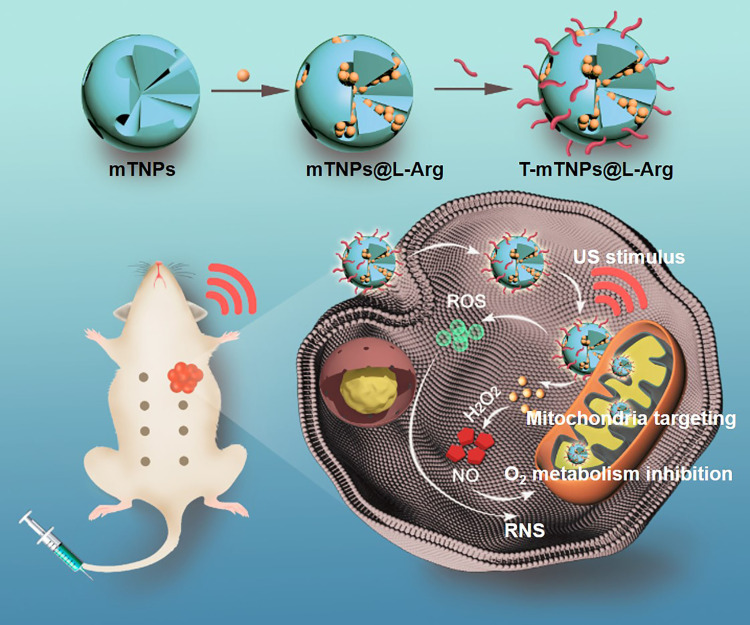
Background: Sonodynamic therapy (SDT) has rapidly advanced as a promising alternative to conventional photodynamic therapy owing to its preferable therapeutic depth. However, single-modal SDT exhibits limited efficacy due to the long-term hypoxia in tumors. Method and Results: To address these issues, we proposed a synergistic SDT strategy that integrates mitochondrial targeting with nitric oxide (NO) gas therapy by using multifunctional nanoplatforms. The nanoplatform, which was named as T-mTNPs@L-Arg, was composed of mesoporous titanium dioxide loaded with the NO donor precursor L-arginine (L-Arg) and modified with triphenyl phosphonium (TPP), a mitochondria-targeting ligand. Therefore, T-mTNPs@L-Arg could efficiently concentrate into mitochondria and release NO gas as well as generate reactive oxygen species (ROS) with ultrasound stimulus. Importantly, the released NO gas exerted multiple synergies with SDT, including inducing NO poisoning, generating more lethal reactive nitrogen species (RNS) by reaction with ROS, and alleviating hypoxia through NO-mediated mitochondrial respiration inhibition. On account of the synergistic effects, T-mTNPs@L-Arg showed an outstanding SDT efficacy and a reduced side effect. Conclusion: This work designed a nanoplatform to integrate mitochondria targeting, SDT and NO gas therapy, providing a new strategy for highly efficient breast cancer therapy.
Overview
The claims and statistics of this paper haven’t been checked yet.
Run the check — free, no account needed. It reads the open-access text and checks the sources it cites.
Integrity signals
Registry
Cites
Statistics
Figures
Verification
Claims the paper makes
The claims and statistics of this paper haven’t been checked yet.
Run the check — free, no account needed. It reads the open-access text and checks the sources it cites.
Claims other papers make about this one
Each is checked against this paper’s own text: does it say what the citing paper says it does?
The cited source says what the claim attributes to it.
In the citing paper
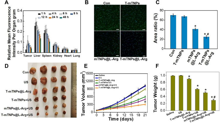
“The T-mTNPs@l-Arg and US (1 MHz, 1 Wcm−2, 1 min) group also showed the most significant inhibitory effect on tumor volume and weight in MCF7-bearing mice (Fig. 10I, J, Table 6).”
From Nanosensitizer-assisted sonodynamic therapy for breast cancer (2025) · cites this paper as [132]
Self-check
Percentages, sums and reported differences re-computed from the paper’s own numbers.
The self-check needs the paper’s full text.
Citations
Cites
Still being checked.
Research works printed but not linked: 34
- Kurian, 2019, J Clin Oncol, 37, 1305, 10.1200/JCO.18.01854 · DOI · Paper page
- Waks, 2019, JAMA, 321, 288, 10.1001/jama.2018.19323 · DOI
- Jain, 2020, J Control Release, 326, 628, 10.1016/j.jconrel.2020.07.003 · DOI
- Khoobchandani, 2020, Int J Nanomedicine, 15, 181, 10.2147/IJN.S219042 · DOI
- Pan, 2018, Sci China Life Sci, 61, 415, 10.1007/s11427-017-9262-x · DOI
- Zhu, 2018, ACS Nano, 12, 3780, 10.1021/acsnano.8b00999 · DOI
- Xu, 2020, Nano Res, 13, 2898, 10.1007/s12274-020-2992-5 · DOI
- Serpe, 2012, Nanotechnol Rev, 1, 173, 10.1515/ntrev-2011-0009 · DOI
- Li, 2017, J Control Release, 260, 12, 10.1016/j.jconrel.2017.05.025 · DOI
- Midden, 1983, J Am Chem Soc, 105, 4129, 10.1021/ja00351a001 · DOI
- McEwan, 2015, J Control Release, 203, 51, 10.1016/j.jconrel.2015.02.004 · DOI
- Chen, 2017, ACS Nano, 11, 12849, 10.1021/acsnano.7b08225 · DOI
- Fu, 2019, Adv Funct Mater, 29, 1906195, 10.1002/adfm.201906195 · DOI
- Fulda, 2010, Nat Rev Drug Discov, 9, 447, 10.1038/nrd3137 · DOI
- Orang, 2019, Mol Metab, 23, 98, 10.1016/j.molmet.2019.01.014 · DOI
- Moreno-Sanchez, 2014, Int J Biochem Cell Biol, 50, 10, 10.1016/j.biocel.2014.01.025 · DOI
- Sang, 2019, Theranostics, 9, 6209, 10.7150/thno.36283 · DOI
- Tiemuer, 2022, Chem Eng J, 430, 132858, 10.1016/j.cej.2021.132858 · DOI
- Chen, 2019, ACS nano, 13, 10887, 10.1021/acsnano.9b04954 · DOI
- Fan, 2018, Angew Chem Int Ed, 57, 8383, 10.1002/anie.201800594 · DOI
- Deng, 2018, Biomaterials, 187, 55, 10.1016/j.biomaterials.2018.09.043 · DOI
- Wan, 2018, Biomaterials, 185, 51, 10.1016/j.biomaterials.2018.09.004 · DOI
- Wang, 2018, Chem Sci, 9, 8103, 10.1039/C8SC03386B · DOI
- Xiang, 2021, Theranostics, 11, 1953, 10.7150/thno.52997 · DOI
- Yang, 2021, Nanoscale, 13, 444, 10.1039/D0NR07484E · DOI
- Soto, 2016, ACS Appl Mater Interfaces, 8, 2220, 10.1021/acsami.5b10942 · DOI
- Yang, 2021, Biomater Sci, 9, 2230, 10.1039/D0BM01932A · DOI
- Han, 2021, Biomater Sci, 9, 7432, 10.1039/D1BM01184G · DOI
- Wang, 2017, J Mater Chem B, 5, 4579, 10.1039/C7TB00938K · DOI
- Shi, 2015, ACS Appl Mater Interfaces, 7, 28554, 10.1021/acsami.5b09937 · DOI
- Qu, 2016, ACS Appl Mater Interfaces, 8, 34261, 10.1021/acsami.6b13974 · DOI
- Zhao, 2022, Angew Chem Int Ed, 61, 10.1002/anie.202113506 · DOI
- Shanei A, Akbari-Zadeh H, Fakhimikabir H, Attaran N. The role of gold nanoparticles in sonosensitization of human cervical carcinoma cell line under ultrasound irradiation: an in vitro study. Paper presented at: Journal of Nano Research; 2019. · DOI
- Yang, 2019, Curr Pharm Des, 25, 401, 10.2174/1381612825666190123114107 · DOI
Cited by
Sorted by citations
The Effectiveness of L-arginine in Clinical Conditions Associated with Hypoxia
Kurhaluk, N. (2023) · International Journal of Molecular Sciences · doi:10.3390/ijms24098205 · Cited by 43
Nanosensitizer-assisted sonodynamic therapy for breast cancer
Yu, J., Hu, J.-R., Tian, Y., Lei, Y.-M., Hu, H.-M., Lei, B.-S., Zhang, G., Sun, Y., & Ye, H.-R. (2025) · Journal of Nanobiotechnology · doi:10.1186/s12951-025-03311-3 · Cited by 43
Metadata
- Authors
- Zuo, S., Zhang, Y., Wang, Z., & Wang, J.
- Affiliations
- Jilin University (CN)
- Corresponding author
- Wang, Jing
- ORCID
- Zhang, Yan
- Venue
- International Journal of Nanomedicine
- Publisher
- Informa UK Limited
- Published
- 2022-03
- Volume
- Volume 17
- Pages
- 989-1002
- ISSN
- 1176-9114 (ISSN-L)
- 1178-2013
- PMC
- PMC8906874
- OpenAlex
- W4214871068
- Cited by
- 41
- References
- 0
- Citations by year
- 2026: 5 · 2025: 9 · 2024: 6 · 2023: 14 · 2022: 7
- FWCI
- 2.62
- Type
- journal article
- Language
- en
- Open access
- gold
- doi.org
- Licence
- Locations
- International Journal of Nanomedicine (publishedVersion, cc-by-nc)
- PubMed Central (publishedVersion, cc-by-nc)
- PubMed (publishedVersion)
- Dove Medical Press (Taylor and Francis Group) (submittedVersion, cc-by-nc)
- Funders
- People's Government of Jilin Province
- Topics
- Physical Sciences › Engineering › Biomedical Engineering › Nanoplatforms for cancer theranostics
- Life Sciences › Biochemistry, Genetics and Molecular Biology › Cancer Research › Photodynamic Therapy Research Studies
- Physical Sciences › Engineering › Biomedical Engineering › Ultrasound and Hyperthermia Applications
- Keywords
- sonodynamic therapy, breast cancer, mitochondrial targeting, mesoporous TiO2, L-arginine, tumor hypoxia, reactive oxygen species, reactive nitrogen species, mitochondrial respiration inhibition, ultrasound-triggered drug release, synergistic cancer therapy
- MeSH terms
- Humans, Mitochondria, Neoplasms, Nitric Oxide, Titanium
About these checks
Registry, retraction, Cites and statistics results are computed, not judged, with one exception: a printed reference that no index lists is read once by a model before it counts as a citation error. Claims are extracted by a model and kept only when their quote appears verbatim in the text; each cited source is then checked, with a model’s help, for whether it says what the claim relies on. Those verdicts can be wrong — read the claim’s quote and the cited source before relying on one.
Statistics and arithmetic are recomputed, not judged.
Confidence bands are shown only for fields calibrated on our annotated test set. None is calibrated yet, so no band is shown.
What counts as a claim ›How we measure confidence ›How we check this ›
Are you an author, or do you see a problem with this page? Request a review
