

The DOI matches its Crossref record.
Current and Future Strategies for the Diagnosis and Treatment of the Alpha-Gal Syndrome (AGS)
Overview
The claims and statistics of this paper haven’t been checked yet.
Run the check — free, no account needed. It reads the open-access text and checks the sources it cites.
Integrity signals
Registry
Cites
Statistics
Figures
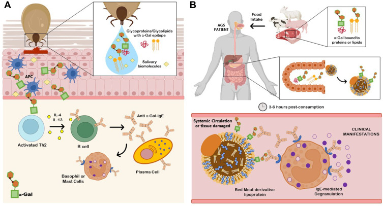
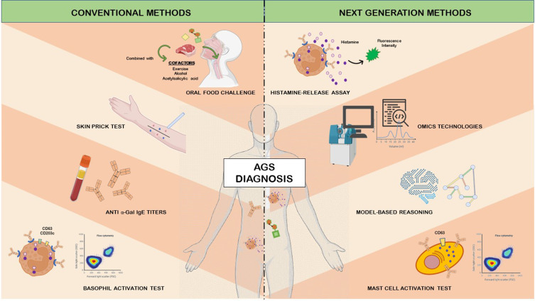
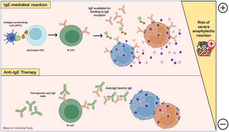
Verification
Claims the paper makes
The claims and statistics of this paper haven’t been checked yet.
Run the check — free, no account needed. It reads the open-access text and checks the sources it cites.
Claims other papers make about this one
Each is checked against this paper’s own text: does it say what the citing paper says it does?
In the citing paper
“Diagnosis of alpha-gal syndrome can be challenging because symptoms are often delayed and clinically heterogeneous.”
From Parasites and allergies: a complex bidirectional relationship from evolutionary origins to modern therapeutics (2026) · cites this paper as Vaz-Rodrigues et al. 2022
In the citing paper
“IgM, IgG, and IgA antibodies specific to α-gal may develop in humans following exposure to α-gal-like epitopes produced by commensal gut bacteria”
From Parasites and allergies: a complex bidirectional relationship from evolutionary origins to modern therapeutics (2026) · cites this paper as Vaz-Rodrigues et al. 2022
Self-check
Percentages, sums and reported differences re-computed from the paper’s own numbers.
The self-check needs the paper’s full text.
Citations
Cites
No references could be resolved yet.
Research works printed but not linked: 139
- Hils, 2020, Front Immunol, 11, 1, 10.3389/fimmu.2020.586924 · DOI
- Hilger, 2019, Curr Allergy and Asthma Rep, 19, 1, 10.1007/s11882-019-0835-9 · DOI
- Galili, 2019, Am J Phys Anthropol, 168, 352, 10.1002/ajpa.23745 · DOI
- Commins, 2009, J Allergy Clin Immunol, 123, 426, 10.1016/j.jaci.2008.10.052 · DOI
- Fischer, 2016, Allergo J Int, 25, 55, 10.1007/s40629-016-0099-z · DOI
- Çelebioğlu, 2021, Turk J Med Sci, 51, 49, 10.3906/sag-2006-95 · DOI
- Fischer, 2022, Hautarzt, 73, 195, 10.1007/s00105-022-04943-4 · DOI
- Chinuki, 2021, J Investig Allergol Clin Immunol, 32, 1, 10.18176/jiaci.0754 · DOI
- Dunkman, 2019, Anesth Analg, 129, 1242, 10.1213/ANE.0000000000003460 · DOI
- Cabezas-Cruz, 2019, Front Immunol, 10, 1, 10.3389/fimmu.2019.01210 · DOI
- Cabezas-Cruz, 2014, Lancet Infect Dis, 14, 916, 10.1016/S1473-3099(14)70902-8 · DOI
- Chung, 2008, N Engl J Med, 358, 1109, 10.1056/nejmoa074943 · DOI · Paper page
- de la Fuente, 2019, Parasit Vectors, 12, 1, 10.1186/s13071-019-3413-z · DOI
- Rodríguez, 2018, J Autoimmun, 88, 21, 10.1016/j.jaut.2017.11.007 · DOI
- Cabezas-Cruz, 2017, Trends Parasitol, 33, 495, 10.1016/j.pt.2017.03.004 · DOI
- Román-Carrasco, 2021, Front Allergy, 2, 1, 10.3389/falgy.2021.783279 · DOI
- Ramasamy, 2021, Parasit Vectors, 14, 1, 10.1186/s13071-021-04801-7 · DOI
- Wilson, 2017, Curr Allergy Asthma Rep, 17, 3, 10.1007/s11882-017-0672-7 · DOI
- Cabezas-Cruz, 2021, Biochem J, 478, 1783, 10.1042/BCJ20200915 · DOI
- Carson, 2022, J Immunol, 208, 267, 10.4049/jimmunol.2100712 · DOI
- Wong, 2018, Int J Dermatol, 57, 1433, 10.1111/ijd.14208 · DOI
- Young, 2021, Ticks Tick Borne Dis, 12, 101674, 10.1016/j.ttbdis.2021.101674 · DOI
- Diaz, 2020, South Med J, 113, 267, 10.14423/SMJ.0000000000001102 · DOI
- van Nunen, 2015, Asia Pac Allergy, 5, 3, 10.5415/apallergy.2015.5.1.3 · DOI
- Platts-Mills, 2020, J Allergy Clin Immunol, 145, 1061, 10.1016/j.jaci.2020.01.047 · DOI
- Joral, 2022, Foods, 11, 466, 10.3390/foods11030466 · DOI
- McGain, 2016, J Allergy Clin Immunol Pract, 2, 309, 10.1080/21674086.1933.11925177 · DOI
- Mullins, 2016, Clin Exp Allergy, 46, 1099, 10.1111/cea.12748 · DOI
- Commins, 2016, Curr Allergy Asthma Rep, 16, 1, 10.1007/s11882-016-0641-6 · DOI
- Levin, 2019, Ann Allergy Asthma Immunol, 122, 598, 10.1016/j.anai.2019.03.021 · DOI · Paper page
- Zurbano-Azqueta, 2021, Rev Clin Esp, 10.1016/j.rceng.2021.06.005 · DOI
- Mateo-Borrega, 2019, J Investig Allergol Clin Immunol, 29, 436, 10.18176/jiaci.0373 · DOI
- Fischer, 2017, Allergy, 72, 1540, 10.1111/all.13156 · DOI
- Villalta, 2016, Clin Exp Allergy, 46, 377, 10.1111/cea.12655 · DOI
- Mitchell, 2020, Parasit Vectors, 13, 1, 10.1186/s13071-020-04343-4 · DOI
- Venturini, 2018, J Investig Allergol Clin Immunol, 28, 106, 10.18176/jiaci.0218 · DOI
- Cabezas-Cruz, 2017, Allergy, 72, 2044, 10.1111/all.13206 · DOI
- Cabezas-Cruz, 2017, Exp Mol Med, 49, e301, 10.1038/emm.2016.164 · DOI
- Yilmaz, 2014, Cell, 159, 1277, 10.1016/j.cell.2014.10.053 · DOI
- Urra, 2021, J Med Virol, 93, 2065, 10.1002/jmv.26575 · DOI
- Steinke, 2015, J Allergy Clin Immunol, 135, 589, 10.1016/j.jaci.2014.12.1947 · DOI
- Commins, 2020, Expert Rev Clin Immunol, 16, 667, 10.1080/1744666X.2020.1782745 · DOI
- Lopes, 2020, Curr Opin Immunol, 66, 57, 10.1016/j.coi.2020.03.014 · DOI
- Antunes, 2009, Allergol Immunopathol, 37, 155, 10.1016/S0301-0546(09)71728-8 · DOI
- Shroba, 2019, Clin Rev Allergy Immunol, 57, 303, 10.1007/s12016-018-8703-2 · DOI
- Jacquenet, 2009, J Allergy Clin Immunol, 124, 603, 10.1016/j.jaci.2009.06.014 · DOI
- Mateos-Hernández, 2017, Oncotarget, 8, 20630, 10.18632/oncotarget.15243 · DOI
- Hocaoglu, 2015, Allergol Immunopathol, 43, 215, 10.1016/j.aller.2013.09.011 · DOI
- Alnæs, 2020, Clin Case Rep, 8, 2366, 10.1002/ccr3.3154 · DOI
- 2020, Middleton’s Allergy: Principles and Practice, 10.1016/B978-0-323-08593-9.00071-1 · DOI
- Calvani, 2019, Medicina, 55, 1, 10.3390/medicina55100651 · DOI
- de la Fuente, 2020, Expert Rev Mol Diagn, 20, 905, 10.1080/14737159.2020.1792781 · DOI
- Mehlich, 2019, J Allergy Clin Immunol, 143, 182, 10.1016/j.jaci.2018.06.049 · DOI
- Mabelane, 2018, Pediatr Allergy Immunol, 29, 841, 10.1111/pai.12969 · DOI
- Eberlein, 2020, J Investig Allergol Clin Immunol, 30, 468, 10.18176/jiaci.0513 · DOI
- Morisset, 2012, Allergy, 67, 699, 10.1111/j.1398-9995.2012.02799.x · DOI
- Fischer, 2016, J Dtsch Dermatol Ges, 14, 38, 10.1111/ddg.12821 · DOI
- Jappe, 2018, Allergy, 73, 93, 10.1111/all.13238 · DOI
- Leung, 2011, J Allergy Clin Immunol, 127, 1131, 10.1016/j.jaci.2011.03.019 · DOI
- Chinuki, 2019, Allergol Int, 68, 296, 10.1016/j.alit.2019.04.001 · DOI
- Mabelane, 2019, Afr J Prim Health Care Fam Med, 11, 1, 10.4102/phcfm.v11i1.1901 · DOI
- Park, 2020, Vaccines, 8, 1, 10.3390/vaccines8010018 · DOI
- Apostolovic, 2021, Clin Transl Allergy, 11, 2, 10.1002/clt2.12087 · DOI
- Gonzalez-Quintela, 2014, Clin Exp Allergy, 44, 1061, 10.1111/cea.12326 · DOI
- Wilson, 2021, J Allergy Clin Immunol, 147, 643, 10.1016/j.jaci.2020.05.034 · DOI
- Passanisi, 2021, Allergy Asthma Proc, 42, 124, 10.2500/AAP.2021.42.200129 · DOI
- Foong, 2021, Pediatr Allergy Immunol, 32, 223, 10.1111/pai.13389 · DOI
- Fernandez-Santamaria, 2021, Curr Treat Options Allergy, 8, 298, 10.1007/s40521-021-00294-y · DOI
- Schmidle, 2021, Allergologie, 44, 288, 10.5414/ALX02185 · DOI
- Santos, 2022, J Allergy Clin Immunol Pract, 10, 1, 10.1016/j.jaip.2021.09.009 · DOI · Paper page
- Santos, 2016, Clin Transl Allergy, 6, 1, 10.1186/s13601-016-0098-7 · DOI
- Wilson, 2018, J Allergy Clin Immunol, 143, 101, 10.1016/j.jaci.2018.10.029 · DOI
- Doña, 2021, J Vis Exp, 171, e62600, 10.3791/62600 · DOI
- Ebo, 2021, J Allergy Clin Immunol, 147, 1143, 10.1016/j.jaci.2020.10.027 · DOI
- Santos, 2021, Allergy, 76, 2420, 10.1111/all.14747 · DOI · Paper page
- Bahri, 2020, Methods Mol Biol, 2163, 227, 10.1007/978-1-0716-0696-4_19 · DOI
- Bahri, 2018, J Allergy Clin Immunol, 142, 485, 10.1016/j.jaci.2018.01.043 · DOI
- Chirumbolo, 2018, Hum Vaccines Immunother, 14, 815, 10.1080/21645515.2017.1417711 · DOI
- Elst, 2021, J Immunol Methods, 495, 113050, 10.1016/j.jim.2021.113050 · DOI
- Elst, 2021, Clin Exp Allergy, 51, 1482, 10.1111/cea.13984 · DOI
- Kirshenbaum, 2019, Int J Mol Sci, 20, 5520, 10.3390/ijms20225520 · DOI
- Chirumbolo, 2018, J Allergy Clin Immunol, 142, 1018, 10.1016/j.jaci.2018.06.020 · DOI
- Gray, 2020, J Asthma Allergy, 13, 51, 10.2147/JAA.S196268 · DOI
- Larsen, 2018, Allergy, 73, 137, 10.1111/all.13243 · DOI
- de la Fuente, 2019, Explor Res Hypothesis Med, 4, 87, 10.14218/erhm.2019.00027 · DOI
- Villar, 2021, Expert Rev Proteomics, 18, 1099, 10.1080/14789450.2021.2018305 · DOI
- Cabezas-Cruz, 2015, Trends Parasitol, 31, 470, 10.1016/j.pt.2015.06.016 · DOI
- Richens, 2020, Nat Commun, 11, 3923, 10.1038/s41467-020-17419-7 · DOI · Paper page
- Geng, 2020, JMIR Med Inform, 8, e23082, 10.2196/23082 · DOI
- Saretta, 2021, Front Pediatr, 9, 10.3389/fped.2021.801753 · DOI
- Wilson, 2019, Curr Opin Allergy Clin Immunol, 19, 229, 10.1097/ACI.0000000000000523 · DOI
- Platts-Mills, 2020, J Allergy Clin Immunol Pract, 8, 15, 10.1016/j.jaip.2019.09.017 · DOI
- Nwamara, 2022, Ticks Tick Borne Dis, 13, 101869, 10.1016/j.ttbdis.2021.101869 · DOI
- Wen, 2021, Leg Med, 51, 101878, 10.1016/j.legalmed.2021.101878 · DOI
- McHugh, 2022, Emerg Med Clin North Am, 40, 19, 10.1016/j.emc.2021.08.004 · DOI
- Lee, 2004, Parasite, 11, 89, 10.1051/parasite/200411189 · DOI
- Daripa, 2022, Cureus, 14, e24551, 10.7759/cureus.24551 · DOI
- Wuerdeman, 2014, Mil Med, 179, e473, 10.7205/MILMED-D-13-00369 · DOI
- Jackson, 2018, Oxf Med Case Rep, 2018, 58, 10.1093/omcr/omx098 · DOI
- Kaplan, 2018, J Am Board Fam Med, 31, 650, 10.3122/jabfm.2018.04.170425 · DOI
- Khoury, 2018, Am J Emerg Med, 36, 10.1016/j.ajem.2017.10.044 · DOI
- Brzozowska, 2021, Cent Eur J Immunol, 46, 398, 10.5114/ceji.2021.108766 · DOI
- Anemüller, 2018, Hautarzt, 69, 848, 10.1007/s00105-018-4224-4 · DOI
- Ghahramani, 2015, Int J Dermatol, 54, e64, 10.1111/ijd.12672 · DOI
- Wen, 2014, Allergy Asthma Immunol Res, 7, 92, 10.4168/aair.2015.7.1.92 · DOI
- Zhang, 2020, IDCases, 22, e00908, 10.1016/j.idcr.2020.e00908 · DOI
- Saleem, 2021, Clin Case Rep, 9, 1, 10.1002/ccr3.4773 · DOI
- Guillier, 2015, Eur J Dermatol, 25, 275, 10.1684/ejd.2015.2533 · DOI
- Brenner, 2021, Am J Gastroenterol, 116, 1587, 10.14309/ajg.0000000000001266 · DOI
- Bircher, 2017, Eur J Dermatol, 27, 3, 10.1684/ejd.2016.2908 · DOI
- Emmanuel, 2013, United Eur Gastroenterol J, 1, 375, 10.1177/2050640613494200 · DOI
- Galili, 2015, J Mol Evol, 80, 1, 10.1007/s00239-014-9652-x · DOI
- Contreras, 2020, Front Cell Infect Microbiol, 10, 10.3389/fcimb.2020.00078 · DOI
- Pacheco, 2020, Vaccines, 8, 195, 10.3390/vaccines8020195 · DOI
- Bryda, 2013, Mo Med, 110, 207
- Fujiwara, 2018, J Cell Physiol, 233, 2889, 10.1002/jcp.26022 · DOI
- Koike, 2002, J Biol Chem, 277, 10114, 10.1074/jbc.M110527200 · DOI
- Dai, 2002, Nat Biotechnol, 20, 251, 10.1038/nbt0302-251 · DOI
- Tearle, 1996, Transplantation, 61, 13, 10.1097/00007890-199601150-00004 · DOI
- Bryant, 2011, Ann N Y Acad Sci, 1245, 31, 10.1111/j.1749-6632.2011.06325.x · DOI · Paper page
- Choudhary, 2021, Immun Inflamm Dis, 9, 984, 10.1002/iid3.457 · DOI
- Araujo, 2016, Int J Parasitol, 46, 213, 10.1016/j.ijpara.2015.12.005 · DOI
- Ayala, 2020, ACS Infect Dis, 6, 1807, 10.1021/acsinfecdis.0c00061 · DOI
- Chandrasekhar, 2019, J Immunol, 203, 813, 10.4049/jimmunol.1801156 · DOI
- Unal, 2017, J Allergy Clin Immunol Pract, 5, 502, 10.1016/j.jaip.2016.12.008 · DOI
- Michelet, 2021, Eur J Immunol, 51, 531, 10.1002/eji.202048833 · DOI
- Yucel, 2019, Pediatr Allergy Immunol, 30, 771, 10.1111/pai.13092 · DOI
- Zhubi-Bakija, 2021, Clin Nutr, 40, 255, 10.1016/j.clnu.2020.05.017 · DOI
- Abenavoli, 2019, Int J Environ Res Public Health, 16, 3011, 10.3390/ijerph16173011 · DOI
- Badina, 2022, Immun Inflamm Dis, 10, e607, 10.1002/iid3.607 · DOI
- Guilleminault, 2022, Clin Rev Allergy Immunol, 62, 216, 10.1007/s12016-021-08902-0 · DOI
- Ramesh, 2018, Human Vaccin Immunother, 14, 2848, 10.1080/21645515.2018.1502126 · DOI
- Long, 2020, Curr Allergy Asthma Rep, 20, 10.1007/s11882-020-0897-8 · DOI
- Chandrasekhar, 2020, Front Immunol, 11, 1, 10.3389/fimmu.2020.01532 · DOI
- de la Fuente, 2016, Parasite Immunol, 38, 754, 10.1111/pim.12339 · DOI
- Hromníková, 2022, Biologia, 77, 1533, 10.1007/s11756-021-00966-9 · DOI
- Narasimhan, 2021, Parasite Immunol, 43, e12808, 10.1111/pim.12808 · DOI
- Karasuyama, 2020, Front Immunol, 11, 601504, 10.3389/fimmu.2020.601504 · DOI
- Kuravi, 2021, J Thorac Cardiovasc Surg, 9, 10.1016/j.jtcvs.2021.03.100 · DOI
Cited by
Sorted by citations
Packi, K., Świerczewski, D., Klimczak, S., Ochab, D., Gałęziewska, J., Gromek, P., Kruczkowska, W., Płuciennik, E., Kałuzińska-Kołat, Ż., & Kołat, D. (2026) · Antonie van Leeuwenhoek · doi:10.1007/s10482-026-02425-4
Metadata
- Authors
- Vaz-Rodrigues, R., Mazuecos, L., & de la Fuente, J.
- Venue
- Journal of Asthma and Allergy
- Publisher
- Informa UK Limited
- Published
- 2022-07
- Volume
- Volume 15
- Pages
- 957-970
- ISSN
- 1178-6965
- PMC
- PMC9307871
- Cited by
- 53
- References
- 0
- Language
- en
About these checks
Registry, retraction, Cites and statistics results are computed, not judged, with one exception: a printed reference that no index lists is read once by a model before it counts as a citation error. Claims are extracted by a model and kept only when their quote appears verbatim in the text; each cited source is then checked, with a model’s help, for whether it says what the claim relies on. Those verdicts can be wrong — read the claim’s quote and the cited source before relying on one.
Statistics and arithmetic are recomputed, not judged.
Confidence bands are shown only for fields calibrated on our annotated test set. None is calibrated yet, so no band is shown.
What counts as a claim ›How we measure confidence ›How we check this ›
Are you an author, or do you see a problem with this page? Request a review
