

The DOI matches its Crossref record; 178 of the 186 references it cites were found in a registry or an index.
Obstructive sleep apnea: current perspectives
Open accessAbstract
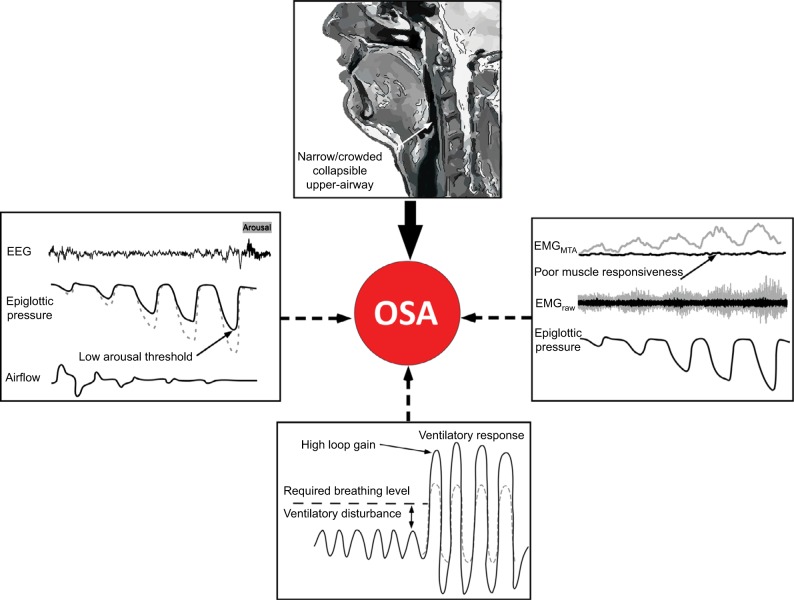
The prevalence of obstructive sleep apnea (OSA) continues to rise. So too do the health, safety, and economic consequences. On an individual level, the causes and consequences of OSA can vary substantially between patients. In recent years, four key contributors to OSA pathogenesis or "phenotypes" have been characterized. These include a narrow, crowded, or collapsible upper airway "anatomical compromise" and "non-anatomical" contributors such as ineffective pharyngeal dilator muscle function during sleep, a low threshold for arousal to airway narrowing during sleep, and unstable control of breathing (high loop gain). Each of these phenotypes is a target for therapy. This review summarizes the latest knowledge on the different contributors to OSA with a focus on measurement techniques including emerging clinical tools designed to facilitate translation of new cause-driven targeted approaches to treat OSA. The potential for some of the specific pathophysiological causes of OSA to drive some of the key symptoms and consequences of OSA is also highlighted.
Overview
The claims and statistics of this paper haven’t been checked yet.
Run the check — free, no account needed. It reads the open-access text and checks the sources it cites.
Integrity signals
Registry
Cites
Statistics
Figures
Verification
Claims the paper makes
The claims and statistics of this paper haven’t been checked yet.
Run the check — free, no account needed. It reads the open-access text and checks the sources it cites.
Claims other papers make about this one
Each is checked against this paper’s own text: does it say what the citing paper says it does?
The source backs part of the claim, not all of it — or backs it for a different population, condition or comparison.
In the citing paper
“Alternatively, insomnia disorder may be associated with a reduced respiratory arousal threshold, which may predispose patients to prematurely awaken to respiratory events [16,105].”
From Co-morbid insomnia and sleep apnea (COMISA): Prevalence, consequences, methodological considerations, and recent randomized controlled trials (2019) · cites this paper as [16]
Experimental — accuracy not yet measuredThe cited source says what the claim attributes to it.
In the citing paper
“OSA is thought to result from a combination of anatomical (e.g., a narrow pharyngeal airway), and non-anatomical factors (e.g., impaired upper airway muscle function, low arousal threshold, and unstable control of breathing) [16].”
From Co-morbid insomnia and sleep apnea (COMISA): Prevalence, consequences, methodological considerations, and recent randomized controlled trials (2019) · cites this paper as [16]
Self-check
Percentages, sums and reported differences re-computed from the paper’s own numbers.
The self-check needs the paper’s full text.
Citations
Cites
Increased Prevalence of Sleep-Disordered Breathing in Adults
Peppard, P. E., Young, T., Barnet, J. H., Palta, M., Hagen, E. W., & Hla, K. M. (2013) · American Journal of Epidemiology · doi:10.1093/aje/kws342 · Cited by 4,618
Prevalence of sleep-disordered breathing in the general population: the HypnoLaus study
Heinzer, R., Vat, S., Marques-Vidal, P., Marti-Soler, H., Andries, D., Tobback, N., Mooser, V, Preisig, M., Malhotra, A., Waeber, G., et al. (2015) · The Lancet Respiratory Medicine · doi:10.1016/s2213-2600(15)00043-0 · Cited by 2,481
The epidemiology of adult obstructive sleep apnea
Punjabi, N. M. (2008) · Proceedings of the American Thoracic Society · doi:10.1513/pats.200709-155mg · Cited by 2,361
Pathogenesis of upper airway occlusion during sleep
Remmers, J. E., deGroot, W. J., Sauerland, E. K., & Anch, A. M. (1978) · Journal of Applied Physiology · doi:10.1152/jappl.1978.44.6.931 · Cited by 1,883
Upper-Airway Stimulation for Obstructive Sleep Apnea
Strollo, P. J., Soose, R. J., Maurer, J. T., de Vries, N., Cornelius, J., Froymovich, O., Hanson, R. D., Padhya, T. A., Steward, D. L., Gillespie, M. B., et al. (2014) · New England Journal of Medicine · doi:10.1056/nejmoa1308659 · Cited by 1,299
Defining Phenotypic Causes of Obstructive Sleep Apnea. Identification of Novel Therapeutic Targets
Eckert, D. J., White, D. P., Jordan, A. S., Malhotra, A., & Wellman, A. (2013) · American Journal of Respiratory and Critical Care Medicine · doi:10.1164/rccm.201303-0448oc · Cited by 1,173
Self-citationCollop, N. A., Anderson, W. M., Boehlecke, B. A., Claman, D. M., Goldberg, R., Gottlieb, D. J., Hudgel, D. W., Sateia, M. J., & Schwab, R. (2007) · PubMed Central · Cited by 1,082
Schwab, R. J., Pasirstein, M., Pierson, R., Mackley, A., Hachadoorian, R., Arens, R., Maislin, G., & Pack, A. I. (2003) · American Journal of Respiratory and Critical Care Medicine · doi:10.1164/rccm.200208-866oc · Cited by 952
Drager, L. F., Togeiro, S. M., Polotsky, V. Y., & Lorenzi-Filho, G. (2013) · Journal of the American College of Cardiology · doi:10.1016/j.jacc.2013.05.045 · Cited by 886
Excess weight and sleep-disordered breathing
Young, T., Peppard, P. E., & Taheri, S. (2005) · Journal of Applied Physiology · doi:10.1152/japplphysiol.00587.2005 · Cited by 854
Cited by
Sorted by citations
Alzheimer's Disease in Patients with Obstructive Sleep Apnea Syndrome.
Siachpazidou, D. I., Stavrou, V. T., Astara, K., Pastaka, C., Gogou, E., Hatzoglou, C., Economou, N.-T., & Gourgoulianis, K. I. (2020) · PubMed Central · Cited by 609
Dynamics of ACTH and Cortisol Secretion and Implications for Disease
Lightman, S. L., Birnie, M. T., & Conway-Campbell, B. L. (2020) · Endocrine Reviews · doi:10.1210/endrev/bnaa002 · Cited by 439
Sweetman, A., Lack, L., & Bastien, C. (2019) · Brain Sciences · doi:10.3390/brainsci9120371 · Cited by 253
“, impaired upper airway muscle function, low arousal threshold, and unstable control of breathing) [16].”
BackgroundBi-directional relationships between co-morbid insomnia and sleep apnea (COMISA)
Sweetman, A., Lack, L., McEvoy, R. D., Smith, S., Eckert, D. J., Osman, A., Carberry, J. C., Wallace, D., Nguyen, P. D., & Catcheside, P. (2021) · Sleep Medicine Reviews · doi:10.1016/j.smrv.2021.101519 · Cited by 203
Self-citationObstructive sleep apnea: focus on myofunctional therapy
de Felício, C. M., Dias, F. V. D. S., & Trawitzki, L. V. V. (2018) · Nature and Science of Sleep · doi:10.2147/nss.s141132 · Cited by 156
Metadata
- Authors
- Osman, A. M., Carter, S. G., Carberry, J. C., & Eckert, D. J.
- Affiliations
- UNSW Sydney (AU)
- Corresponding author
- Eckert, Danny J
- Venue
- Nature and Science of Sleep
- Publisher
- Informa UK Limited
- Published
- 2018-01
- Volume
- Volume 10
- Pages
- 21-34
- ISSN
- 1179-1608 (ISSN-L)
- PMC
- PMC5789079
- OpenAlex
- W2784442221
- Cited by
- 441
- References
- 186
- Citations by year
- 2026: 37 · 2025: 56 · 2024: 49 · 2023: 56 · 2022: 64 · 2021: 76 · 2020: 49 · 2019: 44 · 2018: 9
- FWCI
- 5.85
- Type
- journal article
- Language
- en
- Open access
- gold
- doi.org
- Licence
- Locations
- Nature and Science of Sleep (publishedVersion, cc-by-nc)
- PubMed Central (publishedVersion, cc-by-nc)
- PubMed (publishedVersion)
- UNSWorks (University of New South Wales, Sydney, Australia) (submittedVersion, cc-by-nc-nd)
- Dove Medical Press (Taylor and Francis Group) (submittedVersion, cc-by-nc)
- Topics
- Health Sciences › Medicine › Physiology (medical) › Obstructive Sleep Apnea Research
- Life Sciences › Neuroscience › Endocrine and Autonomic Systems › Neuroscience of respiration and sleep
- Social Sciences › Psychology › Experimental and Cognitive Psychology › Sleep and related disorders
- Keywords
- obstructive sleep apnea, OSA pathogenesis, OSA phenotypes, upper airway anatomy, arousal threshold, loop gain, targeted therapy
About these checks
Registry, retraction, Cites and statistics results are computed, not judged, with one exception: a printed reference that no index lists is read once by a model before it counts as a citation error. Claims are extracted by a model and kept only when their quote appears verbatim in the text; each cited source is then checked, with a model’s help, for whether it says what the claim relies on. Those verdicts can be wrong — read the claim’s quote and the cited source before relying on one.
Statistics and arithmetic are recomputed, not judged.
Confidence bands are shown only for fields calibrated on our annotated test set. None is calibrated yet, so no band is shown.
What counts as a claim ›How we measure confidence ›How we check this ›
Are you an author, or do you see a problem with this page? Request a review
