

The DOI matches its Crossref record, with a small difference in title or year.
Brain abscess caused by Nocardia thailandica infection in systemic lupus erythematosus patient with steroid therapy
Abstract
Background: Nocardia is an aerobic Gram-negative bacillus belonging to actinomycetes and has been reported to be an infectious disease in healthy individuals. However, more than 60% have some underlying illnesses and are said to be opportunistic infections. Case Description: The case was a 69-year-old man who had been on long-term steroids for systemic lupus erythematosus. He developed with nausea and gait disturbance and was suspected of having a brain abscess by imaging. Abscess drainage detects actinomycetes that appear to belong to the genus Nocardia and brain abscess by Nocardia thailandica using matrix-assisted laser desorption/ionization time-of-flight mass analysis (MALDITOFMS) I was diagnosed. He died during the course of the treatment, and his organs did not detect his N. thailandica at necropsy, so he concluded that bacterial death from long-term administration of antibiotics was the cause of death. Conclusion: N. thailandica is a very rare bacterium belonging to Nocardia asteroides and is said to easily form brain lesions. In immunocompromised patients, prophylaxis with antibiotics and detailed examination of lung lesions for surveillance were considered necessary. This paper is the first report of a brain abscess caused by N. thailandica, including a review of the literature.
Overview
The claims and statistics of this paper haven’t been checked yet.
Run the check — free, no account needed. It reads the open-access text and checks the sources it cites.
Integrity signals
Registry
Cites
Statistics
Figures
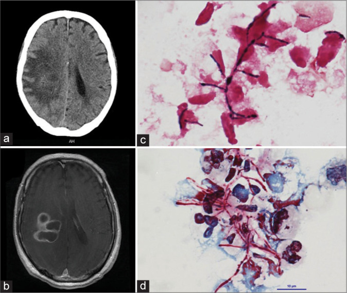
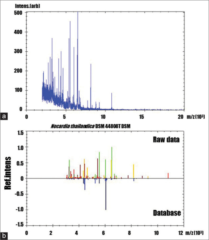
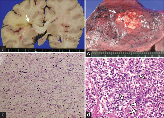
Verification
Claims the paper makes
The claims and statistics of this paper haven’t been checked yet.
Run the check — free, no account needed. It reads the open-access text and checks the sources it cites.
Claims other papers make about this one
Each is checked against this paper’s own text: does it say what the citing paper says it does?
The cited source says what the claim attributes to it.
In the citing paper
“Nocardia can cause infectious disease in healthy individuals, but more than 60% of those affected have some underlying illness [42].”
From Metaproteomic analysis of King Ghezo tomb wall (Abomey, Benin) confirms 19th century voodoo sacrifices (2024) · cites this paper as [42]
The check wasn’t confident either way, so it gives no verdict — never disputed.
In the citing paper
“Although N. thailandica has not previously been reported in Africa, it appears to be distributed worldwide, having been detected in the USA, Japan, Iran, and India.”
From Metaproteomic analysis of King Ghezo tomb wall (Abomey, Benin) confirms 19th century voodoo sacrifices (2024) · cites this paper as [42]
Experimental — accuracy not yet measured
Self-check
Percentages, sums and reported differences re-computed from the paper’s own numbers.
The self-check needs the paper’s full text.
Citations
Cites
No references could be resolved yet.
Research works printed but not linked: 10
- Beaman, 1976, Nocardial infections in the United States, 1972-1974, J Infect Dis, 134, 286, 10.1093/infdis/134.3.286 · DOI
- Edwards, 1989, Isolation and direct complete nucleotide determination of entire genes. Characterization of a gene coding for 16S ribosomal RNA, Nucleic Acids Res, 17, 7843, 10.1093/nar/17.19.7843 · DOI
- Jodlowski, 2007, Linezolid for the treatment of Nocardia spp. infections, Ann Pharmacother, 41, 1694, 10.1345/aph.1K196 · DOI
- Kageyama, 2004, Nocardial infections in Japan from 1992 to 2001, including the first report of infection by Nocardia transvalensis, Eur J Epidemiol, 19, 383, 10.1023/B:EJEP.0000024706.02325.c0 · DOI
- Kageyama, 2004, Nocardia testaceus sp. Nov. and Nocardia senatus sp. Nov., isolated from patients in Japan, Microbiol Immunol, 48, 271, 10.1111/j.1348-0421.2004.tb03523.x · DOI
- Kurahara, 2014, Pulmonary nocardiosis: A clinical analysis of 59 cases, Respir Investig, 52, 160, 10.1016/j.resinv.2013.09.004 · DOI
- Malincarne, 2002, Primary brain abscess with Nocardia farcinica in an immunocompetent patient, Clin Neurol Neurosurg, 104, 132, 10.1016/S0303-8467(01)00201-3 · DOI
- Mamelak, 1994, Nocardial brain abscess: Treatment strategies and factors influencing outcome, Neurosurgery, 35, 622, 10.1227/00006123-199410000-00007 · DOI
- McBride, 2010, Mandell, Douglas, and Bennett’s principles and practice of infectious diseases 7th edition, 7, 218
- Torack, 1957, Fungus infections associated with antibiotic and steroid therapy, Am J Med, 22, 872, 10.1016/0002-9343(57)90023-2 · DOI
Cited by
Charlier, P., Bourdin, V., N'Dah, D., Kielbasa, M., Pible, O., & Armengaud, J. (2024) · PROTEOMICS · doi:10.1002/pmic.202400048 · Cited by 8
Metadata
- Authors
- Yagi, R., Ooi, Y., Nonoguchi, N., & Wanibuchi, M.
- Affiliations
- Department of Neurosurgery, Osaka Medical and Pharmaceutical University, Takatsuki City, Osaka, Japan,
- Department of Infection Control and Prevention, Osaka Medical and Pharmaceutical University, Takatsuki City, Osaka, Japan.
- Venue
- Surgical Neurology International
- Publisher
- Scientific Scholar
- Published
- 2022-04-08
- Volume
- 13
- Pages
- 126
- ISSN
- 2152-7806
- PMC
- PMC9062894
- Cited by
- 5
- References
- 0
- Language
- en
About these checks
Registry, retraction, Cites and statistics results are computed, not judged, with one exception: a printed reference that no index lists is read once by a model before it counts as a citation error. Claims are extracted by a model and kept only when their quote appears verbatim in the text; each cited source is then checked, with a model’s help, for whether it says what the claim relies on. Those verdicts can be wrong — read the claim’s quote and the cited source before relying on one.
Statistics and arithmetic are recomputed, not judged.
Confidence bands are shown only for fields calibrated on our annotated test set. None is calibrated yet, so no band is shown.
What counts as a claim ›How we measure confidence ›How we check this ›
Are you an author, or do you see a problem with this page? Request a review
How to cite
Cite CoChat for the checks on this page, and the paper for its own findings.
- This page
- CoChat. (2026). Citation check: Brain abscess caused by Nocardia thailandica infection in systemic lupus erythematosus patient with steroid therapy. https://app.cochat.ai/papers/10.25259/sni_78_2022
- The paper
- Yagi, R., Ooi, Y., Nonoguchi, N., & Wanibuchi, M. (2022). Brain abscess caused by Nocardia thailandica infection in systemic lupus erythematosus patient with steroid therapy. Surgical Neurology International. https://doi.org/10.25259/sni_78_2022
Data sources
Bibliographic data: Semantic Scholar, OpenAlex, Crossref.
Contains information from the Semantic Scholar Academic Graph Datasets, made available under the ODC Attribution License (ODC-BY 1.0).
Semantic Scholar’s data is described in: Kinney, R., et al. (2023). The Semantic Scholar Open Data Platform. arXiv:2301.10140.
