PreprintCited by 34Immune Response and InflammationComputational Drug Discovery Methodsvaccines and immunoinformatics approaches

The DOI matches its Crossref record; 1 of the 1 references it cites were found in a registry or an index.

Recent advances in studying Toll-like receptors with the use of computational methods

Open access
Bzówka, Maria, Bagrowska, Weronika, Góra, Artur·2023·Journal of Chemical Information and Modeling

Preprint — published as 10.1021/acs.jcim.3c00419 · Paper page

Abstract

Toll-like receptors (TLRs) are transmembrane proteins which recognise various molecular patterns and activate signalling that triggers the immune response. In this review, our goal was to summarise how, in recent years, various computational solutions contributed to a better understanding of TLRs, regarding both their function and mechanism of action. We updated the recent information about small-molecule modulators and expanded the topic towards next- generation vaccine design, as well as studies of the dynamic nature of TLRs. Also, we underlined problems which remain unsolved.

Overview

The claims and statistics of this paper haven’t been checked yet.

Run the check — free, no account needed. It reads the open-access text and checks the sources it cites.

A few free checks a day · papers already verified open instantly · results are public

Integrity signals

Registry

Crossref · 10.26434/chemrxiv-2023-4zp5t-v3

Cites

1 of 1 references verified0 citation errors · 0 not checkable

Statistics

Figures

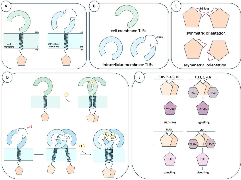
Figure 1 Structural organization and potential Toll-like receptors (TLRs) mechanism of action. (A) The general structure of the TLRs’ monomers. (B) Differences in the TLRs’ LRR domains between the cell membrane and intracellular membrane TLRs. (C) Various orientations (symmetric and asymmetric) of the TIR domain subunits in the TLRs’ TIR dimer. (D) Potential mechanisms of the TLRs activation. The upper panel shows the mechanisms of the cell membrane TLRs activation, while the lower panel presents the mechanisms of the intracellular membrane TLRs containing a Z-loop. (L) indicates the ligand, while the scissors symbol indicates the proteolytic cleavage of the Z-loop. (E) Binding of the adaptor proteins, MyD88 and TRIF, to the respective TLRs’ TIR dimer.
Figure 1 Structural organization and potential Toll-like receptors (TLRs) mechanism of action. (A) The general structure of the TLRs’ monomers. (B) Differences in the TLRs’ LRR domains between the cell membrane and intracellular membrane TLRs. (C) Various orientations (symmetric and asymmetric) of the TIR domain subunits in the TLRs’ TIR dimer. (D) Potential mechanisms of the TLRs activation. The upper panel shows the mechanisms of the cell membrane TLRs activation, while the lower panel presents the mechanisms of the intracellular membrane TLRs containing a Z-loop. (L) indicates the ligand, while the scissors symbol indicates the proteolytic cleavage of the Z-loop. (E) Binding of the adaptor proteins, MyD88 and TRIF, to the respective TLRs’ TIR dimer.CC BY · PubMed Central
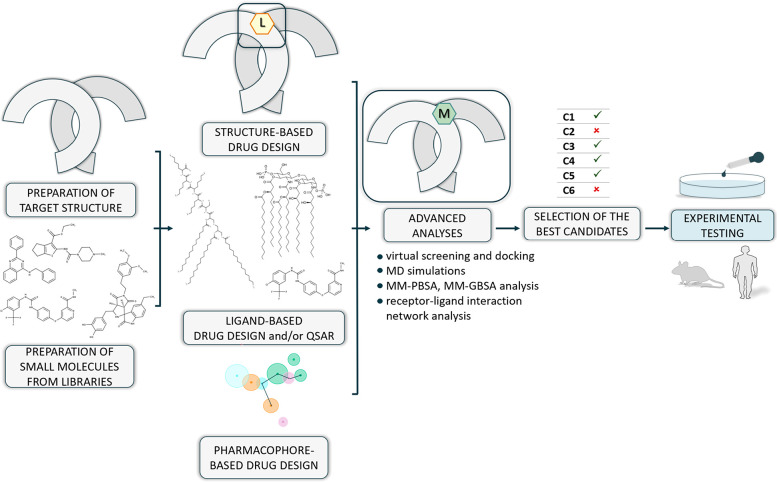
Figure 2 A general protocol for small-molecule modulators design targeting the LRR domain of TLRs. The subunits of the LRR domain are colored gray, indicating both TLRs located in the cell membrane and TLRs located in the intracellular compartments. (L) indicates the location of the ligand binding site, while (M) points out the designed modulator and (C) the selected candidate(s).
Figure 2 A general protocol for small-molecule modulators design targeting the LRR domain of TLRs. The subunits of the LRR domain are colored gray, indicating both TLRs located in the cell membrane and TLRs located in the intracellular compartments. (L) indicates the location of the ligand binding site, while (M) points out the designed modulator and (C) the selected candidate(s).CC BY · PubMed Central
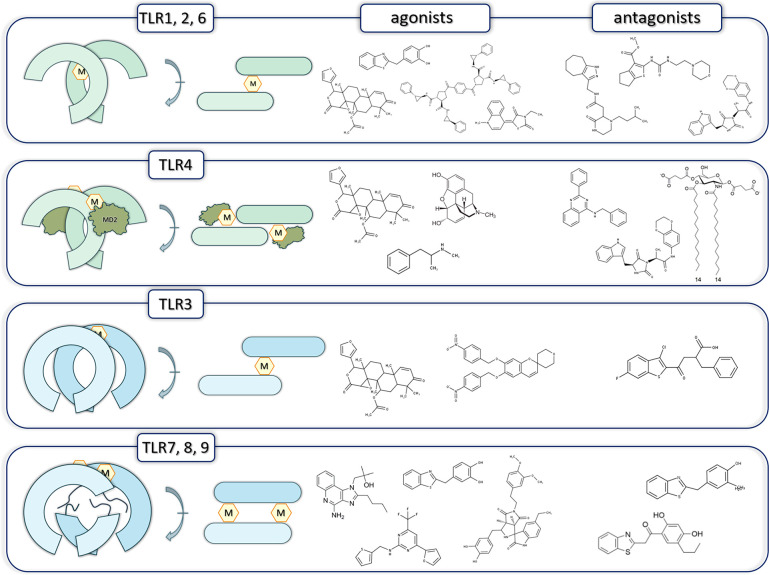
Figure 3 Examples of scaffolds of small-molecule modulators targeting the LRR domain of TLRs. The left panel shows the approximate location of small-molecule modulators (M) with respect to the LRR subunits of the TLR dimers described in this review. Agonists are presented on the middle panel, while antagonists are on the right panel. TLR4 was shown with the associated myeloid differentiation factor 2 (MD2). Please note that one of the agonists’ scaffolds is shown for more than one TLRs. This indicates the possibility of targeting both surface- and endosomal-located TLRs by a given modulator.
Figure 3 Examples of scaffolds of small-molecule modulators targeting the LRR domain of TLRs. The left panel shows the approximate location of small-molecule modulators (M) with respect to the LRR subunits of the TLR dimers described in this review. Agonists are presented on the middle panel, while antagonists are on the right panel. TLR4 was shown with the associated myeloid differentiation factor 2 (MD2). Please note that one of the agonists’ scaffolds is shown for more than one TLRs. This indicates the possibility of targeting both surface- and endosomal-located TLRs by a given modulator.CC BY · PubMed Central
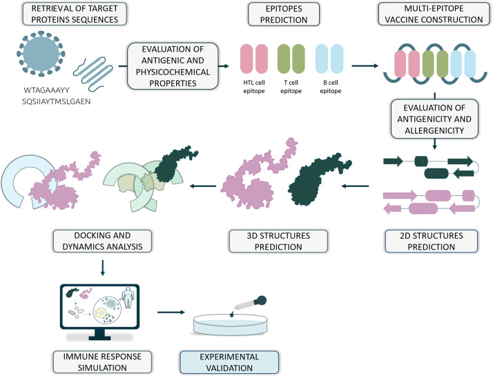
Figure 4 General protocol for next-generation multiepitope vaccine design. The ability of binding different epitopes (shown as dark green and pink shapes, respectively) to LRR subunits of the TLRs located both in the cell membrane (light green) and in the intracellular membrane (light blue) has been shown.
Figure 4 General protocol for next-generation multiepitope vaccine design. The ability of binding different epitopes (shown as dark green and pink shapes, respectively) to LRR subunits of the TLRs located both in the cell membrane (light green) and in the intracellular membrane (light blue) has been shown.CC BY · PubMed Central
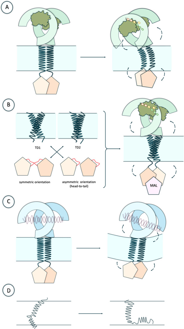
Figure 5 Examples of potential dynamical changes of TLRs observed in cited studies. (A and B) Structural transitions that the particular domains of TLR4 may exhibit (based on Patra et al.116 and Matamoros-Recio et al.117 works). TLR4 was shown with the associated myeloid differentiation factor 2 (MD2) and with the bound lipopolysaccharide LPS (C) Structural rearrangements of TLR3 domains and membrane (based on Patra et al.118 study). TLR3 was shown bound with dsRNA. (D) Differences in the structural organization of the transmembrane helix (TM) and cytoplasmic juxtamembrane (JM) regions that may occur in TLRs (based on Kornilov et al.34 work).
Figure 5 Examples of potential dynamical changes of TLRs observed in cited studies. (A and B) Structural transitions that the particular domains of TLR4 may exhibit (based on Patra et al.116 and Matamoros-Recio et al.117 works). TLR4 was shown with the associated myeloid differentiation factor 2 (MD2) and with the bound lipopolysaccharide LPS (C) Structural rearrangements of TLR3 domains and membrane (based on Patra et al.118 study). TLR3 was shown bound with dsRNA. (D) Differences in the structural organization of the transmembrane helix (TM) and cytoplasmic juxtamembrane (JM) regions that may occur in TLRs (based on Kornilov et al.34 work).CC BY · PubMed Central
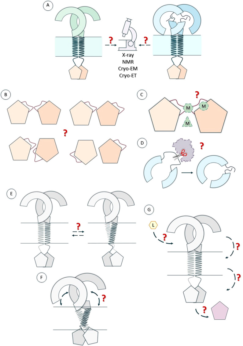
Figure 6 Areas in TLR research that still require further development. (A) Experimental verification of the predicted structures. (B) Studying the orientation of the subunits of the TIR domain dimers of TLRs. (C) Designing small-molecule modulators (M) targeting the TIR domain of TLRs. (D) Studying the proteolytic cleavage of the Z-loop in TLR7–9. (E) Analyzing potential changes in the subunits dynamics in TLRs. (F) Analyzing the conformational changes and structural rearrangements in both TLR receptors and bilayer membrane. (G) Studying the whole process of ligand recognition through the signaling cascade to the immune response.
Figure 6 Areas in TLR research that still require further development. (A) Experimental verification of the predicted structures. (B) Studying the orientation of the subunits of the TIR domain dimers of TLRs. (C) Designing small-molecule modulators (M) targeting the TIR domain of TLRs. (D) Studying the proteolytic cleavage of the Z-loop in TLR7–9. (E) Analyzing potential changes in the subunits dynamics in TLRs. (F) Analyzing the conformational changes and structural rearrangements in both TLR receptors and bilayer membrane. (G) Studying the whole process of ligand recognition through the signaling cascade to the immune response.CC BY · PubMed Central

Verification

Claims the paper makes

The claims and statistics of this paper haven’t been checked yet.

Run the check — free, no account needed. It reads the open-access text and checks the sources it cites.

A few free checks a day · papers already verified open instantly · results are public

Self-check

Percentages, sums and reported differences re-computed from the paper’s own numbers.

The self-check needs the paper’s full text.

Citations

Cites

Metadata

Authors
Bzówka, M., Bagrowska, W., & Góra, A.
Affiliations
Silesian University of Technology (PL)
Corresponding author
Bzówka, Maria
Venue
Journal of Chemical Information and Modeling
Published
2023-03-13
Volume
63
Issue
12
Pages
3669-3687
ISSN
1549-9596 (ISSN-L)
OpenAlex
W4379600654
Cited by
34
References
1
Citations by year
2026: 13 · 2025: 11 · 2024: 9 · 2023: 1
FWCI
0.94
Type
journal article
Language
en
Open access
Locations
  • Journal of Chemical Information and Modeling (publishedVersion, cc-by)
  • ChemRxiv (acceptedVersion, cc-by)
  • PubMed Central (publishedVersion, cc-by)
  • PubMed (publishedVersion)
Funders
Ministerstwo Edukacji i Nauki (DI2018 014148 / 0141/DIA/2019/48)
Topics
  • Life Sciences › Immunology and Microbiology › Immunology › Immune Response and Inflammation
  • Life Sciences › Pharmacology, Toxicology and Pharmaceutics › Drug Discovery › Computational Drug Discovery Methods
  • Life Sciences › Biochemistry, Genetics and Molecular Biology › Molecular Biology › vaccines and immunoinformatics approaches
Keywords
Toll-like receptors, computational methods, Toll-like receptor signaling, pattern recognition, small molecule modulators, vaccine design, innate immunity
MeSH terms
Signal Transduction, Toll-Like Receptors

About these checks

Registry, retraction, Cites and statistics results are computed, not judged, with one exception: a printed reference that no index lists is read once by a model before it counts as a citation error. Claims are extracted by a model and kept only when their quote appears verbatim in the text; each cited source is then checked, with a model’s help, for whether it says what the claim relies on. Those verdicts can be wrong — read the claim’s quote and the cited source before relying on one.

Statistics and arithmetic are recomputed, not judged.

Confidence bands are shown only for fields calibrated on our annotated test set. None is calibrated yet, so no band is shown.

What counts as a claim ›How we measure confidence ›How we check this ›

Are you an author, or do you see a problem with this page? Request a review