

The DOI matches its Crossref record; 18 of the 18 references it cites were found in a registry or an index.
Joubert Syndrome and Related Disorders: Spectrum of Neuroimaging Findings in 75 Patients
Open accessAbstract
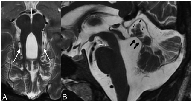
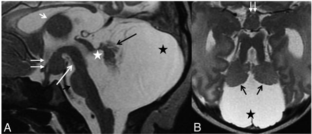
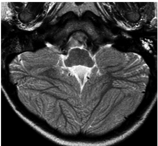
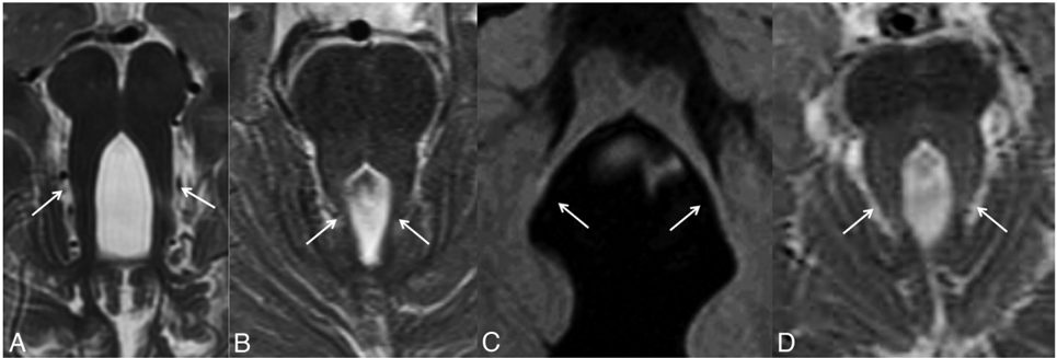
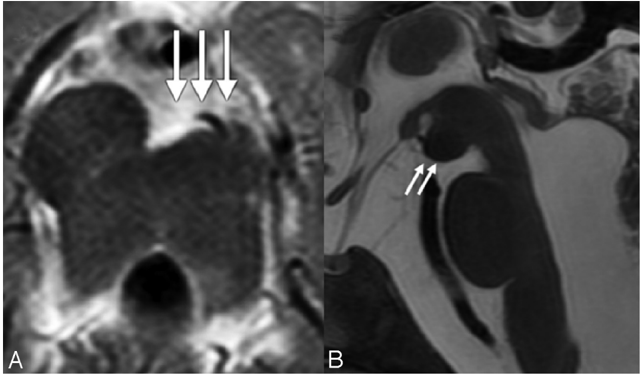
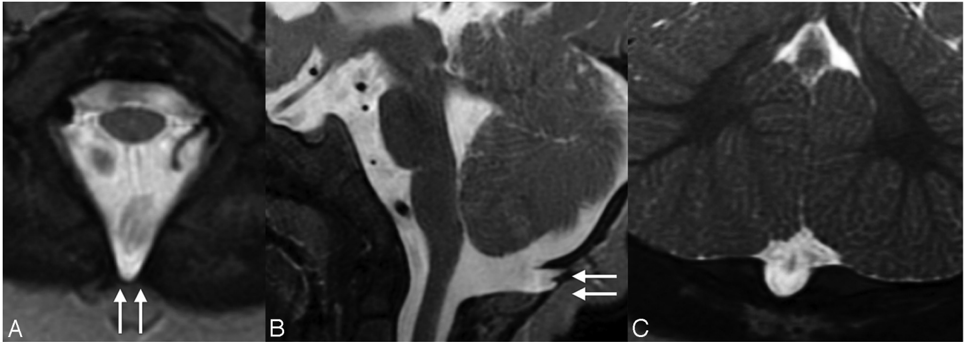
VH and MTS are the neuroimaging hallmarks of JSRD. We aimed to look at the full spectrum of neuroimaging findings in JSRD and reviewed the MR imaging of 75 patients with JSRD, including 13 siblings and 4 patients with OFD VI. All patients had VH and enlargement of the fourth ventricle. The degree of VH and the form of the MTS were variable. In most patients, the cerebellar hemispheres were normal and the PF was enlarged. Brain stem morphology was abnormal in 30% of the patients. Supratentorial findings included hippocampal malrotation, callosal dysgenesis, migration disorders, cephaloceles, and ventriculomegaly. All patients with OFD VI had a similar pattern, including HH in 2 patients. No neuroimaging-genotype correlation could be found. The wide neuroimaging spectrum in our patients supports the heterogeneity of JSRD. Neuroimaging differences in siblings represent intrafamilial heterogeneity. Due to the absence of a correlation with genotype, neuroimaging findings are of limited value in classifying patients with JSRD.
Overview
The claims and statistics of this paper haven’t been checked yet.
Run the check — free, no account needed. It reads the open-access text and checks the sources it cites.
Integrity signals
Registry
Cites
Statistics
Figures
Verification
Claims the paper makes
The claims and statistics of this paper haven’t been checked yet.
Run the check — free, no account needed. It reads the open-access text and checks the sources it cites.
Claims other papers make about this one
Each is checked against this paper’s own text: does it say what the citing paper says it does?
In the citing paper
“Indeed several of the monogenic causes of ASD, including TSC [56] and Joubert Syndrome [57, 58] show manifestations of abnormal axon path finding or migration abnormalities that are known to lead to alterations in brain structural connectivity.”
From Genetics of autism spectrum disorders (2011) · cites this paper as [57]
Self-check
Percentages, sums and reported differences re-computed from the paper’s own numbers.
The self-check needs the paper’s full text.
Citations
Cites
Familial agenesis of the cerebellar vermis
Joubert, M., Eisenring, J.-J., Preston, J., & Andermann, F. (1969) · Neurology · doi:10.1212/wnl.19.9.813 · Cited by 651
Joubert Syndrome and related disorders
Brancati, F., Dallapiccola, B., & Valente, E. M. (2010) · Orphanet Journal of Rare Diseases · doi:10.1186/1750-1172-5-20 · Cited by 468
"Joubert Syndrome" Revisited: Key Ocular Motor Signs With Magnetic Resonance Imaging Correlation
Maria, B. L., Hoang, K. B. N., Tusa, R. J., Mancuso, A., Hamed, L. M., Quisling, R. G., ten Hove, M., Fennell, E. B., Booth‐Jones, M., Ringdahl, D. M., et al. (1997) · Journal of Child Neurology · doi:10.1177/088307389701200703 · Cited by 330
Mutations in the AHI1 Gene, Encoding Jouberin, Cause Joubert Syndrome with Cortical Polymicrogyria
Dixon‐Salazar, T., Silhavy, J. L., Marsh, S., Louie, C. M., Scott, L. C., Gururaj, A., Al‐Gazali, L., Al-Tawari, A. A., Kayserili, H., Sztriha, L., et al. (2004) · The American Journal of Human Genetics · doi:10.1086/425985 · Cited by 293
Clinical and molecular features of Joubert syndrome and related disorders
Parisi, M. A. (2009) · American Journal of Medical Genetics Part C Seminars in Medical Genetics · doi:10.1002/ajmg.c.30229 · Cited by 268
Molar tooth sign of the midbrain–hindbrain junction: Occurrence in multiple distinct syndromes
Gleeson, J. G., Keeler, L. C., Parisi, M. A., Marsh, S., Chance, P. F., Glass, I. A., Graham, J. M., Maria, B. L., Barkovich, A. J., & Dobyns, W. B. (2003) · American Journal of Medical Genetics Part A · doi:10.1002/ajmg.a.20437 · Cited by 246
Joubert Syndrome: Insights Into Brain Development, Cilium Biology, and Complex Disease
Doherty, D. (2009) · Seminars in Pediatric Neurology · doi:10.1016/j.spen.2009.06.002 · Cited by 182
Diffusion Tensor Imaging in Joubert Syndrome
Poretti, A., Boltshauser, E., Loenneker, T., Valente, E. M., Brancati, F., Il'yasov, K. A., & Huisman, T. A. G. M. (2007) · American Journal of Neuroradiology · doi:10.3174/ajnr.a0703 · Cited by 147
Self-citationJoubert, M., Eisenring, J.-J., Robb, J. P., & Andermann, F. (1999) · Journal of Child Neurology · doi:10.1177/088307389901400902 · Cited by 129
Quisling, R. G., Barkovich, A. J., & Maria, B. L. (1999) · Journal of Child Neurology · doi:10.1177/088307389901401002 · Cited by 98
Cited by
Sorted by citations
Genetics of autism spectrum disorders
Geschwind, D. H. (2011) · Trends in Cognitive Sciences · doi:10.1016/j.tics.2011.07.003 · Cited by 696
Joubert syndrome: congenital cerebellar ataxia with the molar tooth
Romani, M., Micalizzi, A., & Valente, E. M. (2013) · The Lancet Neurology · doi:10.1016/s1474-4422(13)70136-4 · Cited by 405
Joubert syndrome: a model for untangling recessive disorders with extreme genetic heterogeneity
Bachmann‐Gagescu, R., Dempsey, J. C., Phelps, I. G., O’Roak, B. J., Knutzen, D. M., Rue, T. C., Ishak, G. E., Isabella, C. R., Gorden, N. T., Adkins, J. R., et al. (2015) · Journal of Medical Genetics · doi:10.1136/jmedgenet-2015-103087 · Cited by 298
Primary cilia in neurodevelopmental disorders
Valente, E. M., Rosti, R. O., Gibbs, E., & Gleeson, J. G. (2014) · Nature Reviews Neurology · doi:10.1038/nrneurol.2013.247 · Cited by 254
Congenital Abnormalities of the Posterior Fossa
Bosemani, T., Orman, G., Boltshauser, E., Tekes, A., Huisman, T. A. G. M., & Poretti, A. (2015) · Radiographics · doi:10.1148/rg.351140038 · Cited by 193
Self-citationMidbrain and hindbrain malformations: advances in clinical diagnosis, imaging, and genetics
Doherty, D., Millen, K. J., & Barkovich, A. J. (2013) · The Lancet Neurology · doi:10.1016/s1474-4422(13)70024-3 · Cited by 140
Cerebellar hypoplasia: Differential diagnosis and diagnostic approach
Poretti, A., Boltshauser, E., & Doherty, D. (2014) · American Journal of Medical Genetics Part C Seminars in Medical Genetics · doi:10.1002/ajmg.c.31398 · Cited by 140
Self-citationGenotype–phenotype correlates in Joubert syndrome: A review
Gana, S., Serpieri, V., & Valente, E. M. (2022) · American Journal of Medical Genetics Part C Seminars in Medical Genetics · doi:10.1002/ajmg.c.31963 · Cited by 122
Cilia, ciliopathies and hedgehog-related forebrain developmental disorders
Andreu‐Cervera, A., Catala, M., & Schneider‐Maunoury, S. (2020) · Neurobiology of Disease · doi:10.1016/j.nbd.2020.105236 · Cited by 118
Tuz, K., Bachmann‐Gagescu, R., O’Day, D. R., Hua, K., Isabella, C. R., Phelps, I. G., Stolarski, A. E., O’Roak, B. J., Dempsey, J. C., Lourenço, C. M., et al. (2013) · The American Journal of Human Genetics · doi:10.1016/j.ajhg.2013.11.019 · Cited by 112
Juric‐Sekhar, G., Adkins, J. R., Doherty, D., & Hevner, R. F. (2012) · Acta Neuropathologica · doi:10.1007/s00401-012-0951-2 · Cited by 92
Weghe, J. C. V. D., Rusterholz, T. D. S., Latour, B. L., Grout, M. E., Aldinger, K. A., Shaheen, R., Dempsey, J. C., Maddirevula, S., Cheng, Y.-H. H., Phelps, I. G., et al. (2017) · The American Journal of Human Genetics · doi:10.1016/j.ajhg.2017.05.010 · Cited by 84
Delineation and Diagnostic Criteria of Oral-Facial-Digital Syndrome Type VI
Poretti, A., Vitiello, G., Hennekam, R. C. M., Arrigoni, F., Bertini, E., Borgatti, R., Brancati, F., D’Arrigo, S., Faravelli, F., Giordano, L., et al. (2012) · Orphanet Journal of Rare Diseases · doi:10.1186/1750-1172-7-4 · Cited by 83
Self-citationPoretti, A., Snow, J., Summers, A. C., Tekes, A., Huisman, T. A. G. M., Aygün, N., Carson, K. A., Doherty, D., Parisi, M. A., Toro, C., et al. (2017) · Journal of Medical Genetics · doi:10.1136/jmedgenet-2016-104425 · Cited by 76
Self-citationBachmann‐Gagescu, R., Ishak, G. E., Dempsey, J. C., Adkins, J. R., O’Day, D. R., Phelps, I. G., Gunay‐Aygun, M., Kline, A. D., Szczałuba, K., Martorell, L., et al. (2012) · Journal of Medical Genetics · doi:10.1136/jmedgenet-2011-100552 · Cited by 73
Diffusion tensor imaging and fiber tractography in brain malformations
Poretti, A., Meoded, A., Rossi, A., Raybaud, C., & Huisman, T. A. G. M. (2013) · Pediatric Radiology · doi:10.1007/s00247-012-2428-9 · Cited by 71
Self-citationThe multifaceted roles of primary cilia in the development of the cerebral cortex
Hasenpusch-Theil, K., & Theil, T. (2021) · Frontiers in Cell and Developmental Biology · doi:10.3389/fcell.2021.630161 · Cited by 66
Brooks, B. P., Zein, W. M., Thompson, A., Mokhtarzadeh, M., Doherty, D., Parisi, M. A., Glass, I. A., Malicdan, M. C. V., Vilboux, T., Vemulapalli, M., et al. (2018) · Ophthalmology · doi:10.1016/j.ophtha.2018.05.026 · Cited by 65
The genetics of cerebellar malformations
Aldinger, K. A., & Doherty, D. (2016) · Seminars in Fetal and Neonatal Medicine · doi:10.1016/j.siny.2016.04.008 · Cited by 63
Mutations in CEP120 cause Joubert syndrome as well as complex ciliopathy phenotypes
Roosing, S., Romani, M., Misra‐Isrie, M., Rosti, R. Ö., Micalizzi, A., Musaev, D., Mazza, T., Al‐Gazali, L., Altunoğlu, U., Boltshauser, E., et al. (2016) · Journal of Medical Genetics · doi:10.1136/jmedgenet-2016-103832 · Cited by 65
Self-citationThe essential role of primary cilia in cerebral cortical development and disorders
Liu, S., Trupiano, M. X., Simon, J. M., Guo, J., & Anton, E. S. (2021) · Current topics in developmental biology/Current Topics in Developmental Biology · doi:10.1016/bs.ctdb.2020.11.003 · Cited by 64
Micalizzi, A., Poretti, A., Romani, M., Ginevrino, M., Mazza, T., Aiello, C., Zanni, G., Baumgartner, B., Borgatti, R., Brockmann, K., et al. (2016) · European Journal of Human Genetics · doi:10.1038/ejhg.2016.19 · Cited by 62
Self-citationCongenital basis of posterior fossa anomalies
Cotes, C., Bonfante, E. E., Lazor, J. W., Jadhav, S. P., Caldas, M., Swischuk, L., & Riascos, R. (2015) · The Neuroradiology Journal · doi:10.1177/1971400915576665 · Cited by 58
Ataxia, Intellectual Disability, and Ocular Apraxia with Cerebellar Cysts: A New Disease?
Poretti, A., Häusler, M., von Moers, A., Baumgartner, B., Zerres, K., Klein, A., Aiello, C., Moro, F., Zanni, G., Santorelli, F. M., et al. (2013) · The Cerebellum · doi:10.1007/s12311-013-0521-8 · Cited by 58
Self-citationChung, E. M., Conran, R. M., Schroeder, J. W., Rohena-Quinquilla, I. R., & Rooks, V. J. (2014) · Radiographics · doi:10.1148/rg.341135179 · Cited by 56
Gazea, M., Tasouri, E., Tolve, M., Bosch, V., Kabanova, A., Gojak, C., Kurtulmus, B., Novikov, O., Spatz, J. P., Pereira, G., et al. (2015) · Developmental Biology · doi:10.1016/j.ydbio.2015.10.033 · Cited by 59
MKS1 regulates ciliary INPP5E levels in Joubert syndrome
Slaats, G. G. G., Isabella, C. R., Kroes, H. Y., Dempsey, J. C., Gremmels, H., Monroe, G. R., Phelps, I. G., Duran, K. J., Adkins, J. R., Kumar, S. A., et al. (2015) · Journal of Medical Genetics · doi:10.1136/jmedgenet-2015-103250 · Cited by 47
Consensus Paper: Radiological Biomarkers of Cerebellar Diseases
Baldarçara, L., Currie, S., Hadjivassiliou, M., Hoggard, N., Jack, A., Jackowski, A. P., Mascalchi, M., Parazzini, C., Reetz, K., Righini, A., et al. (2014) · The Cerebellum · doi:10.1007/s12311-014-0610-3 · Cited by 48
Diagnostic Approach to Cerebellar Hypoplasia
Accogli, A., Addour-Boudrahem, N., & Srour, M. (2021) · The Cerebellum · doi:10.1007/s12311-020-01224-5 · Cited by 46
Andreu‐Cervera, A., Anselme, I., Karam, A., Laclef, C., Catala, M., & Schneider‐Maunoury, S. (2019) · Journal of Neuroscience · doi:10.1523/jneurosci.2199-18.2019 · Cited by 49
CNS involvement in OFD1 syndrome: a clinical, molecular, and neuroimaging study
Giudice, E. D., Macca, M., Imperati, F., D’Amico, A., Parent, P., Pasquier, L., Layet, V., Lyonnet, S., Darmency, V., Thauvin‐Robinet, C., et al. (2014) · Orphanet Journal of Rare Diseases · doi:10.1186/1750-1172-9-74 · Cited by 47
The Molar Tooth Sign Is Pathognomonic for Joubert Syndrome!
Poretti, A., Boltshauser, E., & Valente, E. M. (2013) · Pediatric Neurology · doi:10.1016/j.pediatrneurol.2013.11.003 · Cited by 40
Self-citationNeonatal Head Ultrasonography Today: A Powerful Imaging Tool!
Orman, G., Benson, J. E., Kweldam, C. F., Bosemani, T., Tekes, A., de Jong, M. R., Seyfert, D., Northington, F. J., Poretti, A., & Huisman, T. A. G. M. (2014) · Journal of Neuroimaging · doi:10.1111/jon.12108 · Cited by 39
Self-citationMagnetic Resonance Imaging of Malformations of Midbrain-Hindbrain
Razek, A. A. K. A., & Castillo, M. (2015) · Journal of Computer Assisted Tomography · doi:10.1097/rct.0000000000000340 · Cited by 38
Pre- and Postnatal Neuroimaging of Congenital Cerebellar Abnormalities
Poretti, A., Boltshauser, E., & Huisman, T. A. G. M. (2015) · The Cerebellum · doi:10.1007/s12311-015-0699-z · Cited by 37
Self-citationCerebellar and Brainstem Malformations
Poretti, A., Boltshauser, E., & Huisman, T. A. G. M. (2016) · Neuroimaging Clinics of North America · doi:10.1016/j.nic.2016.03.005 · Cited by 37
Self-citationTerminology in morphological anomalies of the cerebellum does matter
Poretti, A., & Boltshauser, E. (2015) · Cerebellum & Ataxias · doi:10.1186/s40673-015-0027-x · Cited by 35
Self-citationSerpieri, V., D’Abrusco, F., Dempsey, J. C., Cheng, Y.-H. H., Arrigoni, F., Baker, J. A., Battini, R., Bertini, E., Borgatti, R., Christman, A. K., et al. (2021) · Journal of Medical Genetics · doi:10.1136/jmedgenet-2021-108114 · Cited by 34
Self-citationExpanding the spectrum of congenital anomalies of the diencephalic–mesencephalic junction
Severino, M., Tortora, D., Pistorio, A., Ramenghi, L. A., Napoli, F., Mancardi, M. M., Striano, P., Capra, V., & Rossi, A. (2015) · Neuroradiology · doi:10.1007/s00234-015-1601-x · Cited by 34
Inherited Cerebellar Ataxia in Childhood: A Pattern-Recognition Approach Using Brain MRI
Vedolin, L., Gonzalez, G., de Souza, C. F. M., Lourenço, C. M., & Barkovich, A. J. (2012) · American Journal of Neuroradiology · doi:10.3174/ajnr.a3055 · Cited by 31
Hasenpusch‐Theil, K., Laclef, C., Colligan, M., Fitzgerald, E., Howe, K., Carroll, E., Abrams, S. R., Reiter, J. F., Schneider‐Maunoury, S., & Theil, T. (2020) · eLife · doi:10.7554/elife.58162 · Cited by 30
Joubert syndrome: report of 11 cases
İncecik, F., Hergüner, M. Ö., Altunbaşak, Ş., & Gleeson, J. G. (2012) · The Turkish Journal of Pediatrics · doi:10.24953/turkjpediatr.2012.1696 · Cited by 30
Cerebellar Cysts in Children: a Pattern Recognition Approach
Boltshauser, E., Scheer, I., Huisman, T. A. G. M., & Poretti, A. (2014) · The Cerebellum · doi:10.1007/s12311-014-0633-9 · Cited by 27
Self-citationKlein, J. L., Lemmon, M. E., Northington, F. J., Boltshauser, E., Huisman, T. A. G. M., & Poretti, A. (2016) · Cerebellum & Ataxias · doi:10.1186/s40673-016-0039-1 · Cited by 27
Self-citationClinical Applications of Fetal MRI in the Brain
Nagaraj, U. D., & Kline‐Fath, B. M. (2022) · Diagnostics · doi:10.3390/diagnostics12030764 · Cited by 26
The Primary Cilium and Neuronal Migration
Stoufflet, J., & Caillé, I. (2022) · Cells · doi:10.3390/cells11213384 · Cited by 25
Arrigoni, F., Romaniello, R., Peruzzo, D., Luca, A. D., Parazzini, C., Valente, E. M., Borgatti, R., & Triulzi, F. M. (2017) · American Journal of Neuroradiology · doi:10.3174/ajnr.a5360 · Cited by 24
Nosological delineation of congenital ocular motor apraxia type Cogan: an observational study
Wente, S., Schröder, S., Buckard, J. A., Büttel, H., von Deimling, F., Diener, W., Häussler, M., Hübschle, S., Kinder, S., Kurlemann, G., et al. (2016) · Orphanet Journal of Rare Diseases · doi:10.1186/s13023-016-0486-z · Cited by 24
Self-citationEnokizono, M., Aida, N., Niwa, T., Osaka, H., Naruto, T., Kurosawa, K., Ohba, C., Suzuki, T., Saitsu, H., Goto, T., et al. (2017) · Journal of the Neurological Sciences · doi:10.1016/j.jns.2017.02.065 · Cited by 23
Imaging Spectrum of Cerebellar Pathologies: A Pictorial Essay
Arora, R. (2015) · Polish Journal of Radiology · doi:10.12659/pjr.892878 · Cited by 23
Prenatal diagnosis of Joubert syndrome
Lingling, Z., & Xie, L. (2017) · Medicine · doi:10.1097/md.0000000000008626 · Cited by 21
Boltshauser, E., Huisman, T., & Poretti, A. (2014) · Seminars in Neurology · doi:10.1055/s-0034-1386762 · Cited by 20
Self-citationFourth ventricle index: sonographic marker for severe fetal vermian dysgenesis/agenesis
Haratz, K. K., Shulevitz, S. L., Leibovitz, Z., Lev, D. C., Shalev, J., Tomarkin, M., Malinger, G., Lerman‐Sagie, T., & Gindes, L. (2018) · Ultrasound in Obstetrics and Gynecology · doi:10.1002/uog.19034 · Cited by 18
MKS1 mutations cause Joubert syndrome with agenesis of the corpus callosum
Bader, I., Decker, E., Mayr, J. A., Lunzer, V., Koch, J., Boltshauser, E., Sperl, W., Pietsch, P., Ertl-Wagner, B. B., Bolz, H. J., et al. (2016) · European Journal of Medical Genetics · doi:10.1016/j.ejmg.2016.06.007 · Cited by 18
Self-citationMuñoz‐Estrada, J., & Ferland, R. J. (2019) · Journal of Cell Science · doi:10.1242/jcs.230680 · Cited by 17
The relevance of primary cilia in neurological disorders
Serpieri, V., D’Abrusco, F., & Valente, E. M. (2025) · The Lancet Neurology · doi:10.1016/s1474-4422(25)00226-1 · Cited by 15
Clinical and genetic characteristics of 36 children with Joubert syndrome
Dong, Y., Zhang, K., Wang, J., Jia, T., Yao, H., Zhu, D., Xu, F., Cheng, M., Zhao, S., & Shi, X. (2023) · Frontiers in Pediatrics · doi:10.3389/fped.2023.1102639 · Cited by 15
Horizontal head titubation in infants with J oubert syndrome: a new finding
Poretti, A., Christen, H. ‐., Elton, L., Baumgärtner, M., Korenke, G. C., Sukhudyan, B., Hethey, S., Cross, E. L. A., Steinlin, M. I., & Boltshauser, E. (2014) · Developmental Medicine & Child Neurology · doi:10.1111/dmcn.12489 · Cited by 15
Self-citationFetal and neonatal neurogenetics
Jansen, A., & Keymolen, K. (2019) · Handbook of clinical neurology · doi:10.1016/b978-0-444-64029-1.00005-9 · Cited by 15
Meoded, A., Orman, G., & Huisman, T. A. G. M. (2020) · Seminars in Pediatric Neurology · doi:10.1016/j.spen.2020.100797 · Cited by 15
Self-citationDiffusion Tensor Imaging for Brain Malformations
Huisman, T. A. G. M., Bosemani, T., & Poretti, A. (2014) · Neuroimaging Clinics of North America · doi:10.1016/j.nic.2014.07.004 · Cited by 15
Self-citationA novel HYLS1 homozygous mutation in living siblings with Joubert syndrome
Oka, M., Shimojima, K. Y., Yamamoto, T., Hanaoka, Y., Sato, S., Yasuhara, T., Yoshinaga, H., & Kobayashi, K. (2016) · Clinical Genetics · doi:10.1111/cge.12752 · Cited by 15
Huang, L.-X., Lu, X.-G., Liu, J.-X., Xu, L., Shang, N., Li, G., & OuYang, Y.-C. (2022) · Frontiers in Genetics · doi:10.3389/fgene.2022.1038274 · Cited by 14
Hsu, C. C.-T., Kwan, G. N. C., & Bhuta, S. (2015) · Pediatric Neurology · doi:10.1016/j.pediatrneurol.2015.02.027 · Cited by 14
Papanagnu, E., Klaehn, L. D., Bang, G. M., Ghadban, R., Mohney, B. G., & Brodsky, M. C. (2014) · Journal of American Association for Pediatric Ophthalmology and Strabismus · doi:10.1016/j.jaapos.2014.03.006 · Cited by 13
Uncovering cilia function in glial development
Bear, R. M., & Caspary, T. (2023) · Annals of Human Genetics · doi:10.1111/ahg.12519 · Cited by 13
Practical approach to syndromic pediatric retinal dystrophies
Sadagopan, K. A. (2017) · Current Opinion in Ophthalmology · doi:10.1097/icu.0000000000000404 · Cited by 13
Genetic, chromosomal, and syndromic causes of neural tube defects.
Seidahmed, M. Z., Abdelbasit, O. B., Shaheed, M. M., Al-Hussein, K., Miqdad, A. M., Samadi, A. S., Khalil, M. I., Mardawi, E. A., & Salih, M. A. M. (2014) · PubMed Central · Cited by 12
ACR Appropriateness Criteria® Ataxia
Juliano, A. F. T., Policeni, B. A., Agarwal, V., Burns, J., Bykowski, J., Harvey, H. B., Hoang, J. K., Hunt, C. H., Kennedy, T. A., Moonis, G., et al. (2019) · Journal of the American College of Radiology · doi:10.1016/j.jacr.2019.02.021 · Cited by 12
Role of neuroimaging in the diagnosis of hereditary cerebellar ataxias in childhood
Perucca, G., Leboucq, N., Roubertie, A., Rivier, F., de Champfleur, N. M., Valentini, C., & Bonafé, A. (2016) · Journal of Neuroradiology · doi:10.1016/j.neurad.2016.03.006 · Cited by 11
CNS Malformations in the Newborn
Baranano, K. W., & Burd, I. D. (2022) · Maternal Health Neonatology and Perinatology · doi:10.1186/s40748-021-00136-4 · Cited by 11
Congenital Brain Malformations- Update on Newer Classification and Genetic Basis
Sarma, A., & Pruthi, S. (2022) · Seminars in Roentgenology · doi:10.1053/j.ro.2022.11.004 · Cited by 9
Congenital Anomalies of the Central Nervous System
Halevy, A., Konen, O., & Mimouni-Bloch, A. (2016) · Springer eBooks · doi:10.1007/978-3-319-18096-0_81 · Cited by 9
Fiori, S., Poretti, A., Pannek, K., Punta, R. D., Pasquariello, R., Tosetti, M., Guzzetta, A., Rose, S., Cioni, G., & Battini, R. (2015) · American Journal of Neuroradiology · doi:10.3174/ajnr.a4607 · Cited by 8
Self-citationConte, G., Caschera, L., Parazzini, C., Cinnante, C. M., Izzo, G., Talenti, G., Severino, M., Ormitti, F., Palumbo, G., Pinelli, L., et al. (2020) · European Radiology · doi:10.1007/s00330-019-06538-5 · Cited by 9
Sarma, A., Heck, J. M., Ndolo, J. M., Newton, A. T., & Pruthi, S. (2021) · Pediatric Radiology · doi:10.1007/s00247-020-04953-1 · Cited by 8
The Many Faces of Oral-Facial-Digital Syndrome
Sukarova-Angelovska, E., Angelkova, N., Palcevska-Kocevska, S., & Kočova, M. (2012) · Balkan Journal of Medical Genetics · doi:10.2478/v10034-012-0006-y · Cited by 8
Orocraniofacial findings of a Pediatric Patient with Joubert Syndrome
Goswami, M., Rajwar, A. S., & Verma, M. (2016) · International Journal of Clinical Pediatric Dentistry · doi:10.5005/jp-journals-10005-1394 · Cited by 8
Ma̧dry, J., Szlufik, S., Koziorowski, D., Królicki, L., & Friedman, A. (2017) · Neurologia i Neurochirurgia Polska · doi:10.1016/j.pjnns.2017.08.005 · Cited by 8
Koyama, S., Sato, H., Wada, M., Kawanami, T., Emi, M., & Kato, T. (2017) · BMC Medical Genetics · doi:10.1186/s12881-017-0399-2 · Cited by 7
Gafner, M., Garel, C., Leibovitz, Z., Valence, S., Haratz, K. K., Oegema, R., Mancini, G. M. S., Héron, D., Bueltmann, E., Bürglen, L., et al. (2023) · American Journal of Neuroradiology · doi:10.3174/ajnr.a7805 · Cited by 7
Self-citationNeurocognitive Functions and Behavior in Joubert Syndrome
Poretti, A., & Gerner, G. J. (2016) · Pediatric Neurology Briefs · doi:10.15844/pedneurbriefs-30-12-3 · Cited by 6
Self-citationCase 266: Pontine Tegmental Cap Dysplasia
Cadman, C. J., Fraser, L., & McArthur, C. (2019) · Radiology · doi:10.1148/radiol.2018162304 · Cited by 6
Neuroimaging in cerebellar ataxia in childhood: A review
Serrallach, B. L., Orman, G., Boltshauser, E., Hackenberg, A., Desai, N. K., Kralik, S. F., & Huisman, T. A. G. M. (2022) · Journal of Neuroimaging · doi:10.1111/jon.13017 · Cited by 8
Self-citationAbnormalities of the Central Nervous System Across the Ciliopathy Spectrum
Baker, K., & Beales, P. L. (2012) · Springer eBooks · doi:10.1007/978-94-007-5808-7_9 · Cited by 6
Joubert Syndrome: Imaging Features and Illustration of a Case
Arora, R. (2014) · Polish Journal of Radiology · doi:10.12659/pjr.890941 · Cited by 6
Alves, C., Ferraciolli, S. F., Matsui, C., & Lucato, L. T. (2017) · Neuroradiology · doi:10.1007/s00234-017-1940-x · Cited by 6
Coats-like retinopathy in Joubert syndrome
Abouammoh, M. A., Al-Shibani, S. K., Alhawwas, A., & Bosley, T. M. (2016) · Journal of American Association for Pediatric Ophthalmology and Strabismus · doi:10.1016/j.jaapos.2016.03.018 · Cited by 5
Bashford, A. L., & Subramanian, V. (2022) · Human Molecular Genetics · doi:10.1093/hmg/ddac095 · Cited by 5
Neuro-Ophthalmologic Manifestations of Systemic and Intracranial Disease
Brodsky, M. C. (2009) · Springer eBooks · doi:10.1007/978-0-387-69069-8_11 · Cited by 5
Neuronal guidance behaviours: the primary cilium perspective
Atkins, M., Fassier, C., & Nicol, X. (2025) · Frontiers in Cell and Developmental Biology · doi:10.3389/fcell.2025.1612555 · Cited by 4
Joubert Syndrome: A Molar Tooth Sign in Disguise
Shaik, L., RAVALANI, A., Nelekar, S. B., Gorijala, V. K., & Shah, K. (2020) · Cureus · doi:10.7759/cureus.9718 · Cited by 4
Ibrahim, R. S. M., & Hachem, R. H. (2020) · The Egyptian Journal of Radiology and Nuclear Medicine · doi:10.1186/s43055-020-00152-y · Cited by 4
Hevner, R. F., Dobyns, W. B., & Valente, E. M. (2018) · doi:10.1002/9781119013112.ch14 · Cited by 4
The role of primary cilia in physiological and pathological states of the central nervous system
Zhai, D., Zhou, X., Xu, Z., Chen, X., Li, Y., Li, J., Zhou, T., & Xie, S. (2025) · Journal of genetics and genomics/Journal of Genetics and Genomics · doi:10.1016/j.jgg.2025.11.011 · Cited by 4
An unusual association of classical Joubert syndrome with retrocerebellar arachnoid cyst
Baldawa, S. S. (2016) · Child s Nervous System · doi:10.1007/s00381-016-3099-x · Cited by 3
Karegowda, L. H., Shenoy, P. M., Sripathi, S., & Varman, M. (2014) · BMJ Case Reports · doi:10.1136/bcr-2014-203887 · Cited by 3
Hydrocephalus associated with a molar tooth sign: A distinct subtype of Joubert syndrome
Gafner, M., Haddad, L., Gupta, R., Leibovitz, Z., Ron, I. Z., Ben‐Sira, L., Libzon, S., Gindes, L., Boltshauser, E., & Lerman‐Sagie, T. (2024) · Developmental Medicine & Child Neurology · doi:10.1111/dmcn.15845 · Cited by 3
Self-citationNicolas‐Jilwan, M., Alahmari, A., Al‐Owain, M. A., Altuhaini, K. S., & Shail, E. A. (2019) · Child s Nervous System · doi:10.1007/s00381-019-04048-9 · Cited by 3
Siegert, S., Mindler, G. T., Brücke, C., Kranzl, A., Patsch, J., Ritter, M., Janecke, A., & Vodopiutz, J. (2021) · Genes · doi:10.3390/genes12111648 · Cited by 3
Metadata
- Authors
- Poretti, A., Huisman, T. A. G. M., Scheer, I., & Boltshauser, E.
- Affiliations
- University Children's Hospital Zurich (CH)
- Johns Hopkins University (US)
- University of Zurich (CH)
- Venue
- American Journal of Neuroradiology
- Published
- 2011-06-16
- Volume
- 32
- Issue
- 8
- Pages
- 1459-1463
- ISSN
- 0195-6108 (ISSN-L)
- 1936-959X
- PMC
- PMC7964342
- OpenAlex
- W2108545390
- Cited by
- 161
- References
- 18
- Citations by year
- 2026: 8 · 2025: 8 · 2024: 4 · 2023: 10 · 2022: 11 · 2021: 12 · 2020: 9 · 2019: 8 · 2018: 3 · 2017: 12 · 2016: 20 · 2015: 17 · 2014: 20 · 2013: 8 · 2012: 8
- FWCI
- 10.96
- Type
- journal article
- Language
- en
- Open access
- hybrid
- doi.org
- Licence
- cc-by
- Locations
- American Journal of Neuroradiology (publishedVersion, cc-by)
- PubMed Central (publishedVersion)
- PubMed (publishedVersion)
- Funders
- National Science Foundation
- Schweizerischer Nationalfonds zur Förderung der Wissenschaftlichen Forschung
- Topics
- Health Sciences › Medicine › Pediatrics, Perinatology and Child Health › Fetal and Pediatric Neurological Disorders
- Health Sciences › Medicine › Clinical Neurology › Spinal Dysraphism and Malformations
- Life Sciences › Biochemistry, Genetics and Molecular Biology › Genetics › Genetic and Kidney Cyst Diseases
- Keywords
- Joubert syndrome, Joubert syndrome-related disorders, fourth ventricle enlargement, hippocampal malrotation, callosal dysgenesis, ventriculomegaly, cerebellar vermis
- MeSH terms
- Abnormalities, Multiple, Adolescent, Adult, Cerebellar Diseases, Cerebellum, Child, Child, Preschool, Eye Abnormalities, Humans, Infant, Infant, Newborn, Retina, Retrospective Studies, Kidney Diseases, Cystic, Young Adult, Neuroimaging
About these checks
Registry, retraction, Cites and statistics results are computed, not judged, with one exception: a printed reference that no index lists is read once by a model before it counts as a citation error. Claims are extracted by a model and kept only when their quote appears verbatim in the text; each cited source is then checked, with a model’s help, for whether it says what the claim relies on. Those verdicts can be wrong — read the claim’s quote and the cited source before relying on one.
Statistics and arithmetic are recomputed, not judged.
Confidence bands are shown only for fields calibrated on our annotated test set. None is calibrated yet, so no band is shown.
What counts as a claim ›How we measure confidence ›How we check this ›
Are you an author, or do you see a problem with this page? Request a review
