

The DOI matches its DataCite record; 238 of the 240 references it cites were found in a registry or an index.
Salivary gland diseases in children
Open accessAbstract
Salivary gland diseases in children are rare, apart from viral-induced diseases. Nevertheless, it is essential for the otolaryngologist to recognize these uncommon findings in children and adolescents and to diagnose and initiate the proper treatment. The present work provides an overview of the entire spectrum of congenital and acquired diseases of the salivary glands in childhood and adolescence. The current literature was reviewed and the results discussed and summarized. Besides congenital diseases of the salivary glands in children, the main etiologies of viral and bacterial infections, autoimmune diseases and tumors of the salivary glands were considered. In addition to the known facts, new developments in diagnostics, imaging and therapy, including sialendoscopy in obstructive diseases and chronic recurrent juvenile sialadenitis were taken into account. In addition, systemic causes of salivary gland swelling and the treatment of sialorrhoea were discussed. Although salivary gland diseases in children are usually included in the pathology of the adult, they differ in their incidence and sometimes in their symptoms. Clinical diagnostics and especially the surgical treatment are influenced by a stringent indications and a less invasive strategy. Due to the rarity of tumors of the salivary glands in children, it is recommended to treat them in a specialized center with greater surgical experience. Altogether the knowledge of the differential diagnoses in salivary gland diseases in children is important for otolaryngologists, to indicate the proper therapeutic approach.
Overview
The claims and statistics of this paper haven’t been checked yet.
Run the check — free, no account needed. It reads the open-access text and checks the sources it cites.
Integrity signals
Registry
Cites
Statistics
Figures
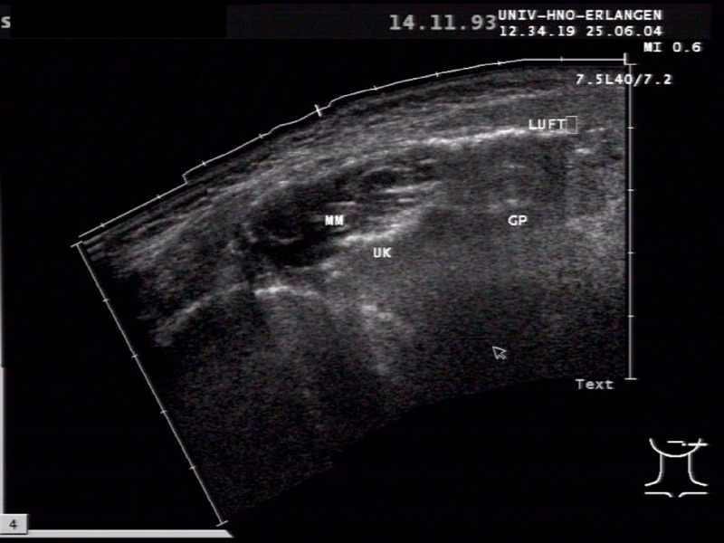
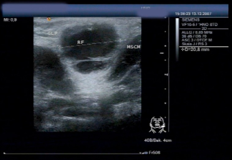
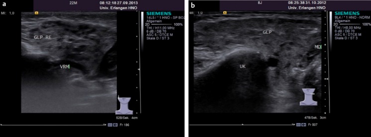
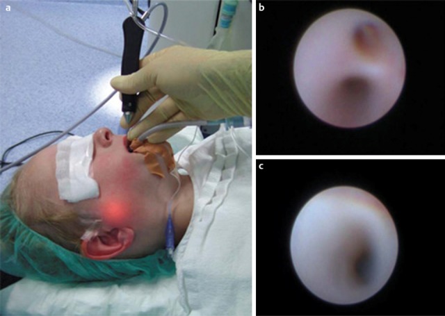
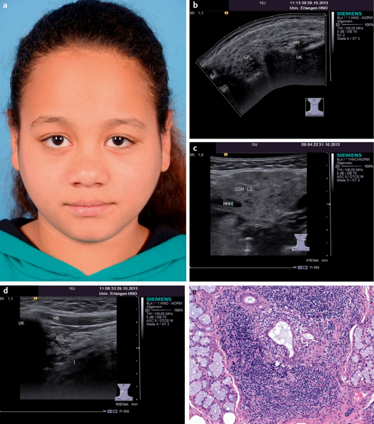
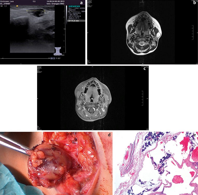
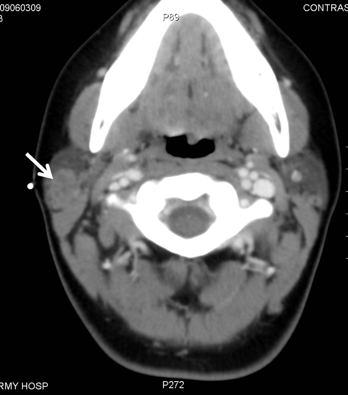
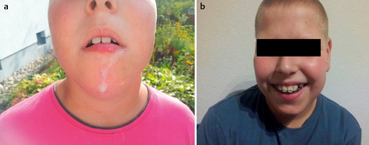
Verification
Claims the paper makes
The claims and statistics of this paper haven’t been checked yet.
Run the check — free, no account needed. It reads the open-access text and checks the sources it cites.
Claims other papers make about this one
Each is checked against this paper’s own text: does it say what the citing paper says it does?
The source backs part of the claim, not all of it — or backs it for a different population, condition or comparison.
In the citing paper
“The involvement of the salivary glands and deep neck spaces is less common but documented in some cases, as literature points out, thus, it should not be completely ruled out in chronic, poorly documented cases of salivary gland pathology [52,53].”
From Head and Neck Tuberculosis: A Rare Diagnosis and the Role of Surgical Biopsy and Histopathological Evaluation in Extrapulmonary Disease (2025) · cites this paper as [53]
Experimental — accuracy not yet measured
Self-check
Percentages, sums and reported differences re-computed from the paper’s own numbers.
The self-check needs the paper’s full text.
Citations
Cites
Vitali, C., Bombardieri, S., Jonsson, R., Moutsopoulos, H. M., Alexander, E. L., Carsons, S. E., Daniels, T. E., Fox, P. C., Fox, R. I., Kassan, S. S., et al. (2002) · Annals of the Rheumatic Diseases · doi:10.1136/ard.61.6.554 · Cited by 5,285
The Worldwide Incidence of Preterm Birth: A Systematic Review of Maternal Mortality and Morbidity
Beck, S., Wojdyla, D., Say, L., Pilar Bertran, A., Meraldi, M., Harris Requejo, J., Rubens, C., Menon, R., & Van Look, P. (2010) · Bulletin of the World Health Organization · doi:10.2471/blt.08.062554 · Cited by 1,523
Hviid, A. P., Rubin, S. A., & Mühlemann, K. (2008) · The Lancet · doi:10.1016/s0140-6736(08)60419-5 · Cited by 558
Treatment of Primary Sjögren Syndrome: A Systematic Review
Ramos‐Casals, M., Tzioufas, A. G., Stone, J. H., Sisó‐Almirall, A., & Bosch, X. (2010) · JAMA · doi:10.1001/jama.2010.1014 · Cited by 386
Allogeneic mesenchymal stem cell treatment alleviates experimental and clinical Sjögren syndrome
Xu, J., Wang, D., Liu, D., Fan, Z., Zhang, H., Liu, O., Ding, G., Gao, R., Zhang, C., Ding, Y., et al. (2012) · Blood · doi:10.1182/blood-2011-11-391144 · Cited by 325
Clinical review of 580 ranulas
Zhao, Y., Jia, Y., Chen, X., & Zhang, W. (2004) · Oral Surgery Oral Medicine Oral Pathology Oral Radiology and Endodontology · doi:10.1016/s1079-2104(04)00080-0 · Cited by 249
Lindeboom, J. A., Kuijper, E. J., Bruijnesteijn van Coppenraet, E. S., Lindeboom, R., & Prins, J. M. (2007) · Clinical Infectious Diseases · doi:10.1086/512675 · Cited by 245
Gottenberg, J., Cinquetti, G., Larroche, C., Combe, B. G., HACHULLA, E., Meyer, O., Pertuiset, É., Kaplanski, G., Chiche, L., Berthelot, J., et al. (2012) · Annals of the Rheumatic Diseases · doi:10.1136/annrheumdis-2012-202293 · Cited by 235
Activin disrupts epithelial branching morphogenesis in developing glandular organs of the mouse
Ritvos, O., Tuuri, T., ERÄMAA, M., Sainio, K., Hildén, K., Saxén, L., & Gilbert, S. F. (1995) · Mechanisms of Development · doi:10.1016/0925-4773(94)00342-k · Cited by 211
Hodgkin and non-Hodgkin lymphoma of the head and neck
Weber, A. L., Rahemtullah, A., & Ferry, J. A. (2003) · Neuroimaging Clinics of North America · doi:10.1016/s1052-5149(03)00039-x · Cited by 206
Baum, B. J., Alevizos, I., Zheng, C., Cotrim, A. P., Liu, S., McCullagh, L., Goldsmith, C. M., Burbelo, P. D., Citrin, D. E., Mitchell, J. B., et al. (2012) · Proceedings of the National Academy of Sciences · doi:10.1073/pnas.1210662109 · Cited by 196
Giess, R. (2000) · Journal of Neurology Neurosurgery & Psychiatry · doi:10.1136/jnnp.69.1.121 · Cited by 196
Richardson, M., Elliman, D. A. C., Maguire, H., SIMPSON, J., & Nicoll, A. (2001) · The Pediatric Infectious Disease Journal · doi:10.1097/00006454-200104000-00004 · Cited by 194
Brannon, R. B., Fowler, C. B., & Hartman, K. S. (1991) · Oral Surgery Oral Medicine Oral Pathology · doi:10.1016/0030-4220(91)90225-2 · Cited by 187
Sialendoscopy in the Diagnosis and Treatment of Sialolithiasis
Zenk, J., Koch, M., Klintworth, N., König, B., Konz, K., Gillespie, M. B., & Iro, H. (2012) · Otolaryngology · doi:10.1177/0194599812452837 · Cited by 180
Self-citationPrimary Sjögren syndrome in the paediatric age: a multicentre survey
Cimaz, R., Casadei, A., Rosé, C. D., Bartůňková, J., Šedivá, A., Falcini, F., Picco, P. P., Taglietti, M., Zulian, F., Cate, R. T., et al. (2003) · European Journal of Pediatrics · doi:10.1007/s00431-003-1277-9 · Cited by 179
Algorithms for Treatment of Salivary Gland Obstructions
Koch, M., Zenk, J., & Iro, H. (2009) · Otolaryngologic Clinics of North America · doi:10.1016/j.otc.2009.08.002 · Cited by 172
Self-citationModern management and pathophysiology of ranula: Literature review
Harrison, J. D. (2010) · Head & Neck · doi:10.1002/hed.21326 · Cited by 170
Summary of the II International Symposium on Cytomegalovirus
de Jong, M. D., Galasso, G. J., Gazzard, B., Griffiths, P., Jabs, D. A., Kern, E. R., & Spector, S. A. (1998) · Antiviral Research · doi:10.1016/s0166-3542(98)00044-8 · Cited by 169
Orvidas, L. J., Kasperbauer, J. L., Lewis, J. E., Olsen, K. D., & Lesnick, T. G. (2000) · Archives of Otolaryngology - Head and Neck Surgery · doi:10.1001/archotol.126.2.177 · Cited by 162
Haverkamp, M. H., Arend, S. M., Lindeboom, J. A. H., Hartwig, N. G., & van Dissel, J. T. (2004) · Clinical Infectious Diseases · doi:10.1086/422319 · Cited by 161
Salivary Gland Neoplasms in Children
Callender, D. L., Frankenthaler, R. A., Luna, M. A., Lee, S. S., & Goepfert, H. H. (1992) · Archives of Otolaryngology - Head and Neck Surgery · doi:10.1001/archotol.1992.01880050018003 · Cited by 160
Sultan, I. Y., Galindo, C. R., Al‐Sharabati, S., Guzzo, M., Casanova, M., & Ferrari, A. (2010) · Head & Neck · doi:10.1002/hed.21629 · Cited by 156
Avşar, A., Elli, M., Darka, Ö., & Pınarlı, F. G. (2007) · Oral Surgery Oral Medicine Oral Pathology Oral Radiology and Endodontology · doi:10.1016/j.tripleo.2007.02.029 · Cited by 130
Jongerius, P. H., Joosten, F. B. M., van den Hoogen, F. J. A., Gabreëls, F. J. M., & Rotteveel, J. J. (2003) · The Laryngoscope · doi:10.1097/00005537-200301000-00020 · Cited by 124
Pérez, D. E. D. C., Pires, F. R., Alves, F. A., de Almeida, O. P., & Kowalski, L. P. (2004) · International Journal of Pediatric Otorhinolaryngology · doi:10.1016/j.ijporl.2004.02.004 · Cited by 123
Ericson, S., Zetterlund, B., & Öhman, J. (1991) · Annals of Otology Rhinology & Laryngology · doi:10.1177/000348949110000702 · Cited by 122
Primary Sjögren's syndrome in children and adolescents: proposal for diagnostic criteria.
Bartůňková, J., Šedivá, A., Vencovský, J., & Tesař, V. (1999) · PubMed · Cited by 120
Bulbar amyotrophic lateral sclerosis: Patterns of progression and clinical management
Hıllel, A. D., & Miller, R. P. (1989) · Head & Neck · doi:10.1002/hed.2880110110 · Cited by 120
Management of Nontuberculous Mycobacteria-Induced Cervical Lymphadenitis With Observation Alone
Zeharia, A., Eidlitz-Markus, T., Haimi-Cohen, Y., Samra, Z., Kaufman, L., & Amir, J. (2008) · Pediatric Infectious Disease Journal · doi:10.1097/inf.0b013e3181734fa3 · Cited by 117
Tumour-like Lesions of the Salivary Glands
Seifert, G. (1992) · Pathology - Research and Practice · doi:10.1016/s0344-0338(11)80241-4 · Cited by 117
Malignant tumors of the head and neck in children a twenty-year review
Cunningham, M. J., Myers, E. N., & Bluestone, C. D. (1987) · International Journal of Pediatric Otorhinolaryngology · doi:10.1016/0165-5876(87)90109-1 · Cited by 115
Ellies, M., Laskawi, R., Rohrbach, S., Arglebe, C., & Beuche, W. (2002) · The Laryngoscope · doi:10.1097/00005537-200201000-00015 · Cited by 113
Everberg, B. G. (1957) · Acta Oto-Laryngologica · doi:10.3109/00016485709126900 · Cited by 113
THE SURGICAL TREATMENT OF DROOLING
Wilkie, T. F., & Brody, G. S. (1977) · Plastic & Reconstructive Surgery · doi:10.1097/00006534-197706000-00001 · Cited by 109
Epithelial salivary glands neoplasms in children and adolescents: A forty‐four‐year experience
Ribeiro, K. B., Kowalski, L. P., Saba, L. M. B., & de Camargo, B. (2002) · Medical and Pediatric Oncology · doi:10.1002/mpo.10168 · Cited by 109
Botulinum toxin A as a treatment for excessive drooling in children
Bothwell, J. E., Clarke, K., Dooley, J. M., Gordon, K., Anderson, R. L., Wood, E. P., Camfield, C. S., & Camfield, P. R. (2002) · Pediatric Neurology · doi:10.1016/s0887-8994(02)00381-8 · Cited by 108
Perceptive Deafness in Connection with Mumps:A Study of 298 Servicemen suffering from Mumps
Vuori, M., LAHIKAINEN, E. A., & Peltonen, T. O. (1962) · Acta Oto-Laryngologica · doi:10.3109/00016486209127357 · Cited by 106
Submandibular Duct Relocation for Drooling: A 10‐Year Experience with 194 Patients
Crysdale, W. S., & White, A. (1989) · Otolaryngology · doi:10.1177/019459988910100114 · Cited by 103
Clinical Characteristics and Survival for Major Salivary Gland Malignancies in Children
Shapiro, N. L., & Bhattacharyya, N. (2006) · Otolaryngology · doi:10.1016/j.otohns.2005.11.018 · Cited by 101
Acute Bacterial Sialadenitis: A Study of 29 Cases and Review
RAAD, I. I., Sabbagh, M. F., & Caranasos, G. J. (1990) · Clinical Infectious Diseases · doi:10.1093/clinids/12.4.591 · Cited by 100
Vitali, C., Bootsma, H., Bowman, S., Dörner, T., Gottenberg, J., Mariette, X., Ramos‐Casals, M., Ravaud, P., Séror, R., Theander, E., et al. (2012) · Annals of the Rheumatic Diseases · doi:10.1136/annrheumdis-2012-202565 · Cited by 98
Outcomes of Pediatric Patients with Malignancies of the Major Salivary Glands
Kupferman, M. E., de la Garza, G. O., Santillan, A. A., Williams, M. D., Varghese, B. T., Huh, W. W., Roberts, D. B., & Weber, R. S. (2010) · Annals of Surgical Oncology · doi:10.1245/s10434-010-1165-2 · Cited by 98
Long-Term Experience With Endoscopic Diagnosis and Treatment of Juvenile Recurrent Parotitis
Shacham, R., Droma, E. B., London, D., Bar, T., & Nahlieli, O. (2008) · Journal of Oral and Maxillofacial Surgery · doi:10.1016/j.joms.2008.09.027 · Cited by 97
Primary Sjögren's syndrome in children and adolescents: are proposed diagnostic criteria applicable?
Houghton, K. M., Malleson, P. N., Cabral, D. A., Petty, R. E., & Tucker, L. B. (2005) · PubMed · Cited by 96
Davidkin, I., Jokinen, S., Paananen, A., Leinikki, P., & Peltola, H. (2005) · The Journal of Infectious Diseases · doi:10.1086/427338 · Cited by 96
Klinische und diagnostische Befunde bei der Sialolithiasis
Zenk, J., Constantinidis, J., Kydles, S., Hornung, J. A., & Iro, H. (1999) · HNO · doi:10.1007/s001060050476 · Cited by 95
Self-citationA systematic review for evidence of efficacy of anticholinergic drugs to treat drooling
Jongerius, P. H. (2003) · Archives of Disease in Childhood · doi:10.1136/adc.88.10.911 · Cited by 94
Quenin, S., Plouin‐Gaudon, I., Marchal, F., Froehlich, P., Disant, F., & Faure, F. (2008) · Archives of Otolaryngology - Head and Neck Surgery · doi:10.1001/archotol.134.7.715 · Cited by 94
Diseases of the salivary glands in infants and adolescents
Ellies, M., & Laskawi, R. (2010) · Head & Face Medicine · doi:10.1186/1746-160x-6-1 · Cited by 94
TUMORS OF THE MAJOR SALIVARY GLANDS IN CHILDREN
Shikhani, A. H., & Johns, M. E. (1988) · Head & Neck Surgery · doi:10.1002/j.1930-2398.1988.tb00009.x · Cited by 93
Cytomegalovirus reinfection in young children
Bale, J. F., Petheram, S. J., Souza, I. E., & Murph, J. R. (1996) · The Journal of Pediatrics · doi:10.1016/s0022-3476(96)70279-2 · Cited by 90
Galili, D., & Marmary, Y. (1986) · Oral Surgery Oral Medicine Oral Pathology · doi:10.1016/0030-4220(86)90091-5 · Cited by 89
Minimally invasive options for salivary calculi
Witt, R. L., Iro, H., Koch, M., McGurk, M., Nahlieli, O., & Zenk, J. (2012) · The Laryngoscope · doi:10.1002/lary.23272 · Cited by 86
Self-citationTherapy of nontuberculous mycobacterial disease
Griffith, D. E. (2007) · Current Opinion in Infectious Diseases · doi:10.1097/qco.0b013e328055d9a2 · Cited by 85
CLINICAL APPEARANCE AND OUTCOME IN MUMPS ENCEPHALITIS IN CHILDREN
Koskiniemi, M., Donner, M., & PETTAY, O. (1983) · Acta Paediatrica · doi:10.1111/j.1651-2227.1983.tb09778.x · Cited by 85
Effect of childhood malnutrition on salivary flow and pH
Psoter, W. J., Spielman, A. L., Gebrian, B., Jean, R. S., & Katz, R. V. (2007) · Archives of Oral Biology · doi:10.1016/j.archoralbio.2007.09.007 · Cited by 84
Cervical lymphadenitis due to non-tuberculous mycobacteria: surgical treatment and review
Flint, D., Mahadevan, M., Barber, C., Grayson, D. E., & Small, R. (2000) · International Journal of Pediatric Otorhinolaryngology · doi:10.1016/s0165-5876(00)82006-6 · Cited by 83
Védrine, P. O., Coffinet, L., Temam, S., Montagne, K., Lapeyre, M., Oberlin, O., Orbach, D. H., Simon, C., & Sommelet, D. (2006) · Head & Neck · doi:10.1002/hed.20429 · Cited by 82
A Study on the Pathogenesis of Recurrent Parotitis in Childhood
Konno, A., & Ito, E. (1979) · Annals of Otology Rhinology & Laryngology · doi:10.1177/00034894790886s301 · Cited by 81
Epithelial Salivary Gland Tumors in Children and Adolescents
Seifert, G., Okabe, H., & Caselitz, J. (2010) · ORL · doi:10.1159/000275859 · Cited by 80
Case 6 Congenital Epithelial Tumor of the Parotid—Sialoblastoma
Taylor, G. P. (1988) · Pediatric Pathology · doi:10.3109/15513818809041583 · Cited by 80
The problem of drooling in cerebral palsy: a surgical approach.
Wilkie, T. F. (1967) · PubMed · Cited by 80
The molecular epidemiology of mumps virus
Mühlemann, K. (2004) · Infection Genetics and Evolution · doi:10.1016/j.meegid.2004.02.003 · Cited by 79
Drool Control: Long‐term Follow‐up
Rapp, D. (1980) · Developmental Medicine & Child Neurology · doi:10.1111/j.1469-8749.1980.tb04349.x · Cited by 79
Management of drooling: 10 years after the Consortium on Drooling, 1990
Blasco, P. A. (2002) · Developmental Medicine & Child Neurology · doi:10.1111/j.1469-8749.2002.tb00286.x · Cited by 79
Mucoepidermoid Carcinoma of the Parotid Gland in Children
Rahbar, R., Grimmer, J. F., Vargas, S. O., Robson, C. D., Mack, J. W., Pérez‐Atayde, A. R., Marcus, K. J., Grier, H. E., Healy, G. B., & McGill, T. J. (2006) · Archives of Otolaryngology - Head and Neck Surgery · doi:10.1001/archotol.132.4.375 · Cited by 76
Plunging Ranula: Congenital or Acquired?
Morton, R. P., Ahmad, Z., & Jain, P. (2009) · Otolaryngology · doi:10.1016/j.otohns.2009.10.014 · Cited by 76
Follow-up Study of Recurrent Parotitis in Children
Geterud, Å., Lindvall, A., & Nyléan, O. (1988) · Annals of Otology Rhinology & Laryngology · doi:10.1177/000348948809700403 · Cited by 76
Surgical Treatment of Drooling
Ekedahl, C. (1974) · Acta Oto-Laryngologica · doi:10.3109/00016487409124619 · Cited by 74
Congenital tumors of the parotid gland.
Gf, V., & Tefft, M. (1966) · PubMed · Cited by 72
SWALLOWING DISTURBANCES ASSOCIATED WITH DROOLING IN CEREBRAL‐PALSIED CHILDREN
Lespargot, A., Langevin, M., Muller, S., & Guillemont, S. (1993) · Developmental Medicine & Child Neurology · doi:10.1111/j.1469-8749.1993.tb11641.x · Cited by 71
Management of drooling in individuals with neurodisability: a surgical experience
Crysdale, W. S., Raveh, E., McCann, C., Roske, L., & Kotler, A.-L. (2001) · Developmental Medicine & Child Neurology · doi:10.1111/j.1469-8749.2001.tb00225.x · Cited by 71
Successful Management of Drooling with Botulinum Toxin A in Neurologically Disabled Children
Ellies, M., Rohrbach, S., Arglebe, C., Wilken, B., Laskawi, R., & Hanefeld, F. A. (2003) · Neuropediatrics · doi:10.1055/s-2002-37084 · Cited by 71
Koch, M., Bozzato, A., Iro, H., & Zenk, J. (2009) · Otolaryngology · doi:10.1016/j.otohns.2009.10.022 · Cited by 69
Self-citationBilateral dysgenetic polycystic parotid Glands
Seifert, G., Thomsen, S., & Donath, K. (1981) · Archiv für Pathologische Anatomie und Physiologie und für Klinische Medicin · doi:10.1007/bf00496559 · Cited by 68
Paediatric Salivary Gland Epithelial Neoplasms
Bradley, P. J., McClelland, L., & Mehta, D. (2007) · ORL · doi:10.1159/000099222 · Cited by 67
Modern management of juvenile recurrent parotitis
Capaccio, P., Sigismund, P. E., LUCA, N. D., Marchisio, P., & Pignataro, L. (2012) · The Journal of Laryngology & Otology · doi:10.1017/s0022215112002319 · Cited by 67
Sialendoscopy‐based diagnosis and classification of parotid duct stenoses
Koch, M., Iro, H., & Zenk, J. (2009) · The Laryngoscope · doi:10.1002/lary.20522 · Cited by 66
Self-citationMaynard, J. D. (1965) · British journal of surgery · doi:10.1002/bjs.1800521021 · Cited by 64
Sialoblastoma: a clinicopathologic and immunohistochemical study of 7 cases
Williams, S. B., Ellis, G. L., & Warnock, G. R. (2006) · Annals of Diagnostic Pathology · doi:10.1016/j.anndiagpath.2006.02.008 · Cited by 64
Tumors of the Salivary Glands in Childhood and Adolescence
Ellies, M., Schaffranietz, F., Arglebe, C., & Laskawi, R. (2006) · Journal of Oral and Maxillofacial Surgery · doi:10.1016/j.joms.2006.03.006 · Cited by 63
Management of Ranula: 9 Years' Clinical Experience in Pediatric and Adult Patients
Sigismund, P. E., Bozzato, A., Schumann, M., Koch, M., Iro, H., & Zenk, J. (2012) · Journal of Oral and Maxillofacial Surgery · doi:10.1016/j.joms.2012.07.042 · Cited by 63
Self-citationSchneider, H., Koch, M., Künzel, J., Gillespie, M. B., Grundtner, P., Iro, H., & Zenk, J. (2013) · The Laryngoscope · doi:10.1002/lary.24291 · Cited by 62
Self-citationLimbrock, G. J., Hoyer, H., & Scheying, H. (1990) · PubMed · Cited by 62
Treatment of chronic sialadenitis by intraductal penicillin or saline
Antoniades, D., Harrison, J. D., Epivatianos, A., & Papanayotou, P. (2004) · Journal of Oral and Maxillofacial Surgery · doi:10.1016/j.joms.2003.07.007 · Cited by 62
Autosomal dominant juvenile recurrent parotitis.
Reid, E., Douglas, F., Crow, Y. J., Hollman, A., & Gibson, J. (1998) · Journal of Medical Genetics · doi:10.1136/jmg.35.5.417 · Cited by 62
Neonatal suppurative parotitis over the last 4 decades: Report of three new cases and review
Ismail, E. A. R., Seoudi, T. M. M., Al‐Amir, M., & Al‐Esnawy, A. A. (2012) · Pediatrics International · doi:10.1111/j.1442-200x.2012.03738.x · Cited by 61
Acute Transient Sialadenopathy during Induction of Anesthesia
ATTAS, M., Sabawala, P. B., & Keats, A. S. (1968) · Anesthesiology · doi:10.1097/00000542-196809000-00036 · Cited by 61
Primary sjögren syndrome in childhood: report of a case and review of the literature
Nikitakis, N. G., Rivera, H., Lariccia, C., Papadimitriou, J. C., & Sauk, J. J. (2003) · Oral Surgery Oral Medicine Oral Pathology Oral Radiology and Endodontology · doi:10.1016/s1079-2104(03)00159-8 · Cited by 60
Pediatric salivary gland lesions
Mehta, D., & Willging, J. P. (2006) · Seminars in Pediatric Surgery · doi:10.1053/j.sempedsurg.2006.02.004 · Cited by 60
Follow‐up of parotid pleomorphic adenomas treated by extracapsular dissection
Iro, H., Zenk, J., Koch, M., & Klintworth, N. (2012) · Head & Neck · doi:10.1002/hed.23032 · Cited by 60
Self-citationDrooling: review of the literature and proposals for management
Nunn, J. (2000) · Journal of Oral Rehabilitation · doi:10.1046/j.1365-2842.2000.00575.x · Cited by 60
Hidas, A., Fux‐Noy, A., Birman, N., Shapira, J., Matot, I., Steinberg, D., & Moskovitz, M. (2011) · Archives of Oral Biology · doi:10.1016/j.archoralbio.2011.03.018 · Cited by 59
Management of pediatric plunging ranula
Mahadevan, M., & Vasan, N. R. (2005) · International Journal of Pediatric Otorhinolaryngology · doi:10.1016/j.ijporl.2005.10.022 · Cited by 59
Neonatal suppurative parotitis.
Leake, D., & Leake, R. (1970) · PubMed · Cited by 58
Subacute necrotizing sialadenitis: Report of 7 cases and a review of the literature
Fowler, C. B., & Brannon, R. B. (2000) · Oral Surgery Oral Medicine Oral Pathology Oral Radiology and Endodontology · doi:10.1067/moe.2000.105943 · Cited by 58
Parotid neoplasms in children: experience of diagnosis and management in a district general hospital
Ethunandan, M., Ethunandan, A., Macpherson, D. W., Conroy, B., & Pratt, C. A. (2003) · International Journal of Oral and Maxillofacial Surgery · doi:10.1054/ijom.2002.0381 · Cited by 57
Treatments of various otolaryngological cystic diseases by OK-4321
Ohta, N., Fukase, S., Suzuki, Y., Ishida, A., & Aoyagi, M. (2010) · The Laryngoscope · doi:10.1002/lary.21141 · Cited by 57
Results of Minimally Invasive Gland-Preserving Treatment in Different Types of Parotid Duct Stenosis
Koch, M., Iro, H., Klintworth, N., Psychogios, G. V., & Zenk, J. (2012) · Archives of Otolaryngology - Head and Neck Surgery · doi:10.1001/archoto.2012.1618 · Cited by 57
Self-citationMichel, M., L’Heureux, N., Auger, F. A., & Germain, L. (1997) · Journal of Cellular Physiology · doi:10.1002/(sici)1097-4652(199705)171:2 · Cited by 56
Swallowing Dysfunction in the Brain-Damaged with Drooling
Ekedahl, C., Månsson, I., & Sandberg, N. (1974) · Acta Oto-Laryngologica · doi:10.3109/00016487409126339 · Cited by 56
Mycobacterium lentiflavum: An Etiologic Agent of Cervical Lymphadenitis
Haase, G., Kentrup, H., Skopnik, H., Springer, B., & Böttger, E. C. (1997) · Clinical Infectious Diseases · doi:10.1086/516958 · Cited by 55
Koheil, R., Sochaniwskyj, A., Bablich, K., Kenny, D. J., & Milner, M. (1987) · Developmental Medicine & Child Neurology · doi:10.1111/j.1469-8749.1987.tb02103.x · Cited by 54
Sialoendoscopy for the Treatment of Pediatric Salivary Gland Disorders
Hackett, A. M., Barañano, C. F., Reed, M., Duvvuri, U., Smith, R. J., & Mehta, D. (2012) · Archives of Otolaryngology - Head and Neck Surgery · doi:10.1001/2013.jamaoto.244 · Cited by 54
Surgical Treatment of Drooling by Bilateral Parotid Duct Ligation and Submandibular Gland Resection
Dundas, D. F., & Peterson, R. A. (1979) · Plastic & Reconstructive Surgery · doi:10.1097/00006534-197907000-00009 · Cited by 53
Natural history of patients with recurrent parotitis and punctate sialectasis
Watkin, G. T., & HOBSLEY, M. (1986) · British journal of surgery · doi:10.1002/bjs.1800730922 · Cited by 53
Lindeboom, J. A. H., Lindeboom, R., van Coppenraet, E. S. B., Kuijper, E. J., Tuk, J. G., & Prins, J. M. (2009) · The Pediatric Infectious Disease Journal · doi:10.1097/inf.0b013e3181aa6411 · Cited by 52
Recurrent Pneumoparotid: Cause and Treatment
Han, S., & Isaacson, G. C. (2004) · Otolaryngology · doi:10.1016/j.otohns.2004.04.035 · Cited by 52
McGurk, M. (2006) · Journal of Oral and Maxillofacial Surgery · doi:10.1016/j.joms.2006.05.033 · Cited by 51
CHRONIC INFLAMMATORY DISORDERS OF THE SALIVARY GLANDS
Rice, D. H. (1999) · Otolaryngologic Clinics of North America · doi:10.1016/s0030-6665(05)70174-2 · Cited by 50
The Role of Parotid Biopsy in the Diagnosis of Pediatric Sjögren Syndrome
McGuirt, W. F., Whang, C., & Moreland, W. S. (2002) · Archives of Otolaryngology - Head and Neck Surgery · doi:10.1001/archotol.128.11.1279 · Cited by 50
Microbiology of recurrent parotitis
Giglio, M., Landaeta, M., & Pinto, M. E. (1997) · The Pediatric Infectious Disease Journal · doi:10.1097/00006454-199704000-00010 · Cited by 49
Zhi, K., Wen, Y., Ren, W., & Zhang, Y. (2008) · International Journal of Pediatric Otorhinolaryngology · doi:10.1016/j.ijporl.2008.02.012 · Cited by 49
Salivary proteins and early childhood caries: A gel electrophoretic analysis
Tandon, S., Bhalla, S., & Satyamoorthy, K. (2010) · Contemporary Clinical Dentistry · doi:10.4103/0976-237x.62515 · Cited by 48
Salivary gland biometry in female patients with eating disorders
Bozzato, A., Burger, P. H. M., Zenk, J., Uter, W. J. C., & Iro, H. (2008) · European Archives of Oto-Rhino-Laryngology · doi:10.1007/s00405-008-0598-8 · Cited by 47
Self-citationPrimacy of surgery in the management of mucoepidermoid carcinoma in children
Ryan, J. T., El‐Naggar, A. K., Huh, W. W., Hanna, E. Y., Weber, R. S., & Kupferman, M. E. (2011) · Head & Neck · doi:10.1002/hed.21675 · Cited by 47
Management options for the drooling patient.
Ws, C. (1989) · PubMed · Cited by 47
The function of the tympanic plexus as related to frey's syndrome
Ross, J. A. T. (1970) · The Laryngoscope · doi:10.1002/lary.5540801204 · Cited by 46
Ranula successfully treated by botulinum toxin type A: report of 3 cases
Chow, T., Chan, S., & Lam, S. (2007) · Oral Surgery Oral Medicine Oral Pathology Oral Radiology and Endodontology · doi:10.1016/j.tripleo.2007.04.007 · Cited by 46
A Non‐surgical Method of Reducing Drooling in Cerebral‐palsied Children
Harris, M. M., & Dignam, P. F. (1980) · Developmental Medicine & Child Neurology · doi:10.1111/j.1469-8749.1980.tb03708.x · Cited by 46
Fischer-Brandies, H., Avalle, C., & Limbrock, G. J. (1987) · European Journal of Orthodontics · doi:10.1093/ejo/9.2.139 · Cited by 46
Self-induced pneumoparotitis in an adolescent. Report of a case and review of the literature
Markowitz-Spence, L., Brodsky, L. S., Seidell, G., & Stanievich, J. F. (1987) · International Journal of Pediatric Otorhinolaryngology · doi:10.1016/0165-5876(87)90021-8 · Cited by 44
Diagnosis and gland‐preserving minimally invasive therapy for wharton's duct stenoses
Koch, M., Iro, H., Künzel, J., Psychogios, G. V., Bozzato, A., & Zenk, J. (2011) · The Laryngoscope · doi:10.1002/lary.22452 · Cited by 44
Self-citationTreatment of lymph node tuberculosis – a randomized clinical trial of two 6‐month regimens
Jawahar, M. S., Rajaram, K., Sivasubramanian, S., Paramasivan, C. N., Chandrasekar, K., Kamaludeen, M. N., Thirithuvathas, A. J., Ananthalakshmi, V., & Prabhakar, R. (2005) · Tropical Medicine & International Health · doi:10.1111/j.1365-3156.2005.01493.x · Cited by 44
Sağ, Ç., Özden, F. O., Açıkgöz, G., & Anlar, F. Y. (2007) · Clinical Therapeutics · doi:10.1016/j.clinthera.2007.10.014 · Cited by 44
Houghton, K. M., Cabral, D. A., Petty, R. E., & Tucker, L. B. (2005) · PubMed · Cited by 44
Cases presenting as parotid abscesses in children
Saarinen, R. T., Kolho, K., & Pitkäranta, A. (2007) · International Journal of Pediatric Otorhinolaryngology · doi:10.1016/j.ijporl.2007.02.011 · Cited by 43
Bohlender, J. E., Rauh, M., Zenk, J., & Gröschl, M. (2003) · Journal of Endocrinology · doi:10.1677/joe.0.1780217 · Cited by 42
Self-citationJuvenile Sjögren syndrome: distinctive age, unique findings.
de Souza, T. R., Silva, I. H. M., Carvalho, A. T., Gomes, V. B., Duarte, Â. L. B. P., Leão, J. C., & Gueiros, L. A. (2012) · PubMed · Cited by 42
Hemenway, W. G. (1971) · The Laryngoscope · doi:10.1288/00005537-197104000-00001 · Cited by 42
Sonographic analysis of recurrent parotitis in children
Shimizu, M., Ußmüller, J., Donath, K., Yoshiura, K., Ban, S., Kanda, S., Ozeki, S., & Shinohara, M. (1998) · Oral Surgery Oral Medicine Oral Pathology Oral Radiology and Endodontology · doi:10.1016/s1079-2104(98)90355-9 · Cited by 41
Reduction of Salivary Flow with Scopoderm Tts
Talmi, Y. P., Zohar, Y., Finkelstein, Y., & Laurian, N. (1988) · Annals of Otology Rhinology & Laryngology · doi:10.1177/000348948809700206 · Cited by 40
Pediatric parotitis: A 5-year review at a tertiary care pediatric institution
Stong, B. C., Sipp, J. A., & Sobol, S. E. (2005) · International Journal of Pediatric Otorhinolaryngology · doi:10.1016/j.ijporl.2005.08.001 · Cited by 40
Cervicofacial nontuberculous mycobacterium lymphadenitis in children: Is surgery always necessary?
Harris, R. L., Modayil, P. C., Adam, J., Sharland, M., Heath, P. T., Planche, T., & Daya, H. (2009) · International Journal of Pediatric Otorhinolaryngology · doi:10.1016/j.ijporl.2009.06.006 · Cited by 39
Bergeron, C., Thiesse, P., Rey, A., Orbach, D. H., Boutard, P., Thomas, C., Schmitt, C., Scopinaro, M. J., Bernard, F., Stevens, M. C. G., et al. (2008) · European Journal of Cancer · doi:10.1016/j.ejca.2007.12.007 · Cited by 39
Hoyer, H., & Limbrock, G. J. (1990) · PubMed · Cited by 39
Salivary Gland Enlargement During Induction of Anesthesia
Bonchek, L. I. (1969) · JAMA · doi:10.1001/jama.1969.03160240072025 · Cited by 39
Parasympathetic Nerve Section for Control of Sialorrhea
Michel, R. G., Johnson, K. A., & Patterson, C. N. (1977) · Archives of Otolaryngology · doi:10.1001/archotol.1977.00780190074008 · Cited by 39
Salivary gland aplasia with cleft lip and palateA case report and review of the literature
Matsuda, C., Matsui, Y., Ohno, K., & Michi, K.-I. (1999) · Oral Surgery Oral Medicine Oral Pathology Oral Radiology and Endodontology · doi:10.1016/s1079-2104(99)70140-x · Cited by 39
Moderne Konzepte zur Diagnostik und Therapie der Sialolithiasis
Iro, H., Zenk, J., & Koch, M. (2010) · HNO · doi:10.1007/s00106-009-2075-0 · Cited by 39
Self-citationRubaltelli, L., Sponga, T., Candiani, F., Pittarello, F., & Andretta, M. (1987) · British Journal of Radiology · doi:10.1259/0007-1285-60-720-1211 · Cited by 39
Aetiological and histological classification of sialadenitis.
Seifert, G. (1997) · PubMed · Cited by 39
Speichelsteine der Glandula submandibularis
Zenk, J., Gottwald, F., Bozzato, A., & Iro, H. (2004) · HNO · doi:10.1007/s00106-004-1110-4 · Cited by 37
Self-citationLieberman, S. M. (2013) · Current Opinion in Rheumatology · doi:10.1097/bor.0b013e328363ed23 · Cited by 37
Bajaj, N. P. S., Rose, P., Clifford-Jones, R. E., & Hughes, P. J. (2001) · Acta Neurologica Scandinavica · doi:10.1034/j.1600-0404.2001.00340.x · Cited by 37
Treatment of metastatic sialoblastoma with chemotherapy and surgery
Scott, J. X., Krishnan, S., Bourne, A. J., Williams, M. P., Agzarian, M., & Révész, T. (2006) · Pediatric Blood & Cancer · doi:10.1002/pbc.20788 · Cited by 36
MANDEL, L., & WITEK, E. L. (2001) · The Journal of the American Dental Association · doi:10.14219/jada.archive.2001.0125 · Cited by 35
Drool control and tongue thrust therapy for the mentally retarded.
McCracken, A. (1978) · PubMed · Cited by 34
Prigent, M., Teissier, N., Peuchmaur, M. M., Elmaleh, M., Philippe‐Chomette, P., Cardin, P., & Orbach, D. H. (2010) · International Journal of Pediatric Otorhinolaryngology · doi:10.1016/j.ijporl.2010.01.026 · Cited by 34
Extracorporeal and Intracorporeal Lithotripsy of Salivary Gland Stones: Basic Investigations
Zenk, J., Koch, M., & Iro, H. (2009) · Otolaryngologic Clinics of North America · doi:10.1016/j.otc.2009.08.005 · Cited by 33
Self-citationSalivary gland pathology as a new finding in Treacher Collins syndrome
Østerhus, I., Skogedal, N., Akre, H., Johnsen, U. L.-H., Nordgarden, H., & Åsten, P. M. (2012) · American Journal of Medical Genetics Part A · doi:10.1002/ajmg.a.35331 · Cited by 33
Intraductal Tetracycline Therapy for the Treatment of Chronic Recurrent Parotitis
Bowling, L. D. M., Rauch, S. D., & Goodman, M. L. (1994) · Ear Nose & Throat Journal · doi:10.1177/014556139407300414 · Cited by 33
Brainstem encephalitis and acute disseminated encephalomyelitis following mumps
Sönmez, F. M., Ödemiş, E., Ahmetoğlu, A., & Ayvaz, A. (2004) · Pediatric Neurology · doi:10.1016/j.pediatrneurol.2003.09.004 · Cited by 32
Cusi, M. G., Bianchi, S., Valassina, M., Santini, L., Arnetoli, M., & Valensin, P. E. (1996) · Research in Virology · doi:10.1016/0923-2516(96)89653-1 · Cited by 32
Benign Pleomorphic Adenomas in Children
Malone, B. N., & Baker, S. R. (1984) · Annals of Otology Rhinology & Laryngology · doi:10.1177/000348948409300304 · Cited by 32
Acute and Chronic Inflammatory Diseases of the Salivary Glands: Diagnosis and Management
Travis, L. W., & Hecht, D. W. (1977) · Otolaryngologic Clinics of North America · doi:10.1016/s0030-6665(20)32618-9 · Cited by 32
Treatment of chronic recurrent parotitis.
Diamant, H., & Enfors, B. (1965) · The Laryngoscope · doi:10.1288/00005537-196501000-00017 · Cited by 31
Pneumoparotid during dental treatment
Piette, E., & Walker, R. T. (1991) · Oral Surgery Oral Medicine Oral Pathology · doi:10.1016/0030-4220(91)90550-v · Cited by 31
Acute Parotitis Following Sitting Position Neurosurgical Procedures
Berker, M., Şahin, A., Aypar, Ü., & Özgen, T. (2003) · Journal of Neurosurgical Anesthesiology · doi:10.1097/00008506-200401000-00007 · Cited by 31
Submandibular sialolithiasis in children
Bodner, L., & Azaz, B. (1982) · Journal of Oral and Maxillofacial Surgery · doi:10.1016/0278-2391(82)90281-6 · Cited by 31
PAROTID ABSCESS CAUSED BY MYCOBACTERIUM TUBERCULOSIS
Chatterjee, A., Varman, M., & Quinlan, T. W. (2001) · The Pediatric Infectious Disease Journal · doi:10.1097/00006454-200109000-00019 · Cited by 30
Mylohyoid herniation of the sublingual gland diagnosed by magnetic resonance imaging
Hopp, E., Mortensen, B., & Kolbenstvedt, A. (2004) · Dentomaxillofacial Radiology · doi:10.1259/dmfr/31454077 · Cited by 30
Ericson, S., & Sjöbäck, I. (1996) · PubMed · Cited by 29
Mumps epidemiology in Germany 2007-11
Takla, A., Wichmann, O., Klinc, C., Hautmann, W., Rieck, T., & Koch, J. (2013) · Eurosurveillance · doi:10.2807/1560-7917.es2013.18.33.20557 · Cited by 29
Xerostomia: aetiology and management in cancer patients
Holmes, S. (1998) · Supportive Care in Cancer · doi:10.1007/s005200050176 · Cited by 29
Surgical treatment of ranula with carbon dioxide laser radiation
Niccoli‐Filho, W., & Morosolli, A. R. C. (2004) · Lasers in Medical Science · doi:10.1007/s10103-004-0293-y · Cited by 28
Ußmüller, J., & Donath, K. (1999) · Klinische Pädiatrie · doi:10.1055/s-2008-1043780 · Cited by 28
Sialometaplasia of the soft palate in a 2-year-old girl.
Ylikontiola, L. P., Siponen, M., Salo, T. A., & Sándor, G. K. B. (2007) · PubMed · Cited by 28
Saunders, H. F. (1973) · New England Journal of Medicine · doi:10.1056/nejm197309272891322 · Cited by 28
Benign and malignant salivary gland diseases in children
Muenscher, A., Diegel, T., Jaehne, M., Ußmüller, J., Koops, S., & Sánchez-Hanke, M. (2008) · Auris Nasus Larynx · doi:10.1016/j.anl.2008.07.006 · Cited by 27
Increased low-level chromosome 21 mosaicism in older individuals with Down syndrome
Jenkins, E. C., Schupf, N., Genovese, M., Ye, L., Kapell, D., Canto, B., Harris, M. S., Devenny, D. A., Lee, J. H., & Brown, W. T. (1997) · American Journal of Medical Genetics · doi:10.1002/(sici)1096-8628(19970120)68:2 · Cited by 27
Minor salivary gland malignancies in the pediatric population
Galer, C. E., Santillan, A. A., Chelius, D. C., Huh, W. W., El‐Naggar, A. K., Hanna, E. Y., Weber, R. S., & Kupferman, M. E. (2012) · Head & Neck · doi:10.1002/hed.21980 · Cited by 27
Pediatric submandibular triangle masses: a fifteen‐year experience
Hockstein, N. G., Samadi, D. S., Gendron, K. B., Carpentieri, D. F., & Wetmore, R. F. (2004) · Head & Neck · doi:10.1002/hed.20019 · Cited by 27
Salivary Gland Neoplasms in Children
Deng, R., Huang, X., Hao, J., Ding, J. F., & Hu, Q. (2013) · Journal of Craniofacial Surgery · doi:10.1097/scs.0b013e3182801866 · Cited by 26
Benign lymphoepithelial cysts of the parotid: long-term surgical results
Steehler, M. K., Davison, S. P., & Steehler, M. W. (2012) · HIV/AIDS - Research and Palliative Care · doi:10.2147/hiv.s27755 · Cited by 26
Syebele, K., & Bütow, K.-W. (2011) · Oral Surgery Oral Medicine Oral Pathology Oral Radiology and Endodontology · doi:10.1016/j.tripleo.2010.09.067 · Cited by 26
Children with sialorrhoea in the absence of neurological abnormalities
Johnson, H., King, J. A., & Reddihough, D. S. (2001) · Child Care Health and Development · doi:10.1046/j.1365-2214.2001.00233.x · Cited by 26
Die Durchmesser des Stenon- und Wharton-Ganges
Zenk, J., Zikarsky, B., Hosemann, W. G., & Iro, H. (1998) · HNO · doi:10.1007/s001060050345 · Cited by 26
Self-citationSekretionshemmung exokriner Drüsen des Kopf-Hals-Bereiches durch Applikation von Botulinum Toxin A
Ellies, M., Laskawi, R., Rohrbach, S., Rödel, R. M. W., & Beuche, W. (2001) · HNO · doi:10.1007/s001060170028 · Cited by 25
Faulconbridge, R. V. L., Tranter, R., Moffat, V., & Green, E. (2001) · Clinical Otolaryngology · doi:10.1046/j.1365-2273.2001.00434.x · Cited by 25
Andrade, R. E., Hagen, K. A., & Manivel, J. C. (1988) · Human Pathology · doi:10.1016/s0046-8177(88)80009-1 · Cited by 25
Extracorporeal Shockwave Lithotripsy for Salivary Calculi in Pediatric Patients
Ottaviani, P. M. F. (2001) · Acta Oto-Laryngologica · doi:10.1080/00016480152602366 · Cited by 25
Parotidectomy in children: indications and complications
Xie, C. M., & Kubba, H. (2010) · The Journal of Laryngology & Otology · doi:10.1017/s0022215110001209 · Cited by 24
Recurrent parotitis and isolated IgG3 subclass deficiency
Marsman, W. A., & Sukhai, R. N. (1999) · European Journal of Pediatrics · doi:10.1007/s004310051177 · Cited by 24
Ex utero intrapartum treatment (EXIT) for congenital giant ranula
Chan, D. F. Y., Lee, C. H., Fung, T. Y., Chan, D. L., Abdullah, V. J., & Ng, P. C. (2006) · Acta Paediatrica · doi:10.1080/08035250600580545 · Cited by 24
Congenital aplasia of the major salivary glands: literature review and case report.
TAJI, S. S., Savage, N. W., HOLCOMBE, T. F., Khan, F., & Seow, W. K. (2011) · PubMed · Cited by 23
On minor salivary gland secretion in children, adolescents and adults.
Sonesson, M. (2011) · PubMed · Cited by 23
Parotid Swelling in a Premature Neonate
Chevalier, J., & Jadcherla, S. R. (2002) · American Journal of Perinatology · doi:10.1055/s-2002-36842 · Cited by 23
Mumps Virus Infection in Adults: Three Cases of Supraglottic Edema
Ishida, M., Fushiki, H., Morijiri, M., Maruyama, M., Motoshima, H., Asai, M., & Watanabe, Y. (2006) · The Laryngoscope · doi:10.1097/01.mlg.0000246189.38777.2c · Cited by 23
Clinical features of pediatric parotid tumors: 10-year experience of a single institute
Lee, D. H., Yoon, T. M., Lee, J. K., & Lim, S. C. (2013) · Acta Oto-Laryngologica · doi:10.3109/00016489.2013.822554 · Cited by 23
Paediatric salivary gland cancer in Finland
Aro, K. L. S., Leivo, I. V. V., Grenman, R. A., & Mäkitie, A. A. (2012) · International Journal of Pediatric Otorhinolaryngology · doi:10.1016/j.ijporl.2012.05.024 · Cited by 23
Limbrock, G. J., Hoyer, H., & Scheying, H. (1990) · PubMed · Cited by 22
Harris, M., Blewitt, R. W., DAVIES, V. J., & Steward, W. P. (1989) · Histopathology · doi:10.1111/j.1365-2559.1989.tb01586.x · Cited by 21
Bono, V. D., Pretolesi, F., Pontali, E., Martinoli, C., Bassetti, M., Mazzarello, G., Chiaramondia, M., Derchi, L. E., & Bassetti, D. (2000) · Clinical Infectious Diseases · doi:10.1086/313813 · Cited by 21
Die chronisch rezidivierende Parotitis
Zenk, J., Koch, M., Klintworth, N., & Iro, H. (2010) · HNO · doi:10.1007/s00106-009-2079-9 · Cited by 20
Self-citationNontuberculous mycobacterial cervicofacial adenitis in children: epidemiologic study.
Tremblay, V., Ayad, T., Lapointe, A., Giguàre, C., Quintal, M., Arcand, P., & Abela, A. (2008) · PubMed · Cited by 20
Stenosen und andere nicht steinbedingte Obstruktionen der Speicheldrüsenausführungsgänge
Koch, M., Iro, H., & Zenk, J. (2010) · HNO · doi:10.1007/s00106-009-2076-z · Cited by 20
Self-citationIdiopathic recurrent pneumoparotitis
McCormick, M. E., Bawa, G., & Shah, R. K. (2013) · American Journal of Otolaryngology · doi:10.1016/j.amjoto.2012.11.005 · Cited by 20
Chang, C., & Wong, M. (2001) · Plastic & Reconstructive Surgery · doi:10.1097/00006534-200104010-00001 · Cited by 19
Research works printed but not linked: 8
- [4]Seifert G Miehlke A Haubrich J Chilla R Speicheldrüsenkrankheiten. Pathologie, Klinik, Therapie, Fazialischirurgie 1984 Stuttgart Thieme 91 97
- [15]Gruber W Congenitaler Mangel beider Glandulae submaxillares bei einem wohlgebildeten, erwachsenen Subjecte Arch Pathol Anat Physiol Klin Med 1885 102 1 9 11 10.1007/BF01932760 Available from: http://dx.doi.org/10.1007/BF01932760 · DOI
- [17]Ferreira AP Gomez RS Castro WH Calixto NS Silva RA Aguiar MJ Congenital absence of lacrimal puncta and salivary glands: report of a Brazilian family and review Am J Med Genet 4 9 2000 94 1 32 34 10982479
- [18]Wiedemann HR Salivary gland disorders and heredity Am J Med Genet 20 1 1997 68 2 222 224 9028463
- [35]Webster RG Granoff A Rima BK Mumps virus Encyclopedia of Virology. Vol 2 1994 New York Academic Press 876 883
- [133]Wu AJ Lafrenie RM Park C Apinhasmit W Chen ZJ Birkedal-Hansen H Yamada KM Stetler-Stevenson WG Baum BJ Modulation of MMP-2 (gelatinase A) and MMP-9 (gelatinase B) by interferon-gamma in a human salivary gland cell line J Cell Physiol 5 1997 171 2 117 124 9130458
- [141]Iro H Uttenweiler V Zenk J Kopf-Hals-Sonographie. Eine Anleitung zur praxisbezogenen Ultraschalluntersuchung 2000 Berlin Springer 10.1007/978-3-642-57012-4 Available from: http://dx.doi.org/10.1007/978-3-642-57012-4 · DOI
- [192]Orvidas LJ Kasperbauer JL Lewis JE Olsen KD Lesnick TG Pediatric parotid masses Arch Otolaryngol Head Neck Surg 2 2000 126 2 177 184 10.1001/archotol.126.2.177 Available from: http://dx.doi.org/10.1001/archotol.126.2.177 10680869 · DOI
Cited by
Ogle, O. E. (2019) · Dental Clinics of North America · doi:10.1016/j.cden.2019.08.007 · Cited by 84
Sialendoscopy for non‐stone disorders: The current evidence
Erkul, E., & Gillespie, M. B. (2016) · Laryngoscope Investigative Otolaryngology · doi:10.1002/lio2.33 · Cited by 58
Sialendoscopy in treating pediatric salivary gland disorders: a systematic review
Schwarz, Y., Bezdjian, A., & Daniel, S. J. (2017) · European Archives of Oto-Rhino-Laryngology · doi:10.1007/s00405-017-4830-2 · Cited by 44
Chronic malnutrition and oral health status in children aged 1 to 5 years
Vieira, K. A., Júnior, L. D. S. R., Souza, M. A., dos Santos, N. B., Florêncio, T. M. D. M. T., & Bussadori, S. K. (2020) · Medicine · doi:10.1097/md.0000000000019595 · Cited by 38
Surun, A., Schneider, D. T., Ferrari, A., Stachowicz‐Stencel, T., Rascon, J., Synakiewicz, A., Agaimy, A., Мартинова, К., Kachanov, D. Y., Roganović, J., et al. (2021) · Pediatric Blood & Cancer · doi:10.1002/pbc.29058 · Cited by 32
Metadata
- Authors
- Iro, H., & Zenk, J.
- Affiliations
- Friedrich-Alexander-Universität Erlangen-Nürnberg (DE)
- Venue
- German Medical Science GMS Publishing House
- Publisher
- German Medical Science GMS Publishing House
- Published
- 2014
- Volume
- 13
- Pages
- Doc06-Doc06
- PMC
- PMC4273167
- OpenAlex
- W38431779
- Cited by
- 54
- References
- 240
- Citations by year
- 2026: 2 · 2025: 6 · 2024: 2 · 2023: 6 · 2022: 4 · 2021: 6 · 2020: 5 · 2019: 7 · 2018: 5 · 2017: 4 · 2016: 7
- FWCI
- 2.44
- Type
- journal article
- Language
- en
- Open access
- green
- www.ncbi.nlm.nih.gov
- Licence
- cc-by-nc-nd
- Locations
- Europe PMC (PubMed Central) (publishedVersion, cc-by-nc-nd)
- PubMed Central (publishedVersion, cc-by-nc-nd)
- PubMed (publishedVersion)
- German Medical Science (German Research Foundation) (publishedVersion, cc-by-nc-nd)
- OPUS (Augsburg University) (submittedVersion, cc-by-nc-nd)
- German Medical Science (German Research Foundation)
- Topics
- Health Sciences › Medicine › Physiology (medical) › Salivary Gland Disorders and Functions
- Health Sciences › Medicine › Surgery › Salivary Gland Tumors Diagnosis and Treatment
- Health Sciences › Dentistry › General Dentistry › HIV/AIDS oral health manifestations
- Keywords
- salivary gland diseases, salivary gland tumors, sialendoscopy, drooling, differential diagnosis, salivary gland surgery, adolescents
About these checks
Registry, retraction, Cites and statistics results are computed, not judged, with one exception: a printed reference that no index lists is read once by a model before it counts as a citation error. Claims are extracted by a model and kept only when their quote appears verbatim in the text; each cited source is then checked, with a model’s help, for whether it says what the claim relies on. Those verdicts can be wrong — read the claim’s quote and the cited source before relying on one.
Statistics and arithmetic are recomputed, not judged.
Confidence bands are shown only for fields calibrated on our annotated test set. None is calibrated yet, so no band is shown.
What counts as a claim ›How we measure confidence ›How we check this ›
Are you an author, or do you see a problem with this page? Request a review
Data sources
Bibliographic data: Semantic Scholar, OpenAlex, Crossref.
Contains information from the Semantic Scholar Academic Graph Datasets, made available under the ODC Attribution License (ODC-BY 1.0).
Semantic Scholar’s data is described in: Kinney, R., et al. (2023). The Semantic Scholar Open Data Platform. arXiv:2301.10140.
