Cited by 18

The DOI matches its Crossref record; 1 of the 1 references it cites were found in a registry or an index.

Specific inhibition of the transporter MRP4/ABCC4 affects multiple signaling pathways and thrombus formation in human platelets

Wolf, Robert, Grammbauer, Sophie, Palankar, Raghavendra, Tolksdorf, Céline, Moritz, Eileen, Böhm, Andreas, Hasan, Mahmoud, Hafkemeyer, Annika, Greinacher, Andreas, Tzvetkov, Mladen V., Rauch, Bernhard H., Jedlitschky, Gabriele·2022·Haematologica

Abstract

The multidrug resistance protein 4 (MRP4) is highly expressed in platelets and several lines of evidence point to an impact on platelet function. MRP4 represents a transporter for cyclic nucleotides as well as for certain lipid mediators. The aim of the present study was to comprehensively characterize the effect of a short-time specific pharmacological inhibition of MRP4 on signaling pathways in platelets. Transport assays in isolated membrane vesicles showed a concentrationdependent inhibition of MRP4-mediated transport of cyclic nucleotides, thromboxane (Tx)B2 and fluorescein (FITC)- labeled sphingosine-1-phosphate (S1P) by the selective MRP4 inhibitor Ceefourin-1. In ex vivo aggregometry studies in human platelets, Ceefourin-1 significantly inhibited platelet aggregation by about 30-50% when ADP or collagen was used as activating agents, respectively. Ceefourin-1 significantly lowered the ADP-induced activation of integrin aIIbb3, indicated by binding of FITC-fibrinogen (about 50% reduction at 50 mM Ceefourin-1), and reduced calcium influx. Furthermore, pre-incubation with Ceefourin-1 significantly increased PGE1- and cinaciguat-induced vasodilatorstimulated phosphoprotein (VASP) phosphorylation, indicating increased cytosolic cAMP as well as cGMP concentrations, respectively. The release of TxB2 from activated human platelets was also attenuated. Finally, selective MRP4 inhibition significantly reduced both the total area covered by thrombi and the average thrombus size by about 40% in a flow chamber model. In conclusion, selective MRP4 inhibition causes reduced platelet adhesion and thrombus formation under flow conditions. This finding is mechanistically supported by inhibition of integrin aIIbb3 activation, elevated VASP phosphorylation and reduced calcium influx, based on inhibited cyclic nucleotide and thromboxane transport as well as possible further mechanisms.

Overview

The claims and statistics of this paper haven’t been checked yet.

Run the check — free, no account needed. It reads the open-access text and checks the sources it cites.

A few free checks a day · papers already verified open instantly · results are public

Integrity signals

Registry

Crossref · 10.3324/haematol.2021.279761

Cites

1 of 1 references verified0 citation errors · 0 not checkable

Statistics

Figures

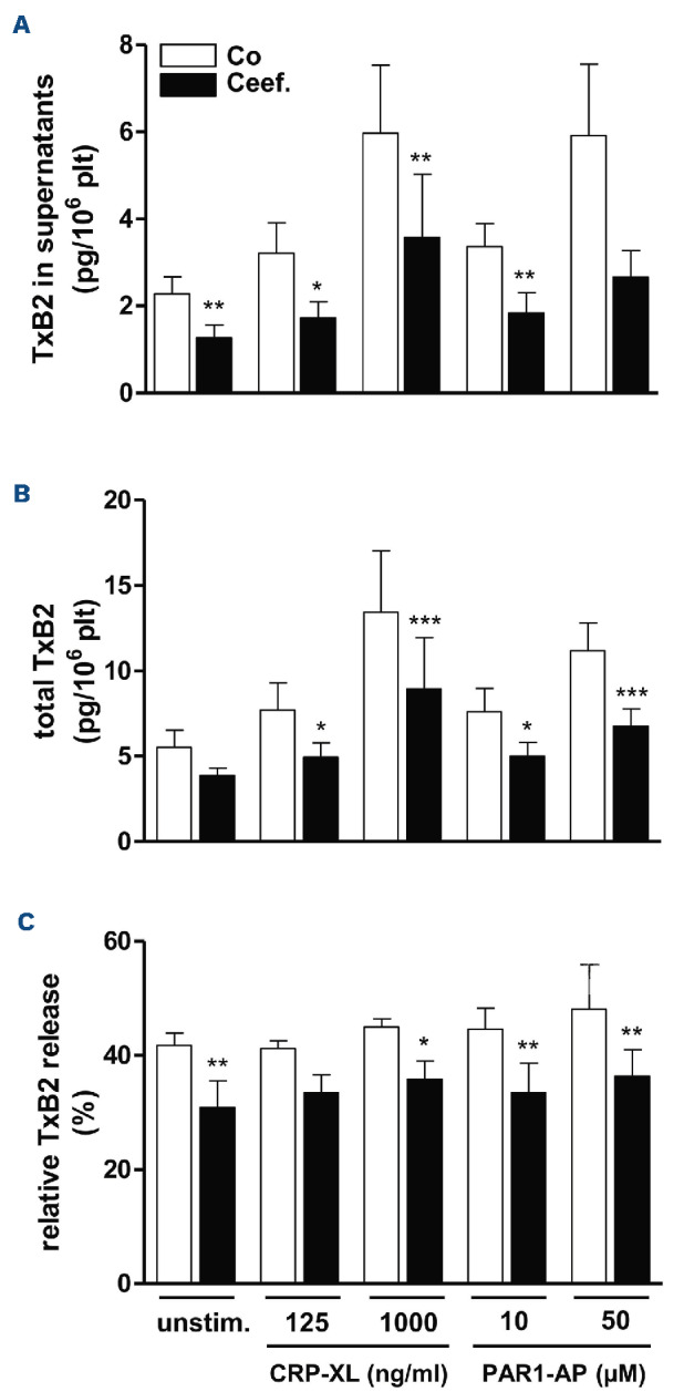
Figure 1. Inhibition of TxB2 release from human platelets by Ceefourin-1. Washed platelets were incubated for 15 minutes at 37°C with Ceefourin-1 (Ceef., 50 mM, black bars) or the respective solvent control (Co, 0.5% dimethyl sulfoxide [DMSO], white bars) and then either left unstimulated (unstim.) or activated with collagen-related peptide CRP-XL (125 ng/mL and 1,000 ng/mL) and the thrombin receptor-activating peptide PAR1-AP (10 mM and 50 mM). After 15 min, the platelets were pelleted and TxB2 was analyzed by liquid chromatography-tandem mass spectrometry (LC-MS/MS) in the supernatants and pellets. (A and B) Absolute TxB2 concentrations in platelet supernatants (A) and in platelet pellets (B) in pg/1x106 platelets (plt). (C) Platelet TxB2 release in % of the total amount formed. Values represent means + standard error of the mean from n=6 different donors. *P<0.05; **P<0.01 vs. the respective solvent control.
Figure 1. Inhibition of TxB2 release from human platelets by Ceefourin-1. Washed platelets were incubated for 15 minutes at 37°C with Ceefourin-1 (Ceef., 50 mM, black bars) or the respective solvent control (Co, 0.5% dimethyl sulfoxide [DMSO], white bars) and then either left unstimulated (unstim.) or activated with collagen-related peptide CRP-XL (125 ng/mL and 1,000 ng/mL) and the thrombin receptor-activating peptide PAR1-AP (10 mM and 50 mM). After 15 min, the platelets were pelleted and TxB2 was analyzed by liquid chromatography-tandem mass spectrometry (LC-MS/MS) in the supernatants and pellets. (A and B) Absolute TxB2 concentrations in platelet supernatants (A) and in platelet pellets (B) in pg/1x106 platelets (plt). (C) Platelet TxB2 release in % of the total amount formed. Values represent means + standard error of the mean from n=6 different donors. *P<0.05; **P<0.01 vs. the respective solvent control.CC BY-NC · PubMed Central
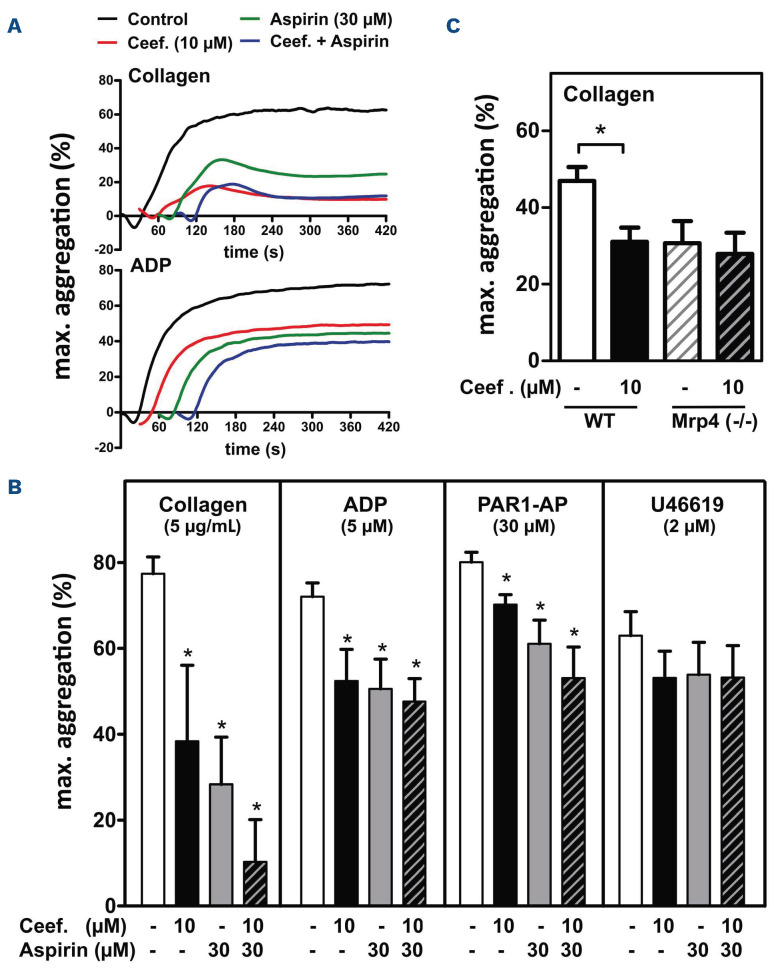
Figure 2. Effect of Ceefourin-1 on platelet aggregation ex vivo. (A and B) Human platelet-rich plasma (PRP) was pretreated with either only the solvent (control, (-)), or Ceefourin-1 (Ceef.; 10 mM for 20 minutes), or aspirin (30 mM; for 5 minutes), or the combination of both, and then stimulated with the following agonists: collagen (5 mg/mL), ADP (5 mM), PAR1-AP (30 mM) or U46619 (2 mM). Platelet aggregation was determined by light transmission aggregometry. Representative curves of platelet aggregation stimulated by collagen and ADP are given in (A). The inhibition of the maximal aggregation is summarized in (B) (means + standard error of the mean [SEM]; n=6-11; *P<0.05 vs. control). (C) PRP (diluted 1:2 with Tyrode’s buffer) from age- and sex-matched wild-type (WT) or Mrp4-deficient (Mrp4(-/-)) mice was pretreated with either only the solvent (control, (-)) or Ceefourin-1 (Ceef.; 10 mM) and then stimulated with collagen (10 mg/mL). Aggregation curves were monitored and the maximal extent of aggregation (%) was calculated (mean + SEM; n=5-7). Ceefourin-1 led to a significant reduction of the aggregation only in WT platelets (*P<0.05).
Figure 2. Effect of Ceefourin-1 on platelet aggregation ex vivo. (A and B) Human platelet-rich plasma (PRP) was pretreated with either only the solvent (control, (-)), or Ceefourin-1 (Ceef.; 10 mM for 20 minutes), or aspirin (30 mM; for 5 minutes), or the combination of both, and then stimulated with the following agonists: collagen (5 mg/mL), ADP (5 mM), PAR1-AP (30 mM) or U46619 (2 mM). Platelet aggregation was determined by light transmission aggregometry. Representative curves of platelet aggregation stimulated by collagen and ADP are given in (A). The inhibition of the maximal aggregation is summarized in (B) (means + standard error of the mean [SEM]; n=6-11; *P<0.05 vs. control). (C) PRP (diluted 1:2 with Tyrode’s buffer) from age- and sex-matched wild-type (WT) or Mrp4-deficient (Mrp4(-/-)) mice was pretreated with either only the solvent (control, (-)) or Ceefourin-1 (Ceef.; 10 mM) and then stimulated with collagen (10 mg/mL). Aggregation curves were monitored and the maximal extent of aggregation (%) was calculated (mean + SEM; n=5-7). Ceefourin-1 led to a significant reduction of the aggregation only in WT platelets (*P<0.05).CC BY-NC · PubMed Central
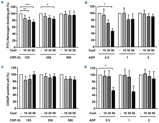
Figure 3. Effect of MRP4 inhibition on platelet fibrinogen binding and CD62P surface exposure. Binding of FITC-labeled fibrinogen (A and B) and R-phycoerythrin-labeled anti-CD62P antibody (C and D) to platelets (plt) was assessed by flow cytometry as a measure for integrin aIIbb3 activation and a-granule release, respectively. Platelet-rich plasma was preincubated with Ceefourin-1 (Ceef.) (10–50 mM) or the solvent control (0.5% dimethyl sulfoxide [DMSO]) for 15 minutes (min) and then stimulated with the collagen-related peptide CRP-XL (125–500 ng/mL) (A and C) or ADP (0.5–5 mM) (B and D) for 10 min before FITC-fibrinogen or anti-CD63P antibody were added. After fixation in formaldehyde the samples were subjected to flow cytometric analysis. Data are given in % of the respective solvent control (means + standard error of the mean, n=4-6 platelet-rich plasma samples of different donors, performed in duplicates). Significant differences with *P<0.05, **P<0.01, and **P<0.005 vs. the respective solvent control.
Figure 3. Effect of MRP4 inhibition on platelet fibrinogen binding and CD62P surface exposure. Binding of FITC-labeled fibrinogen (A and B) and R-phycoerythrin-labeled anti-CD62P antibody (C and D) to platelets (plt) was assessed by flow cytometry as a measure for integrin aIIbb3 activation and a-granule release, respectively. Platelet-rich plasma was preincubated with Ceefourin-1 (Ceef.) (10–50 mM) or the solvent control (0.5% dimethyl sulfoxide [DMSO]) for 15 minutes (min) and then stimulated with the collagen-related peptide CRP-XL (125–500 ng/mL) (A and C) or ADP (0.5–5 mM) (B and D) for 10 min before FITC-fibrinogen or anti-CD63P antibody were added. After fixation in formaldehyde the samples were subjected to flow cytometric analysis. Data are given in % of the respective solvent control (means + standard error of the mean, n=4-6 platelet-rich plasma samples of different donors, performed in duplicates). Significant differences with *P<0.05, **P<0.01, and **P<0.005 vs. the respective solvent control.CC BY-NC · PubMed Central
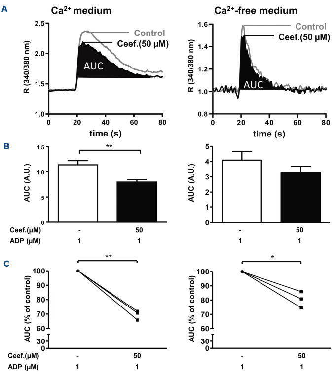
Figure 4. Effect of MRP4 inhibition on intracellular calcium levels in platelets. Cytosolic free calcium was measured spectrophotofluorometrically in Fura-2-loaded washed platelets. Measurements were performed either in Ca2+ medium (Tyrode’s buffer with 2 mM CaCl2) (left panels) or Ca2+-free medium (Tyrode’s buffer with 0.2 mM EGTA) (right panels). In (A) the time-dependent increase in cytosolic calcium (indicated by R (340/380 nm)) after stimulation with ADP (1 mM) and the effect of Ceefourin-1 (50 mM) in representative experiments are depicted. In (B) the area under the fluorescence curve (area under the curve [AUC]) as a measure for the total change in calcium concentration was calculated (means + standard error of the mean from n=3 different donors, each measured in duplicates or quadruplicates). The reduction after Ceefourin-1 treatment is also given in % of the respective solvent control for each donor (each separate experiment) in (C) (significant differences with *P<0.05, and **P<0.01).
Figure 4. Effect of MRP4 inhibition on intracellular calcium levels in platelets. Cytosolic free calcium was measured spectrophotofluorometrically in Fura-2-loaded washed platelets. Measurements were performed either in Ca2+ medium (Tyrode’s buffer with 2 mM CaCl2) (left panels) or Ca2+-free medium (Tyrode’s buffer with 0.2 mM EGTA) (right panels). In (A) the time-dependent increase in cytosolic calcium (indicated by R (340/380 nm)) after stimulation with ADP (1 mM) and the effect of Ceefourin-1 (50 mM) in representative experiments are depicted. In (B) the area under the fluorescence curve (area under the curve [AUC]) as a measure for the total change in calcium concentration was calculated (means + standard error of the mean from n=3 different donors, each measured in duplicates or quadruplicates). The reduction after Ceefourin-1 treatment is also given in % of the respective solvent control for each donor (each separate experiment) in (C) (significant differences with *P<0.05, and **P<0.01).CC BY-NC · PubMed Central
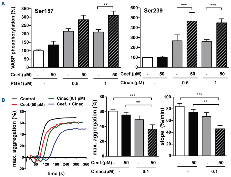
Figure 5. Effect of Ceefourin-1 on VASP phosphorylation (A) and cyclic nucleotide-dependent platelet inhibition (B). The phosphorylation of VASP at two different serine residues, serine-157 (the preferred substrate of PKA) (A, left panel) and serine-239 (the preferred substrate of PKG) (A, right panel) was determined by flow cytometry in human platelets as a measure for the cytosolic levels of cAMP and cGMP, respectively. Ceefourin-1 (Ceef., 50 µM) significantly amplified cAMP- and cGMP-dependent VASP phosphorylation, induced by PGE1 (1 µM) and cinaciguat (Cinac., 0.5 and 1 µM), respectively (means + standard error of the mean, n=6). (B) Platelet aggregation in the presence of cinaciguat (Cinac., 0.1 µM) and Ceefourin-1 (Ceef., 50 µM) was measured by light transmission aggregometry, to confirm the enhancement of cGMP-mediated effects by MRP4 inhibition. The aggregation was here stimulated by 1.25 µg/mL collagen. The left panel shows the aggregation curves of a representative experiment. Maximum platelet aggregation (middle panel) and the slope of platelet aggregation (right panel) were markedly decreased when both compounds were combined (means + standard error of the mean; n=20 measurements with platelet-rich plasma from 4 different donors).
Figure 5. Effect of Ceefourin-1 on VASP phosphorylation (A) and cyclic nucleotide-dependent platelet inhibition (B). The phosphorylation of VASP at two different serine residues, serine-157 (the preferred substrate of PKA) (A, left panel) and serine-239 (the preferred substrate of PKG) (A, right panel) was determined by flow cytometry in human platelets as a measure for the cytosolic levels of cAMP and cGMP, respectively. Ceefourin-1 (Ceef., 50 µM) significantly amplified cAMP- and cGMP-dependent VASP phosphorylation, induced by PGE1 (1 µM) and cinaciguat (Cinac., 0.5 and 1 µM), respectively (means + standard error of the mean, n=6). (B) Platelet aggregation in the presence of cinaciguat (Cinac., 0.1 µM) and Ceefourin-1 (Ceef., 50 µM) was measured by light transmission aggregometry, to confirm the enhancement of cGMP-mediated effects by MRP4 inhibition. The aggregation was here stimulated by 1.25 µg/mL collagen. The left panel shows the aggregation curves of a representative experiment. Maximum platelet aggregation (middle panel) and the slope of platelet aggregation (right panel) were markedly decreased when both compounds were combined (means + standard error of the mean; n=20 measurements with platelet-rich plasma from 4 different donors).CC BY-NC · PubMed Central
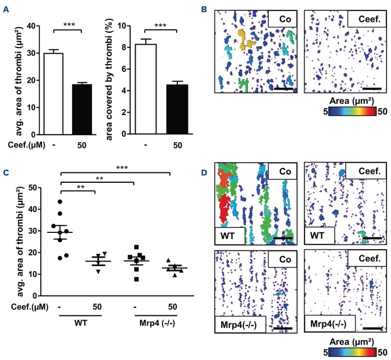
Figure 6. Role of MRP4 in platelet adhesion and thrombus formation under flow. (A and B) Whole human blood with FITC-anti-CD42a-labeled platelets (from n=3 different donors) was pre-incubated with Ceefourin-1 (Ceef., 50 mM) or the respective solvent (Co, 0.5 % dimethyl sulfoxide [DMSO]) and perfused through collagen-coated microchannels under high arterial shear conditions (1,800-1 ) for 5 minutes. Micrographs were taken at the beginning, the end, and in the center of the channel using a confocal laser scanning microscope (Zeiss LSM 780) with a 40x objective. The average thrombus area (A, left panel) and surface area coverage by thrombi (A, right panel; means + standard error of the mean [SEM]) were analyzed with the ImageJ software. In (B) representative images are shown. (C and D) The effect of Ceefourin-1 was also evaluated in perfusions performed with whole blood from age- and sex-matched wild-type (WT) or Mrp4(-/-) mice (with X488-labeled platelets). The average thrombus area (C, means ± SEM and values of individual animals; n=4-8) was markedly reduced in Mrp4(-/-) and Ceefourin-1-treated WT mice with no significant additional Ceefourin-1 effect in Mrp4(-/-) animals. (D) Representative micrographs. Scale bars correspond to 50 µm.
Figure 6. Role of MRP4 in platelet adhesion and thrombus formation under flow. (A and B) Whole human blood with FITC-anti-CD42a-labeled platelets (from n=3 different donors) was pre-incubated with Ceefourin-1 (Ceef., 50 mM) or the respective solvent (Co, 0.5 % dimethyl sulfoxide [DMSO]) and perfused through collagen-coated microchannels under high arterial shear conditions (1,800-1 ) for 5 minutes. Micrographs were taken at the beginning, the end, and in the center of the channel using a confocal laser scanning microscope (Zeiss LSM 780) with a 40x objective. The average thrombus area (A, left panel) and surface area coverage by thrombi (A, right panel; means + standard error of the mean [SEM]) were analyzed with the ImageJ software. In (B) representative images are shown. (C and D) The effect of Ceefourin-1 was also evaluated in perfusions performed with whole blood from age- and sex-matched wild-type (WT) or Mrp4(-/-) mice (with X488-labeled platelets). The average thrombus area (C, means ± SEM and values of individual animals; n=4-8) was markedly reduced in Mrp4(-/-) and Ceefourin-1-treated WT mice with no significant additional Ceefourin-1 effect in Mrp4(-/-) animals. (D) Representative micrographs. Scale bars correspond to 50 µm.CC BY-NC · PubMed Central

Verification

Claims the paper makes

The claims and statistics of this paper haven’t been checked yet.

Run the check — free, no account needed. It reads the open-access text and checks the sources it cites.

A few free checks a day · papers already verified open instantly · results are public

Claims other papers make about this one

Each is checked against this paper’s own text: does it say what the citing paper says it does?

Self-check

Percentages, sums and reported differences re-computed from the paper’s own numbers.

The self-check needs the paper’s full text.

Citations

Cites

Show all 1 references

Research works printed but not linked: 29

  1. 10.1182/blood-2003-12-4330 · DOI
  2. 10.1097/FPC.0b013e32833997b0 · DOI
  3. 10.1016/j.jacc.2011.03.049 · DOI
  4. 10.1182/blood.2019002320 · DOI
  5. 10.1081/DMR-200047984 · DOI
  6. 10.1182/blood-2011-09-336933 · DOI
  7. 10.1172/JCI35067 · DOI
  8. 10.1074/jbc.M110.166868 · DOI
  9. 10.1182/blood-2015-02-631044 · DOI
  10. 10.1182/blood-2014-08-595942 · DOI
  11. 10.1016/j.phrs.2016.04.002 · DOI
  12. 10.1073/pnas.1033060100 · DOI · Paper page
  13. 10.1124/jpet.107.131342 · DOI
  14. 10.1160/TH17-04-0291 · DOI
  15. 10.1160/TH16-04-0316 · DOI
  16. 10.1055/s-0038-1629920 · DOI
  17. 10.1016/j.jacbts.2017.10.005 · DOI
  18. 10.1016/S0076-6879(98)92047-X · DOI
  19. 10.1160/TH12-04-0232 · DOI
  20. 10.1016/j.ejphar.2014.05.001 · DOI
  21. 10.1016/j.bcp.2014.05.023 · DOI · Paper page
  22. 10.1038/nmeth.2089 · DOI · Paper page
  23. 10.1182/bloodadvances.2020002372 · DOI
  24. 10.1111/jth.14717 · DOI
  25. 10.1016/j.prostaglandins.2015.07.004 · DOI
  26. Tamargo J, Duarte J, Caballero R, Delpón E. Cinaciguat, a soluble guanylate cyclase activator for the potential treatment of acute heart failure. Curr Opin Investig Drugs. 2010; 11(9):1039-1047.
  27. 10.3390/ijms21030750 · DOI
  28. 10.3390/molecules26010018 · DOI
  29. 10.1182/blood-2017-05-743187 · DOI

Cited by

Sorted by citations

Metadata

Authors
Wolf, R., Grammbauer, S., Palankar, R., Tolksdorf, C., Moritz, E., Böhm, A., Hasan, M., Hafkemeyer, A., Greinacher, A., Tzvetkov, M. V., Rauch, B. H., & Jedlitschky, G.
Venue
Haematologica
Published
2022-03-17
Volume
107
Issue
9
Pages
2206-2217
ISSN
  • 1592-8721
  • 0390-6078
Cited by
18
References
1

About these checks

Registry, retraction, Cites and statistics results are computed, not judged, with one exception: a printed reference that no index lists is read once by a model before it counts as a citation error. Claims are extracted by a model and kept only when their quote appears verbatim in the text; each cited source is then checked, with a model’s help, for whether it says what the claim relies on. Those verdicts can be wrong — read the claim’s quote and the cited source before relying on one.

Statistics and arithmetic are recomputed, not judged.

Confidence bands are shown only for fields calibrated on our annotated test set. None is calibrated yet, so no band is shown.

What counts as a claim ›How we measure confidence ›How we check this ›

Are you an author, or do you see a problem with this page? Request a review