

The DOI matches its Crossref record; 205 of the 210 references it cites were found in a registry or an index.
The PD1:PD-L1/2 Pathway from Discovery to Clinical Implementation
Open accessAbstract
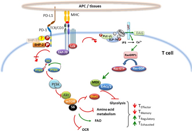
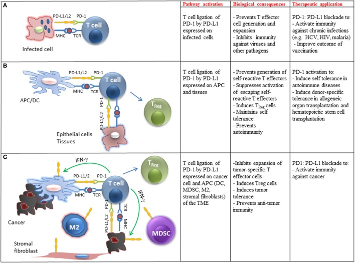
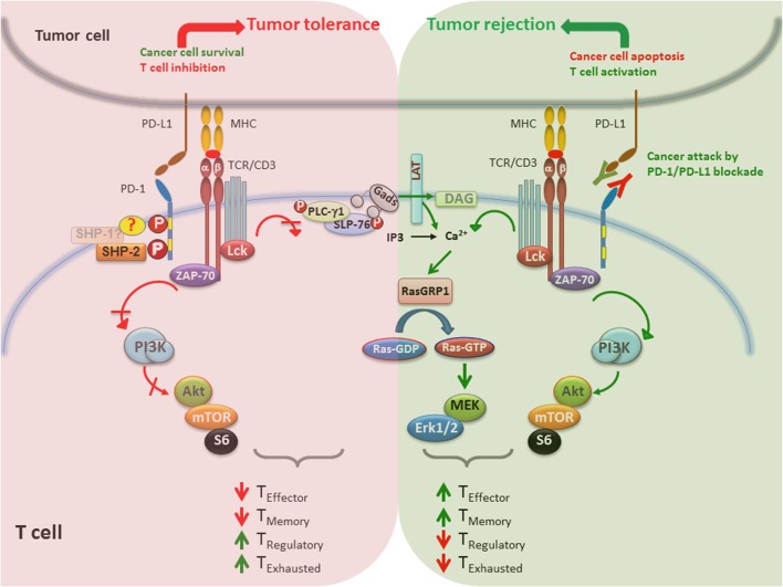
The immune system has the difficult challenge of discerning and defending against a diversity of microbial pathogens, while simultaneously avoiding self-reactivity. T lymphocytes function as effectors and regulators of the immune system. While central tolerance mechanism results in deletion of the majority of self-reactive T lymphocytes during thymic selection, a fraction of self reactive lymphocytes escapes to the periphery and retains the potential to inflict destructive autoimmune pathology. The immune system evolved various mechanisms to restrain such auto-reactive T cells and maintain peripheral tolerance, including T cell anergy, deletion, and suppression by regulatory T cells (TRegs). These effects are regulated by a complex network of stimulatory and inhibitory receptors expressed on T cells and their ligands, which deliver cell-to-cell signals that dictate the outcome of T cell encountering with cognate antigens. Among the inhibitory immune mediators, the pathway consisting of the programmed cell death 1 (PD-1) receptor (CD279) and its ligands PD-L1 (B7-H1, CD274) and PD-L2 (B7-DC; CD273) plays a vital role in the induction and maintenance of peripheral tolerance and for the maintenance of T cell homeostasis. In contrast to its beneficial role in self-tolerance, the PD-1: PD-L1/L2 pathway mediates potent inhibitory signals that prevent the expansion and function of T effector cells and have detrimental effects on antiviral and antitumor immunity. Therapeutic targeting of this pathway has resulted in successful enhancement of T cell immunity against viral pathogens and tumors. Here, we will provide a brief overview on the properties of the components of the PD-1 pathway, the signaling events that are regulated by PD-1 triggering, and their consequences on the function of T effector cells.
Overview
The claims and statistics of this paper haven’t been checked yet.
Run the check — free, no account needed. It reads the open-access text and checks the sources it cites.
Integrity signals
Registry
Cites
Statistics
Figures
Verification
Claims the paper makes
The claims and statistics of this paper haven’t been checked yet.
Run the check — free, no account needed. It reads the open-access text and checks the sources it cites.
Claims other papers make about this one
Each is checked against this paper’s own text: does it say what the citing paper says it does?
In the citing paper
“As a result, PD-1 signaling suppresses T cell functions, including activation, proliferation, and cytokine production [6].”
From Combination strategies with PD-1/PD-L1 blockade: current advances and future directions (2022) · cites this paper as [6]
Self-check
Percentages, sums and reported differences re-computed from the paper’s own numbers.
The self-check needs the paper’s full text.
Citations
Cites
PD-1 and PD-L1 Immune Checkpoint Blockade to Treat Breast Cancer
Hartkopf, A. D., Taran, F., Wallwiener, M., Walter, C. B., Krämer, B., Grischke, E., & Brucker, S. Y. (2016) · Breast Care · doi:10.1159/000453569 · Cited by 30,989
Adverse Renal Effects of Immune Checkpoint Inhibitors: A Narrative Review
Wanchoo, R., Karam, S., Uppal, N. N., Barta, V. S., Deray, G., Devoe, C., Launay‐Vacher, V., Jhaveri, K. D., & on behalf of Cancer and Kidney International Network Workgroup on Immune Checkpoint Inhibitors (2017) · American Journal of Nephrology · doi:10.1159/000455014 · Cited by 15,320
Improved Survival with Ipilimumab in Patients with Metastatic Melanoma
Hodi, F. S., O'Day, S. J., McDermott, D. F., Weber, R. W., Sosman, J. A., Haanen, J. B., Gonzalez, R., Robert, C., Schadendorf, D., Hassel, J. C., et al. (2010) · New England Journal of Medicine · doi:10.1056/nejmoa1003466 · Cited by 15,155
Safety, activity, and immune correlates of anti–PD-1 antibody in cancer
Topalian, S. L., Hodi, F. S., Brahmer, J. R., Gettinger, S. N., Smith, D. C., McDermott, D. F., Powderly, J. D., Carvajal, R. D., Sosman, J. A., Atkins, M. B., et al. (2012) · New England Journal of Medicine · doi:10.1056/nejmoa1200690 · Cited by 12,628
PD-1 Blockade in Tumors with Mismatch-Repair Deficiency
Le, D. T., Uram, J. N., Wang, H., Bartlett, B. R., Kemberling, H., Eyring, A. D., Skora, A. D., Luber, B. S., Azad, N. S., Laheru, D., et al. (2015) · New England Journal of Medicine · doi:10.1056/nejmoa1500596 · Cited by 9,377
Nivolumab versus docetaxel in advanced nonsquamous non-small-cell lung cancer
Borghaei, H., Paz-Ares, L., Horn, L., Spigel, D. R., Steins, M., Ready, N. E., Chow, L. Q., Vokes, E. E., Felip, E., Holgado, E., et al. (2015) · New England Journal of Medicine · doi:10.1056/nejmoa1507643 · Cited by 9,335
Nivolumab versus Docetaxel in Advanced Squamous-Cell Non–Small-Cell Lung Cancer
Brahmer, J., Reckamp, K. L., Baas, P., Crinò, L., Eberhardt, W. E. E., Poddubskaya, E., Antonia, S., Pluzanski, A., Vokes, E. E., Holgado, E., et al. (2015) · New England Journal of Medicine · doi:10.1056/nejmoa1504627 · Cited by 8,447
Control of Regulatory T Cell Development by the Transcription Factor Foxp3
Hori, S., Nomura, T., & Sakaguchi, S. (2003) · Science · doi:10.1126/science.1079490 · Cited by 8,187
Combined Nivolumab and Ipilimumab or Monotherapy in Untreated Melanoma
Larkin, J., Chiarion-Sileni, V., Gonzalez, R., Grob, J. J., Cowey, C. L., Lao, C. D., Schadendorf, D., Dummer, R., Smylie, M., Rutkowski, P., et al. (2015) · New England Journal of Medicine · doi:10.1056/nejmoa1504030 · Cited by 8,157
Safety and activity of anti-PD-L1 antibody in patients with advanced cancer
Brahmer, J. R., Tykodi, S. S., Chow, L. Q. M., Hwu, W.-J., Topalian, S. L., Hwu, P., Drake, C. G., Camacho, L. H., Kauh, J., Odunsi, K., et al. (2012) · New England Journal of Medicine · doi:10.1056/nejmoa1200694 · Cited by 7,945
Research works printed but not linked: 1
- Carter, 2002, PD-1:PD-L inhibitory pathway affects both CD4(+) and CD8(+) T cells and is overcome by IL-2, Eur J Immunol, 32, 634, 10.1002/1521-4141(200203)32:3634::AID-IMMU6343.0.CO;2-9 · DOI
Cited by
Sorted by citations
Combination strategies with PD-1/PD-L1 blockade: current advances and future directions
Yi, M., Zheng, X., Niu, M., Zhu, S., Ge, H., & Wu, K. (2022) · Molecular Cancer · doi:10.1186/s12943-021-01489-2 · Cited by 1,445
Regulatory mechanisms of PD-1/PD-L1 in cancers
Lin, X., Kang, K., Chen, P., Zeng, Z., Li, G., Xiong, W., Yi, M., & Xiang, B. (2024) · Molecular Cancer · doi:10.1186/s12943-024-02023-w · Cited by 909
Regulation of PD-L1 expression in the tumor microenvironment
Yi, M., Niu, M., Xu, L., Luo, S., & Wu, K. (2021) · Journal of Hematology & Oncology · doi:10.1186/s13045-020-01027-5 · Cited by 681
Regulatory mechanisms of immune checkpoints PD-L1 and CTLA-4 in cancer
Zhang, H., Dai, Z., Wu, W., Wang, Z., Zhang, N., Zhang, L., Zeng, W., Liu, Z., & Cheng, Q. (2021) · Journal of Experimental & Clinical Cancer Research · doi:10.1186/s13046-021-01987-7 · Cited by 551
The Role of Interleukin 6 During Viral Infections
Velázquez-Salinas, L., Verdugo-Rodríguez, A., Rodriguez, L. L., & Borca, M. (2019) · Frontiers in Microbiology · doi:10.3389/fmicb.2019.01057 · Cited by 488
Metadata
- Authors
- Bardhan, K., Anagnostou, T., & Boussiotis, V. A.
- Affiliations
- Beth Israel Deaconess Medical Center (US)
- Mayo Clinic (US)
- Corresponding author
- Boussiotis, Vassiliki A.
- Venue
- Frontiers in Immunology
- Publisher
- Frontiers Media SA
- Published
- 2016-12-12
- Volume
- 7
- Pages
- 550-550
- ISSN
- 1664-3224 (ISSN-L)
- PMC
- PMC5149523
- OpenAlex
- W2560827811
- Cited by
- 606
- References
- 210
- Citations by year
- 2026: 38 · 2025: 53 · 2024: 83 · 2023: 60 · 2022: 63 · 2021: 100 · 2020: 69 · 2019: 74 · 2018: 45 · 2017: 20
- FWCI
- 4.66
- Type
- journal article
- Language
- en
- Open access
- gold
- doi.org
- Licence
- cc-by
- Locations
- Frontiers in Immunology (publishedVersion, cc-by)
- PubMed Central (publishedVersion, cc-by)
- PubMed (publishedVersion)
- Digital Access to Scholarship at Harvard (DASH) (Harvard University) (submittedVersion, cc-by)
- DOAJ (DOAJ: Directory of Open Access Journals) (submittedVersion, cc-by-sa)
- Funders
- Foundation for the National Institutes of Health (CA183605, CA183605S1, AI098129-01)
- U.S. Department of Defense (PC140571)
- Topics
- Health Sciences › Medicine › Oncology › Cancer Immunotherapy and Biomarkers
- Life Sciences › Immunology and Microbiology › Immunology › T-cell and B-cell Immunology
- Life Sciences › Immunology and Microbiology › Immunology › Immunotherapy and Immune Responses
- Keywords
- PD-1, PD-L1, PD-L2, immune checkpoint, T cell tolerance, peripheral tolerance, T cell homeostasis, T cell anergy, regulatory T cells, antitumor immunity, antiviral immunity, immune checkpoint blockade, T cell signaling
About these checks
Registry, retraction, Cites and statistics results are computed, not judged, with one exception: a printed reference that no index lists is read once by a model before it counts as a citation error. Claims are extracted by a model and kept only when their quote appears verbatim in the text; each cited source is then checked, with a model’s help, for whether it says what the claim relies on. Those verdicts can be wrong — read the claim’s quote and the cited source before relying on one.
Statistics and arithmetic are recomputed, not judged.
Confidence bands are shown only for fields calibrated on our annotated test set. None is calibrated yet, so no band is shown.
What counts as a claim ›How we measure confidence ›How we check this ›
Are you an author, or do you see a problem with this page? Request a review
