

The DOI matches its Crossref record; 2 of the 2 references it cites were found in a registry or an index.
YTHDF1 Negatively Regulates Treponema pallidum-Induced Inflammation in THP-1 Macrophages by Promoting SOCS3 Translation in an m6A-Dependent Manner
Abstract
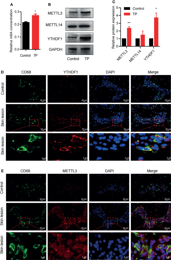
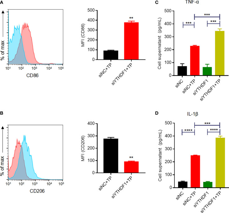
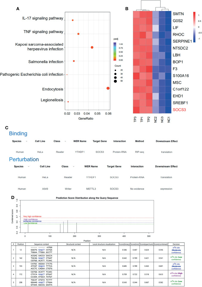
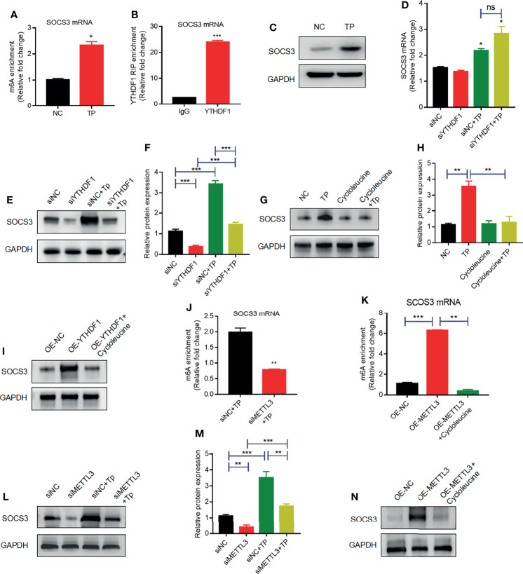
Background: Previous studies have confirmed that the bacterium Treponema pallidum (TP) or its proteins provide signals to macrophages that induce an inflammatory response; however, little is known about the negative regulation of this macrophage-mediated inflammatory response during syphilis infection or the underlying mechanism. Recent evidence suggests the role of the RNA modification, N 6-adenosine methylation (m6A), in regulating the inflammatory response and pathogen-host cell interactions. Therefore, we hypothesized that m6A plays a role in the regulation of the inflammatory response in macrophages exposed to TP. Methods: We first assessed m6A levels in TP-infected macrophages differentiated from the human monocyte cell line THP-1. The binding and interaction between the m6A “writer” methyltransferase-like 3 (METTL3) or the m6A “reader” YT521-B homology (YTH) domain-containing protein YTHDF1 and the suppressor of cytokine signaling 3 (SOCS3), as a major regulator of the inflammatory response, were explored in differentiated TP-infected THP-1 cells as well as in secondary syphilitic lesions from patients. The mechanisms by which YTHDF1 and SOCS3 regulate the inflammatory response in macrophages were assessed. Results and Conclusion: After macrophages were stimulated by TP, YTHDF1 was upregulated in the cells. YTHDF1 was also upregulated in the syphilitic lesions compared to adjacent tissue in patients. YTHDF1 recognizes and binds to the m6A methylation site of SOCS3 mRNA, consequently promoting its translation, thereby inhibiting the JAK2/STAT3 pathway, and reducing the secretion of inflammatory factors, which results in anti-inflammatory regulation. This study provides the first demonstration of the role of m6A methylation in the pathological process of syphilis and further offers new insight into the pathogenesis of TP infection.
Overview
The claims and statistics of this paper haven’t been checked yet.
Run the check — free, no account needed. It reads the open-access text and checks the sources it cites.
Integrity signals
Registry
Cites
Statistics
Figures
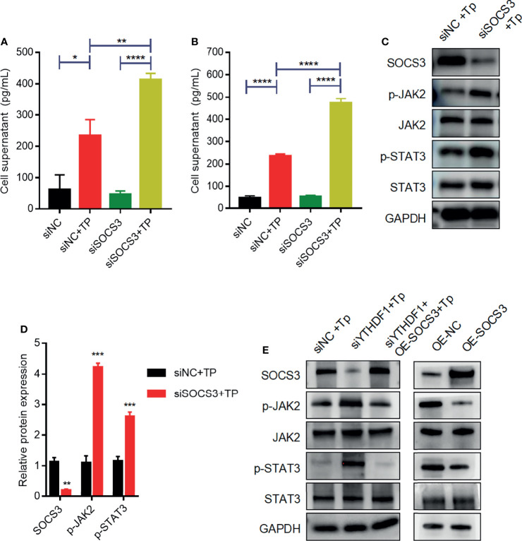
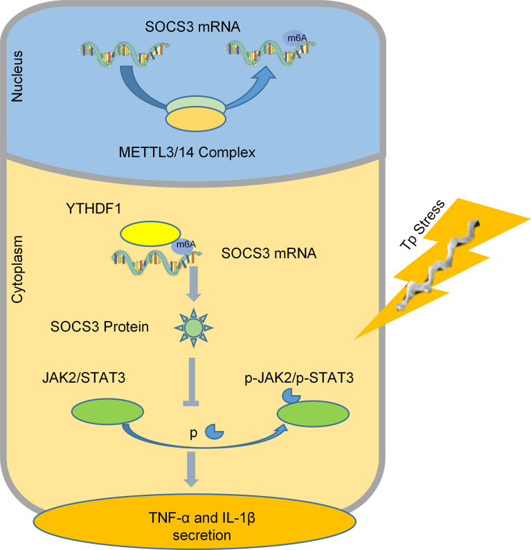
Verification
Claims the paper makes
The claims and statistics of this paper haven’t been checked yet.
Run the check — free, no account needed. It reads the open-access text and checks the sources it cites.
Claims other papers make about this one
Each is checked against this paper’s own text: does it say what the citing paper says it does?
In the citing paper
“Epigenetic regulation and m6A modification, along with dysregulation of related regulators like YTHDF1, play crucial roles in tumor onset, progression, and chemotherapy tolerance [232, 233].”
From Emerging role of exosomes in cancer therapy: progress and challenges (2025) · cites this paper as [233]
Self-check
Percentages, sums and reported differences re-computed from the paper’s own numbers.
The self-check needs the paper’s full text.
Citations
Cites
Recognition of RNA N6-methyladenosine by IGF2BP proteins enhances mRNA stability and translation
Huang, H., Weng, H., Sun, W., Qin, X., Shi, H., Wu, H., Zhao, B. S., Mesquita, A., Liu, C., Yuan, C. L., et al. (2018) · Nature Cell Biology · doi:10.1038/s41556-018-0045-z · Cited by 2,997
SOCS1/JAB Is a Negative Regulator of LPS-Induced Macrophage Activation
Kinjyo, I., Hanada, T., Inagaki-Ohara, K., Mori, H., Aki, D., Ohishi, M., Yoshida, H., Kubo, M., & Yoshimura, A. (2002) · Immunity · doi:10.1016/s1074-7613(02)00446-6 · Cited by 630
Research works printed but not linked: 32
- Keuning, 2020, Congenital Syphilis, the Great Imitator-Case Report and Review, Lancet Infect Dis, 20, 10.1016/S1473-3099(20)30268-1 · DOI
- Xu, 2017, Treponema Pallidum Flagellins Elicit Proinflammatory Cytokines From Human Monocytes via TLR5 Signaling Pathway, Immunobiology, 222, 10.1016/j.imbio.2017.01.002 · DOI
- Xu, 2020, The P2X7 Receptor Mediates NLRP3-Dependent IL-1beta Secretion and Promotes Phagocytosis in the Macrophage Response to Treponema Pallidum, Int Immunopharmacol, 82, 10.1016/j.intimp.2020.106344 · DOI
- Lin, 2018, Development of Tissue Inflammation Accompanied by NLRP3 Inflammasome Activation in Rabbits Infected With Treponema Pallidum Strain Nichols, BMC Infect Dis, 18, 101, 10.1186/s12879-018-2993-0 · DOI
- Carlson, 2011, The Immunopathobiology of Syphilis: The Manifestations and Course of Syphilis Are Determined by the Level of Delayed-Type Hypersensitivity, Am J Dermatopathol, 33, 10.1097/DAD.0b013e3181e8b587 · DOI
- Sell, 1983, The Biology, Pathology, and Immunology of Syphilis, Int Rev Exp Pathol, 24
- Huang, 2020, MicroRNA-101-3p Downregulates TLR2 Expression, Leading to Reduction in Cytokine Production by Treponema Pallidum-Stimulated Macrophages, J Invest Dermatol, 140, 10.1016/j.jid.2019.12.012 · DOI
- Han, 2021, ALKBH5 Regulates Cardiomyocyte Proliferation and Heart Regeneration by Demethylating the mRNA of YTHDF1, Theranostics, 11, 10.7150/thno.47354 · DOI
- Hu, 2021, YTHDF1 Regulates Pulmonary Hypertension Through Translational Control of MAGED1, Am J Respir Crit Care Med, 203, 10.1164/rccm.202009-3419OC · DOI
- Xu, 2020, N6-Methyladenosine RNA Modification in Cancer Therapeutic Resistance: Current Status and Perspectives, Biochem Pharmacol, 182, 10.1016/j.bcp.2020.114258 · DOI
- Zhao, 2017, Post-Transcriptional Gene Regulation by mRNA Modifications, Nat Rev Mol Cell Biol, 18, 31, 10.1038/nrm.2016.132 · DOI
- Liu, 2020, The M6a Reader YTHDF1 Promotes Ovarian Cancer Progression via Augmenting EIF3C Translation, Nucleic Acids Res, 48, 10.1093/nar/gkaa048 · DOI
- Zong, 2021, The N6-Methyladenosine RNA-Binding Protein YTHDF1 Modulates the Translation of TRAF6 to Mediate the Intestinal Immune Response, Nucleic Acids Res, 49, 10.1093/nar/gkab343 · DOI
- Hook, 2017, Syphilis, Lancet, 389, 10.1016/S0140-6736(16)32411-4 · DOI · Paper page
- Peeling, 2017, Syphilis, Nat Rev Dis Primers, 3, 10.1038/nrdp.2017.73 · DOI · Paper page
- Fu, 2020, Understanding SARS-CoV-2-Mediated Inflammatory Responses: From Mechanisms to Potential Therapeutic Tools, Virol Sin, 35, 10.1007/s12250-020-00207-4 · DOI
- Channappanavar, 2016, Dysregulated Type I Interferon and Inflammatory Monocyte-Macrophage Responses Cause Lethal Pneumonia in SARS-CoV-Infected Mice, Cell Host Microbe, 19, 10.1016/j.chom.2016.01.007 · DOI
- Du, 2020, N(6)-Adenosine Methylation of Socs1 mRNA Is Required to Sustain the Negative Feedback Control of Macrophage Activation, Dev Cell, 55, 10.1016/j.devcel.2020.10.023 · DOI
- Lafond, 2006, Biological Basis for Syphilis, Clin Microbiol Rev, 19, 29, 10.1128/CMR.19.1.29-49.2006 · DOI · Paper page
- Cruz, 2012, Immune Evasion and Recognition of the Syphilis Spirochete in Blood and Skin of Secondary Syphilis Patients: Two Immunologically Distinct Compartments, PloS Negl Trop Dis, 6, 10.1371/journal.pntd.0001717 · DOI
- Wu, 2020, Interplay of m(6)A and H3K27 Trimethylation Restrains Inflammation During Bacterial Infection, Sci Adv, 6, a647, 10.1126/sciadv.aba0647 · DOI
- Liu, 2019, N (6)-Methyladenosine RNA Modification-Mediated Cellular Metabolism Rewiring Inhibits Viral Replication, Science, 365, 10.1126/science.aax4468 · DOI
- Wang, 2019, METTL3 Attenuates LPS-Induced Inflammatory Response in Macrophages via NF-kappaB Signaling Pathway, Mediators Inflamm, 2019, 10.1155/2019/3120391 · DOI
- Wang, 2019, Transcriptome-Wide Reprogramming of N(6)-Methyladenosine Modification by the Mouse Microbiome, Cell Res, 29, 10.1038/s41422-018-0127-2 · DOI
- Klepsch, 2019, Intragenic Regulation of SOCS3 Isoforms, Cell Commun Signal, 17, 70, 10.1186/s12964-019-0379-6 · DOI
- Gao, 2018, The Roles of SOCS3 and STAT3 in Bacterial Infection and Inflammatory Diseases, Scand J Immunol, 88, 10.1111/sji.12727 · DOI
- Durham, 2019, Targeting SOCS Proteins to Control JAK-STAT Signalling in Disease, Trends Pharmacol Sci, 40, 298, 10.1016/j.tips.2019.03.001 · DOI · Paper page
- Zhang, 2018, Macrophage/microglial Ezh2 Facilitates Autoimmune Inflammation Through Inhibition of Socs3, J Exp Med, 215, 10.1084/jem.20171417 · DOI
- Zhang, 2020, SOCS3 Protects Against Neonatal Necrotizing Enterocolitis via Suppressing NLRP3 and AIM2 Inflammasome Activation and P65 Nuclear Translocation, Mol Immunol, 122, 10.1016/j.molimm.2020.03.019 · DOI
- Li, 2017, m(6)A mRNA Methylation Controls T Cell Homeostasis by Targeting the IL-7/STAT5/SOCS Pathways, Nature, 548, 10.1038/nature23450 · DOI
- Li, 2021, TRPV4-Induced Müller Cell Gliosis and TNF-α Elevation-Mediated Retinal Ganglion Cell Apoptosis in Glaucomatous Rats via JAK2/STAT3/NF-κB Pathway, J Neuroinflamm, 18, 271, 10.1186/s12974-021-02315-8 · DOI
- Yin, 2018, Manganese Exposure Facilitates Microglial JAK2-STAT3 Signaling and Consequent Secretion of TNF-A and IL-1β to Promote Neuronal Death, Neurotoxicology, 64, 195, 10.1016/j.neuro.2017.04.001 · DOI
Cited by
Sorted by citations
Emerging role of exosomes in cancer therapy: progress and challenges
Li, J., Wang, J., & Chen, Z. (2025) · Molecular Cancer · doi:10.1186/s12943-024-02215-4 · Cited by 297
Metadata
- Authors
- Li, Z., Teng, M., Jiang, Y., Zhang, L., Luo, X., Liao, Y., & Yang, B.
- Venue
- Frontiers in Immunology
- Publisher
- Frontiers Media SA
- Published
- 2022-04-04
- Volume
- 13
- ISSN
- 1664-3224
- PMC
- PMC9013966
- Cited by
- 39
- References
- 2
- Funders
- National Natural Science Foundation of China (81772240)
About these checks
Registry, retraction, Cites and statistics results are computed, not judged, with one exception: a printed reference that no index lists is read once by a model before it counts as a citation error. Claims are extracted by a model and kept only when their quote appears verbatim in the text; each cited source is then checked, with a model’s help, for whether it says what the claim relies on. Those verdicts can be wrong — read the claim’s quote and the cited source before relying on one.
Statistics and arithmetic are recomputed, not judged.
Confidence bands are shown only for fields calibrated on our annotated test set. None is calibrated yet, so no band is shown.
What counts as a claim ›How we measure confidence ›How we check this ›
Are you an author, or do you see a problem with this page? Request a review
