

The DOI matches its Crossref record.
Functional characterization and stability improvement of a ‘thermophilic-like’ ene-reductase from Rhodococcus opacus 1CP
Open accessAbstract
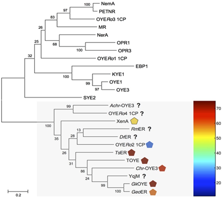
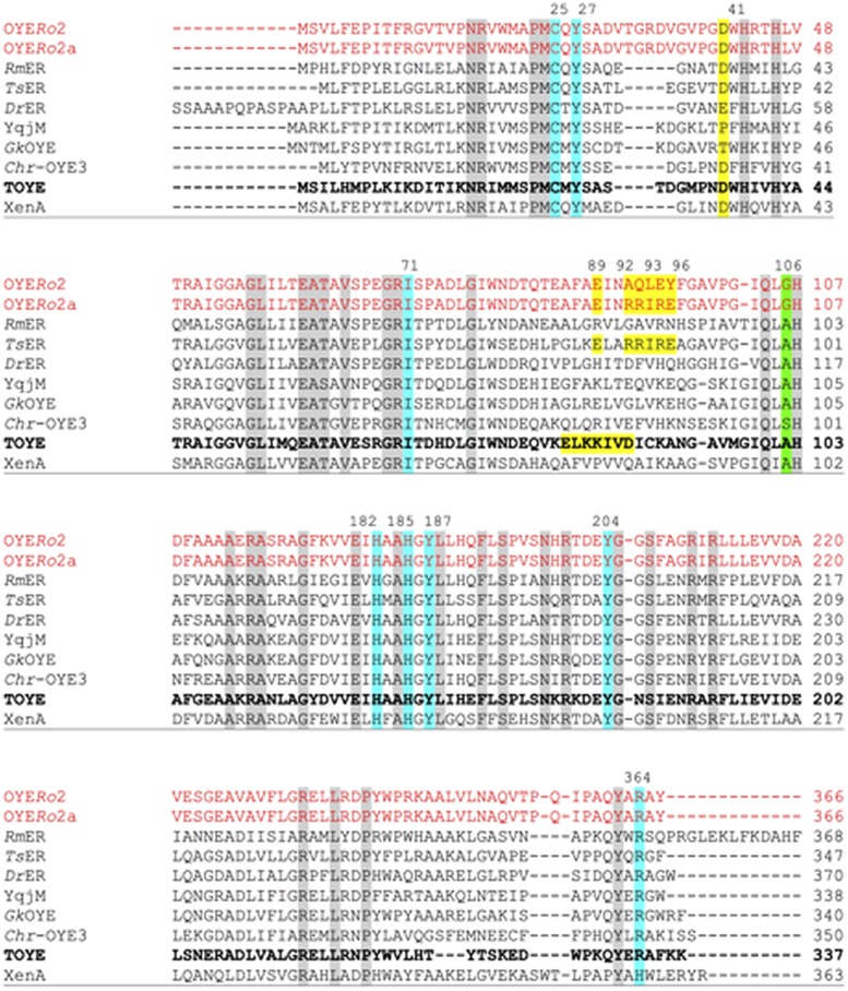
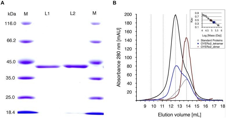
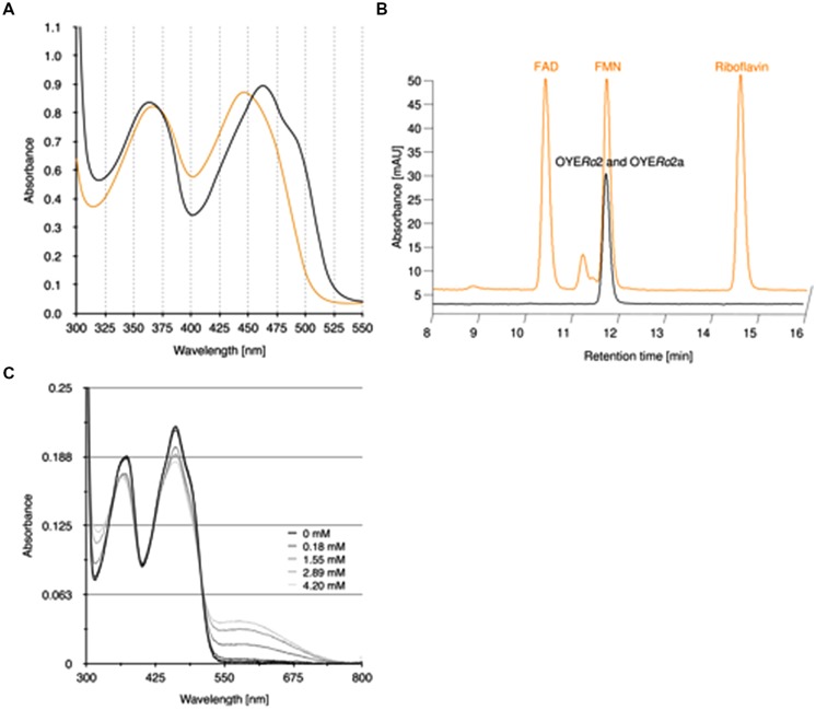
Ene-reductases (ERs) are widely applied for the asymmetric synthesis of relevant industrial chemicals. A novel ER OYERo2 was found within a set of 14 putative old yellow enzymes (OYEs) obtained by genome mining of the actinobacterium Rhodococcus opacus 1CP. Multiple sequence alignment suggested that the enzyme belongs to the group of 'thermophilic-like' OYEs. OYERo2 was produced in Escherichia coli and biochemically characterized. The enzyme is strongly NADPH dependent and uses non-covalently bound FMNH2 for the reduction of activated α,β-unsaturated alkenes. In the active form OYERo2 is a dimer. Optimal catalysis occurs at pH 7.3 and 37°C. OYERo2 showed highest specific activities (45-50 U mg(-1)) on maleimides, which are efficiently converted to the corresponding succinimides. The OYERo2-mediated reduction of prochiral alkenes afforded the (R)-products with excellent optical purity (ee > 99%). OYERo2 is not as thermo-resistant as related OYEs. Introduction of a characteristic intermolecular salt bridge by site-specific mutagenesis raised the half-life of enzyme inactivation at 32°C from 28 to 87 min and improved the tolerance toward organic co-solvents. The suitability of OYERo2 for application in industrial biocatalysis is discussed.
Overview
The claims and statistics of this paper haven’t been checked yet.
Run the check — free, no account needed. It reads the open-access text and checks the sources it cites.
Integrity signals
Registry
Cites
Statistics
Figures
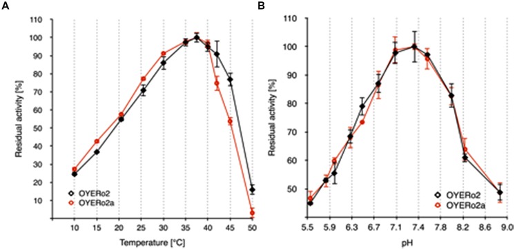
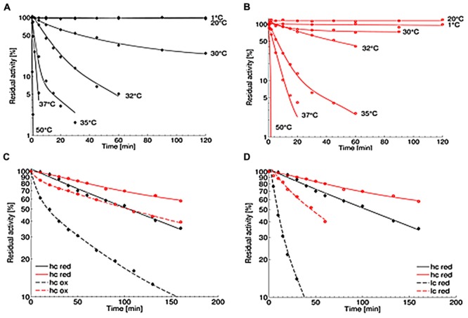
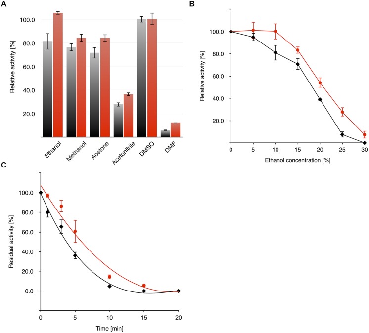
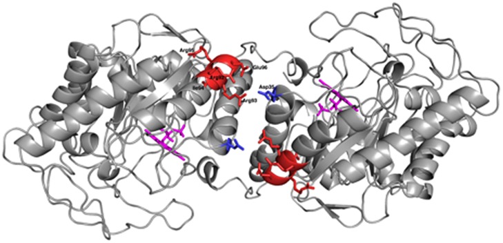
Verification
Claims the paper makes
The claims and statistics of this paper haven’t been checked yet.
Run the check — free, no account needed. It reads the open-access text and checks the sources it cites.
Claims other papers make about this one
Each is checked against this paper’s own text: does it say what the citing paper says it does?
The check wasn’t confident either way, so it gives no verdict — never disputed.
In the citing paper
“sugar is the second main component of jelly candy (sucrose, dextrose and glucose or fructose syrups) and the mixture is boiling at high temperatures, then flavoring and coloring agents were added [7].”
From Functional strawberry and red beetroot jelly candies rich in fibers and phenolic compounds (2021) · cites this paper as [7]
Experimental — accuracy not yet measuredThe check wasn’t confident either way, so it gives no verdict — never disputed.
In the citing paper
“Jelly candies have a poor nutritional value due to their primary ingredients, which include gelling agents and sugar.”
From Functional strawberry and red beetroot jelly candies rich in fibers and phenolic compounds (2021) · cites this paper as [7]
Experimental — accuracy not yet measured
Self-check
Percentages, sums and reported differences re-computed from the paper’s own numbers.
The self-check needs the paper’s full text.
Citations
Cites
No references could be resolved yet.
Research works printed but not linked: 55
- Abramovitz, 1976, Interaction of phenols with old yellow enzyme. Physical evidence for charge-transfer complexes., J. Biol. Chem., 251, 5327, 10.1016/S0021-9258(17)33165-4 · DOI
- Adalbjörnsson, 2010, Biocatalysis with thermostable enzymes: structure and properties of a thermophilic ‘ene’-reductase related to old yellow enzyme., Chembiochem, 11, 197, 10.1002/cbic.200900570 · DOI
- Altschul, 1990, Basic local alignment search tool., J. Mol. Biol., 215, 403, 10.1016/S0022-2836(05)80360-2 · DOI · Paper page
- Altschul, 1997, Gapped BLAST and PSI-BLAST: a new generation of protein database search programs., Nucleic Acids Res., 25, 3389, 10.1093/nar/25.17.3389 · DOI · Paper page
- Barna, 2001, Crystal structure of pentaerythritol tetranitrate reductase: “flipped” binding geometries for steroid substrates in different redox states of the enzyme., J. Mol. Biol., 310, 433, 10.1006/jmbi.2001.4779 · DOI
- Barna, 2002, Crystal structure of bacterial morphinone reductase and properties of the C191A mutant enzyme., J. Biol. Chem., 277, 30976, 10.1074/jbc.M202846200 · DOI
- Breithaupt, 2001, X-ray structure of 12-oxophytodienoate reductase 1 provides structural insight into substrate binding and specificity within the family of OYE., Structure, 9, 419, 10.1016/S0969-2126(01)00602-5 · DOI
- Chaparro-Riggers, 2007, Comparison of three enoate reductases and their potential use for biotransformations., Adv. Synth. Catal., 349, 1521, 10.1002/adsc.200700074 · DOI
- Chauhan, 2013, Asymmetric organocatalytic addition reactions of maleimides: a promising approach towards the synthesis of chiral succinimide derivatives., Chem. Asian J., 8, 328, 10.1002/asia.201200684 · DOI
- Eswar, 2006, Comparative protein structure modeling using Modeller., Curr. Protoc. Bioinformatics, 15(suppl.), 5.6.1, 10.1002/0471250953.bi0506s15 · DOI
- Fox, 1994, Old yellow enzyme at 2 Å Resolution: overall structure, ligand binding, and comparison with related flavoproteins., Structure, 2, 1089, 10.1016/S0969-2126(94)00111-1 · DOI
- Fryszkowska, 2009, Asymmetric reduction of activated alkenes by pentaerythritol tetranitrate reductase: specificity and control of stereochemical outcome by reaction optimisation., Adv. Synth. Catal., 351, 2976, 10.1002/adsc.200900574 · DOI
- Fu, 2012, A novel ene-reductase from Synechococcus sp. PCC 7942 for the asymmetric reduction of alkenes., Process. Biochem., 47, 1988, 10.1016/j.procbio.2012.07.009 · DOI
- Gröning, 2014, Gene redundancy of two-component (chloro)phenol hydroxylases in Rhodococcus opacus 1CP., FEMS Microbiol. Lett., 361, 68, 10.1111/1574-6968.12616 · DOI
- Haas, 1938, Isolierung eines neuen gelben Ferments., Biochem. Z., 6, 378
- Hall, 2007, Asymmetric bioreduction of activated alkenes using cloned 12-oxophytodienoate reductase isoenzymes OPR-1 and OPR-3 from Lycopersicon esculentum (tomato): a striking change of stereoselectivity., Angew. Chem. Int. Ed., 46, 3934, 10.1002/anie.200605168 · DOI
- Kataoka, 2002, Old Yellow Enzyme from Candida macedoniensis catalyzes the stereospecific reduction of the C = C bond of ketoisophorone., Biosci. Biotechnol. Biochem., 66, 2651, 10.1271/bbb.66.2651 · DOI
- Kataoka, 2004, Cloning and overexpression of the old yellow enzyme gene of Candida macedoniensis, and its application to the production of a chiral compound., J. Biotechnol., 114, 1, 10.1016/j.jbiotec.2004.04.033 · DOI
- Kitzing, 2005, The 1.3 Å crystal structure of the flavoprotein YqjM reveals a novel class of old yellow enzymes., J. Biol. Chem., 280, 27904, 10.1074/jbc.M502587200 · DOI
- Kokotos, 2013, An asymmetric Michael addition of α, α-disubstituted aldehydes to maleimides leading to a one-pot enantioselective synthesis of lactones catalyzed by amino acids., Org. Lett., 15, 2406, 10.1021/ol4008662 · DOI
- Kurata, 2004, Asymmetric reduction of 2-chloroacrylic acid to (S)-2-chloropropionic acid by a novel reductase from Burkholderia sp. WS., Tetrahedron Asymmetry, 15, 2837, 10.1016/j.tetasy.2004.06.035 · DOI
- Leuenberger, 1976, Synthesis of optically active natural carotenoids and structurally related compounds. 1. Synthesis of the chiral key compound (4R, 6R)-4-hydroxy-2,2,6-trimethylcyclohexanone., Helv. Chim. Acta., 59, 1832, 10.1002/hlca.19760590541 · DOI
- Litthauer, 2014, Heterologous expression and characterization of the ene-reductase from Deinococcus radiodurans and Ralstonia metallidurans., J. Mol. Catal. B Enzym., 99, 89, 10.1016/j.molcatb.2013.10.020 · DOI
- McLeod, 2006, The complete genome of Rhodococcus sp. RHA1 provides insights into a catabolic powerhouse., Proc. Natl. Acad. Sci. U.S.A., 103, 15582, 10.1073/pnas.0607048103 · DOI
- Mueller, 2010, The substrate spectra of pentaerythritol tetranitrate reductase, morphinone reductase, N-ethylmaleimide reductase and estrogen-binding protein in the asymmetric bioreduction of activated alkenes., Adv. Synth. Catal., 325, 387, 10.1002/adsc.200900832 · DOI
- Nivinskas, 2008, Reduction of aliphatic nitroesters and N-nitramines by Enterobacter cloacae PB2 pentaerythritol tetranitrate reductase: quantitative structure-activity relationships., FEBS J., 24, 6192, 10.1111/j.1742-4658.2008.06744.x · DOI
- Oelschlägel, 2015, Production of a recombinant membrane protein in an Escherichia coli strain for the whole cell biosynthesis of phenylacetic acids., Biotechnol. Rep., 7, 38, 10.1016/j.btre.2015.05.002 · DOI
- Oganesyan, 2007, Effect of osmotic stress and heat shock in recombinant protein overexpression and crystallization., Protein Expr. Purif., 52, 280, 10.1016/j.pep.2006.09.015 · DOI
- Opperman, 2008, A novel chromate reductase from Thermus scotoductus SA-01 related to old yellow enzyme., J. Bacteriol., 190, 3076, 10.1128/JB.01766-07 · DOI
- Opperman, 2010, Crystal structure of a thermostable old yellow enzyme from Thermus scotoductus SA-01., Biochem. Biophys. Res. Commun., 393, 426, 10.1016/j.bbrc.2010.02.011 · DOI
- Paul, 2013, Mimicking nature: synthetic nicotinamide cofactors for C = C bioreduction using enoate reductases., Org. Lett., 15, 180, 10.1021/ol303240a · DOI
- Rice, 2000, EMBOSS: the european molecular biology open software suite., Trends Genet., 16, 276, 10.1016/S0168-9525(00)02024-2 · DOI · Paper page
- Riedel, 2015, Catalytic and hydrodynamic properties of styrene monooxygenases from Rhodococcus opacus 1CP are modulated by cofactor binding., AMB Express, 5, 10.1186/s13568-015-0112-9 · DOI
- Sali, 1993, Comparative protein modelling by satisfaction of spatial restraints., J. Mol. Biol., 234, 779, 10.1006/jmbi.1993.1626 · DOI · Paper page
- Schittmayer, 2011, Old yellow enzyme-catalyzed dehydrogenation of saturated ketones., Adv. Synth. Catal., 353, 268, 10.1002/adsc.201000862 · DOI
- Spiegelhauer, 2010, Cysteine as a modulator residue in the active site of xenobiotic reductase A: a structural, thermodynamic and kinetic study., J. Mol. Biol., 398, 66, 10.1016/j.jmb.2010.02.044 · DOI
- Steinkellner, 2014, Identification of promiscuous ene-reductase activity by mining structural databases using active site constellations., Nat. Commun., 5, 10.1038/ncomms5150 · DOI
- Stott, 1993, Old yellow enzyme – the discovery of multiple isozymes and a family of related proteins., J. Biol. Chem., 268, 6097, 10.1016/S0021-9258(18)53224-5 · DOI
- Straßner, 1999, A homolog of old yellow enzyme in tomato. Spectral properties and substrate specificity of the recombinant protein., J. Biol. Chem., 274, 35067, 10.1074/jbc.274.49.35067 · DOI
- Stückler, 2007, Stereocomplementary bioreduction of α,β-unsaturated dicarbolic acids and dimethyl esters using enoate reductases: enzyme- and substrate-based stereocontrol., Org. Lett., 26, 5409, 10.1021/ol7019185 · DOI
- Stürmer, 2007, Asymmetric bioreduction of activated C = C bonds using enoate reductases from the old yellow enzyme family., Curr. Opin. Chem. Biol., 11, 203, 10.1016/j.cbpa.2007.02.025 · DOI
- Takabe, 1992, Baker’s yeast reduction of 3-phenylthiomethyl-2-butenolide and its derivatives: synthesis of versatile chiral C5-building blocks for terpenoid synthesis., Tetrahedron Asymmetry, 3, 1399, 10.1016/0957-4166(92)80016-P · DOI
- Tao, 2008, Oxidant-induced formation of a neutral flavosemiquinone in the Na+-translocating NADH: quinone oxidoreductase (Na+-NQR) from Vibrio cholera., Biochim. Biophys. Acta, 1777, 696, 10.1016/j.bbabio.2008.04.006 · DOI
- Tischler, 2009, Identification of a novel self-sufficient styrene monooxygenase from Rhodococcus opacus 1CP., J. Bacteriol., 191, 4996, 10.1128/JB.00307-09 · DOI
- Tischler, 2010, StyA1 and StyA2B from Rhodococcus opacus 1CP: a multifunctional styrene monooxygenase system., J. Bacteriol., 192, 5220, 10.1128/JB.00723-10 · DOI
- Toogood, 2010, Biocatalytic reductions and chemical versatility of the Old Yellow Enzyme family of flavoprotein oxidoreductases., ChemCatChem, 2, 892, 10.1002/cctc.201000094 · DOI
- Tsuji, 2014, Isolation and characterization of a thermotolerant ene reductase from Geobacillus sp. 30 and its heterologous expression in Rhodococcus opacus., Appl. Microbiol. Biotechnol., 98, 5925, 10.1007/s00253-014-5668-9 · DOI
- Vaz, 1995, Old yellow enzymes: aromatisation of cyclic enones and the mechanism of a novel dismutation reaction., Biochemistry, 34, 4246, 10.1021/bi00013a014 · DOI
- Vízcaíno-Milla, 2015, Primary amine–2-aminopyrimidine chiral organocatalysts for the enantioselective conjugate addition of branched aldehydes to maleimides., Synthesis, 47, 2199, 10.1055/s-0034-1380718 · DOI
- Warburg, 1932, Über das neue Oxydationsferment., Naturwissenschaften, 20, 980, 10.1007/BF01504728 · DOI
- Whitby, 1953, A new method for preparing flavin-adenine dinucleotide., Biochem. J., 54, 10.1042/bj0540437 · DOI
- Williams, 2002, ‘New uses for an Old Enzyme’ – the Old Yellow Enzyme familiy of flavoenzymes., Microbiology, 148, 1607, 10.1099/00221287-148-6-1607 · DOI
- Xu, 2014, Identification and characterization of a novel ‘thermophilic-like’ Old Yellow Enzyme from the genome of Chryseobacterium sp. CA49., J. Mol. Catal. B Enzym., 108, 64, 10.1016/j.molcatb.2014.07.002 · DOI
- Yanto, 2010, Characterization of xenobiotic reductase A (XenA): study of active site residues, substrate spectrum and stability., Chem. Commun., 46, 8809, 10.1039/c0cc02354j · DOI
- Zhang, 2014, Enzymatic hydrogenation of diverse activated alkenes. identification of two Bacillus old yellow enzymes with broad substrate profiles., J. Mol. Catal. B Enzym., 105, 118, 10.1016/j.molcatb.2014.04.004 · DOI
Cited by
Sorted by citations
Functional strawberry and red beetroot jelly candies rich in fibers and phenolic compounds
Ali, M. R., Mohamed, R. M., & Abedelmaksoud, T. G. (2021) · Food systems · doi:10.21323/2618-9771-2021-4-1-82-88 · Cited by 44
“sugar is the second main component of jelly candy (sucrose, dextrose and glucose or fructose syrups) and the mixture is boiling at high temperatures, then flavoring and coloring agents were added [7].”
Background
Metadata
- Authors
- Riedel, A., Mehnert, M., Paul, C. E., Westphal, A. H., van Berkel, W. J. H., & Tischler, D.
- Affiliations
- TU Bergakademie Freiberg (DE)
- Delft University of Technology (NL)
- Wageningen University & Research (NL)
- Corresponding author
- Riedel, Anika
- Tischler, Dirk
- Venue
- Frontiers in Microbiology
- Publisher
- Frontiers Media SA
- Published
- 2015-10-01
- Volume
- 6
- Pages
- 1073-1073
- ISSN
- 1664-302X (ISSN-L)
- PMC
- PMC4589676
- OpenAlex
- W2178030934
- Cited by
- 38
- References
- 0
- Citations by year
- 2026: 2 · 2025: 2 · 2024: 7 · 2023: 1 · 2022: 4 · 2021: 5 · 2020: 3 · 2019: 5 · 2018: 3 · 2017: 3 · 2016: 3
- FWCI
- 1.30
- Type
- journal article
- Language
- en
- Open access
- gold
- doi.org
- Licence
- cc-by
- Locations
- Frontiers in Microbiology (publishedVersion, cc-by)
- Europe PMC (PubMed Central) (publishedVersion, cc-by)
- PubMed Central (publishedVersion, cc-by)
- PubMed (publishedVersion)
- Socio-Environmental Systems Modeling (acceptedVersion, cc-by)
- DOAJ (DOAJ: Directory of Open Access Journals) (submittedVersion, cc-by-sa)
- Data Archiving and Networked Services (DANS) (submittedVersion, cc-by)
- Funders
- Technische Universität Bergakademie Freiberg
- European Social Fund (100101363)
- Topics
- Life Sciences › Biochemistry, Genetics and Molecular Biology › Molecular Biology › Enzyme Catalysis and Immobilization
- Life Sciences › Biochemistry, Genetics and Molecular Biology › Biotechnology › Enzyme Production and Characterization
- Physical Sciences › Chemistry › Inorganic Chemistry › Asymmetric Hydrogenation and Catalysis
- Keywords
- ene-reductases, old yellow enzyme, asymmetric synthesis, succinimide synthesis, NADPH-dependent enzyme, enzyme dimerization, enzyme thermostability, salt bridges, site-directed mutagenesis, industrial biocatalysis
About these checks
Registry, retraction, Cites and statistics results are computed, not judged, with one exception: a printed reference that no index lists is read once by a model before it counts as a citation error. Claims are extracted by a model and kept only when their quote appears verbatim in the text; each cited source is then checked, with a model’s help, for whether it says what the claim relies on. Those verdicts can be wrong — read the claim’s quote and the cited source before relying on one.
Statistics and arithmetic are recomputed, not judged.
Confidence bands are shown only for fields calibrated on our annotated test set. None is calibrated yet, so no band is shown.
What counts as a claim ›How we measure confidence ›How we check this ›
Are you an author, or do you see a problem with this page? Request a review
