

The DOI matches its Crossref record; 221 of the 221 references it cites were found in a registry or an index.
The Emerging Role of Central and Peripheral Immune Systems in Neurodegenerative Diseases
Open accessAbstract
For decades, it has been widely believed that the blood–brain barrier (BBB) provides an immune privileged environment in the central nervous system (CNS) by blocking peripheral immune cells and humoral immune factors. This view has been revised in recent years, with increasing evidence revealing that the peripheral immune system plays a critical role in regulating CNS homeostasis and disease. Neurodegenerative diseases are characterized by progressive dysfunction and the loss of neurons in the CNS. An increasing number of studies have focused on the role of the connection between the peripheral immune system and the CNS in neurodegenerative diseases. On the one hand, peripherally released cytokines can cross the BBB, cause direct neurotoxicity and contribute to the activation of microglia and astrocytes. On the other hand, peripheral immune cells can also infiltrate the brain and participate in the progression of neuroinflammatory and neurodegenerative diseases. Neurodegenerative diseases have a high morbidity and disability rate, yet there are no effective therapies to stop or reverse their progression. In recent years, neuroinflammation has received much attention as a therapeutic target for many neurodegenerative diseases. In this review, we highlight the emerging role of the peripheral and central immune systems in neurodegenerative diseases, as well as their interactions. A better understanding of the emerging role of the immune systems may improve therapeutic strategies for neurodegenerative diseases.
Overview
The claims and statistics of this paper haven’t been checked yet.
Run the check — free, no account needed. It reads the open-access text and checks the sources it cites.
Integrity signals
Registry
Cites
Statistics
Figures
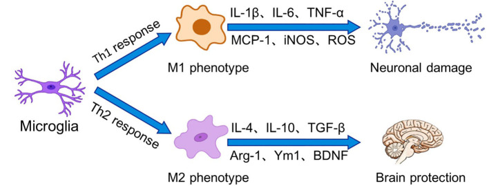
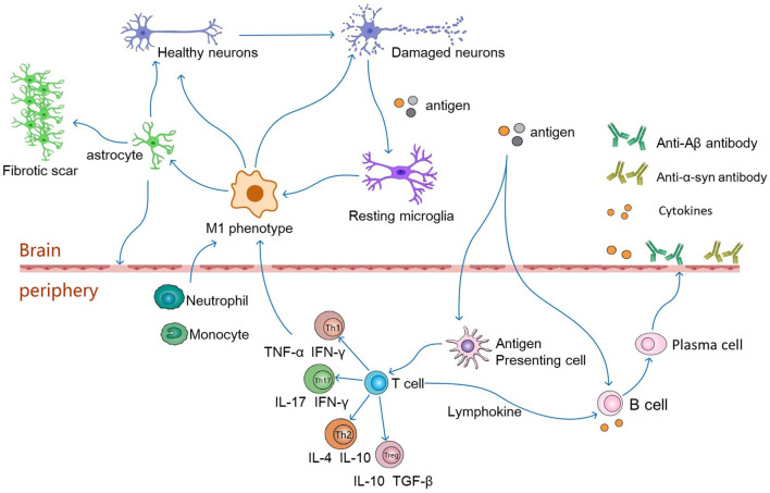
Verification
Claims the paper makes
The claims and statistics of this paper haven’t been checked yet.
Run the check — free, no account needed. It reads the open-access text and checks the sources it cites.
Claims other papers make about this one
Each is checked against this paper’s own text: does it say what the citing paper says it does?
The source backs part of the claim, not all of it — or backs it for a different population, condition or comparison.
In the citing paper
“but when acute, act as messengers to brain immune cells and signals to other local immune cells [44–46].”
From Alzheimer’s disease-associated protective variant Plcg2-P522R modulates peripheral macrophage function in a sex-dimorphic manner (2024) · cites this paper as [46]
Experimental — accuracy not yet measured
Self-check
Percentages, sums and reported differences re-computed from the paper’s own numbers.
The self-check needs the paper’s full text.
Citations
Cites
1 retracted
RETRACTED ARTICLE: Pericyte loss influences Alzheimer-like neurodegeneration in mice
Sagare, A. P., Bell, R. D., Zhao, Z., Ma, Q., Winkler, E. A., Ramanathan, A., & Zlokovic, B. V. (2013) · Nature Communications · doi:10.1038/ncomms3932 · Cited by 589
RetractedNeurotoxic reactive astrocytes are induced by activated microglia
Liddelow, S. A., Guttenplan, K. A., Clarke, L. E., Bennett, F. C., Bohlen, C. J., Schirmer, L., Bennett, M. L., Münch, A. E., Chung, W.-S., Peterson, T. C., et al. (2017) · Nature · doi:10.1038/nature21029 · Cited by 7,971
Resting microglial cells are highly dynamic surveillants of brain parenchyma in vivo
Nimmerjahn, A., Kirchhoff, F., & Helmchen, F. (2005) · Science · doi:10.1126/science.1110647 · Cited by 5,827
Neutrophil recruitment and function in health and inflammation
Kolaczkowska, E., & Kubes, P. (2013) · Nature Reviews Immunology · doi:10.1038/nri3399 · Cited by 5,472
A Hexanucleotide Repeat Expansion in C9ORF72 Is the Cause of Chromosome 9p21-Linked ALS-FTD
Renton, A. E., Majounie, E., Waite, A., Simón-Sánchez, J., Rollinson, S., Gibbs, J. R., Schymick, J. C., Laaksovirta, H., van Swieten, J. C., Myllykangas, L., et al. (2011) · Neuron · doi:10.1016/j.neuron.2011.09.010 · Cited by 4,409
McGeer, P. L., Itagaki, S., Boyes, B. E., & McGeer, E. G. (1988) · Neurology · doi:10.1212/wnl.38.8.1285 · Cited by 2,990
TDP-43 Mutations in Familial and Sporadic Amyotrophic Lateral Sclerosis
Sreedharan, J., Blair, I. P., Tripathi, V. B., Hu, X., Vance, C., Rogelj, B., Ackerley, S., Durnall, J. C., Williams, K. L., Buratti, E., et al. (2008) · Science · doi:10.1126/science.1154584 · Cited by 2,630
Mutations in FUS, an RNA Processing Protein, Cause Familial Amyotrophic Lateral Sclerosis Type 6
Vance, C. A., Rogelj, B., Hortobágyi, T., Vos, K. J. D., Nishimura, A. L., Sreedharan, J., Hu, X., Smith, B. N., Ruddy, D., Wright, P. D., et al. (2009) · Science · doi:10.1126/science.1165942 · Cited by 2,625
Brown, R. H., & Al‐Chalabi, A. (2017) · New England Journal of Medicine · doi:10.1056/nejmra1603471 · Cited by 2,580
Tissue-resident macrophages originate from yolk-sac-derived erythro-myeloid progenitors
Gomez Perdiguero, E., Klapproth, K., Schulz, C., Busch, K., Azzoni, E., Crozet, L., Garner, H., Trouillet, C., de Bruijn, M. F., Geissmann, F., et al. (2015) · Nature · doi:10.1038/nature13989 · Cited by 2,339
Cited by
Sorted by citations
Adamu, A., Li, S., Gao, F., & Xue, G. (2024) · Frontiers in Aging Neuroscience · doi:10.3389/fnagi.2024.1347987 · Cited by 564
COVID-19 and risk of neurodegenerative disorders: A Mendelian randomization study
Li, C., Liu, J., Lin, J., & Shang, H. (2022) · Translational Psychiatry · doi:10.1038/s41398-022-02052-3 · Cited by 150
Kim, S., Jung, U. J., & Kim, S. R. (2025) · Journal of Clinical Medicine · doi:10.3390/jcm14020386 · Cited by 129
Polyphenols: immunonutrients tipping the balance of immunometabolism in chronic diseases
Ferreira, C., Vieira, P., Sá, H., Malva, J. O., Castelo-Branco, M. S., Reis, F., & Viana, S. (2024) · Frontiers in Immunology · doi:10.3389/fimmu.2024.1360065 · Cited by 110
Kim, M. E., & Lee, J. S. (2024) · Current Issues in Molecular Biology · doi:10.3390/cimb47010008 · Cited by 82
New Advances in the Pharmacology and Toxicology of Lithium: A Neurobiologically Oriented Overview
Bortolozzi, A. A., Fico, G., Berk, M., Solmi, M., Fornaro, M., Quevedo, J., Zarate, C. A., Kessing, L. V., Vieta, E., & Carvalho, A. F. (2024) · Pharmacological Reviews · doi:10.1124/pharmrev.120.000007 · Cited by 68
Systemic Inflammation in Asthma: What Are the Risks and Impacts Outside the Airway?
Tattersall, M. C., Jarjour, N. N., & Busse, P. J. (2024) · The Journal of Allergy and Clinical Immunology In Practice · doi:10.1016/j.jaip.2024.02.004 · Cited by 58
Advances in the study of the glymphatic system and aging
Xiong, Y., Yu, Q., Zhi, H., Peng, H., Xie, M., Li, R., Li, K., Ma, Y., & Sun, P. (2024) · CNS Neuroscience & Therapeutics · doi:10.1111/cns.14803 · Cited by 55
The Importance of Phosphoinositide 3-Kinase in Neuroinflammation
Wright, B., King, S. P., & Suphioglu, C. (2024) · International Journal of Molecular Sciences · doi:10.3390/ijms252111638 · Cited by 55
Blossom, V., Ullal, S. D., D’Souza, M. M., Ranade, A. V., Kumar, N. A., & Rai, R. (2025) · 3 Biotech · doi:10.1007/s13205-025-04468-2 · Cited by 57
Neuroinflammation: targeting microglia for neuroprotection and repair after spinal cord injury
Cavalcanti, R. R., de Almeida, F. M., Martinez, A. M. B., & Freria, C. M. (2025) · Frontiers in Immunology · doi:10.3389/fimmu.2025.1670650 · Cited by 55
Anti-Inflammatory Effects of Flavonoids in Common Neurological Disorders Associated with Aging
Martínez‐Coria, H., Arrieta‐Cruz, I., Juárez, R. G., & López-Valdés, H. E. (2023) · International Journal of Molecular Sciences · doi:10.3390/ijms24054297 · Cited by 51
Neuroinflammaging: A Tight Line Between Normal Aging and Age-Related Neurodegenerative Disorders
Soraci, L., Corsonello, A., Paparazzo, E., Montesanto, A., Piacenza, F., Olivieri, F., Gambuzza, M. E., Savedra, E. V., Marino, S., Lattanzio, F., et al. (2024) · Aging and Disease · doi:10.14336/ad.2023.1001 · Cited by 49
Deng, J., Feng, X., Zhou, L., He, C., Li, H., Xia, J., Ge, Y., Zhao, Y., Song, C., Chen, L., et al. (2022) · Food Research International · doi:10.1016/j.foodres.2022.111576 · Cited by 39
Gut Microbiota and Immune System Dynamics in Parkinson’s and Alzheimer’s Diseases
Kuštrimović, N. Z., Balkhi, S., Bilato, G., & Mortara, L. (2024) · International Journal of Molecular Sciences · doi:10.3390/ijms252212164 · Cited by 39
Papadopoulou, P., Polissidis, A., Kythreoti, G., Sagnou, M., Stefanatou, A., & Theoharides, T. C. (2024) · International Journal of Molecular Sciences · doi:10.3390/ijms252011040 · Cited by 36
Fang, M., Zhou, Y., He, K., Lu, Y., Tao, F., & Huang, H. (2025) · Molecular Neurobiology · doi:10.1007/s12035-025-04775-y · Cited by 33
Neuronal plasticity and its role in Alzheimer’s disease and Parkinson’s disease
Jahan, I., Harun‐Ur‐Rashid, M., Islam, M. A., Sharmin, F., Jaouni, S. K. A., Kaki, A. M., & Selim, S. (2024) · Neural Regeneration Research · doi:10.4103/nrr.nrr-d-24-01019 · Cited by 31
Microglial dysfunction in Alzheimer's disease: Mechanisms, emerging therapies, and future directions
Azmal, M., Paul, J. K., Prima, F. S., Haque, A. N. M. S. N. B., Meem, M., & Ghosh, A. (2025) · Experimental Neurology · doi:10.1016/j.expneurol.2025.115374 · Cited by 30
Leal, A. S., Hung, P., Chowdhury, A. S., & Liby, K. T. (2023) · Pharmacology & Therapeutics · doi:10.1016/j.pharmthera.2023.108561 · Cited by 27
Neuroinflammation and Alzheimer's disease: Unravelling the molecular mechanisms
Kakkar, A., Singh, H., Singh, B. K., Kumar, A., Mishra, A. K., & Chopra, H. (2025) · Journal of Alzheimer s Disease · doi:10.1177/13872877251374353 · Cited by 26
Neutrophils: a subgroup of neglected immune cells in ALS
Cao, W., & Fan, D. (2023) · Frontiers in Immunology · doi:10.3389/fimmu.2023.1246768 · Cited by 23
Zaccai, S., Nemirovsky, A., Lerner, L., Alfahel, L., Eremenko, E., Israelson, A., & Monsonego, A. (2024) · Journal of Neuroinflammation · doi:10.1186/s12974-023-03007-1 · Cited by 23
Ding, Y., Palecek, S. P., & Shusta, E. V. (2024) · Fluids and Barriers of the CNS · doi:10.1186/s12987-024-00580-2 · Cited by 22
Single-cell RNA sequencing of CSF reveals neuroprotective RAC1+ NK cells in Parkinson’s disease
Guan, Q., Liu, W., Mu, K., Hu, Q., Xie, J., Cheng, L., & Wang, X. (2022) · Frontiers in Immunology · doi:10.3389/fimmu.2022.992505 · Cited by 24
Beyond the blood–brain barrier: unraveling T cell subsets in CNS immunity and disease
Shokr, M. M. (2025) · Inflammopharmacology · doi:10.1007/s10787-025-01955-z · Cited by 21
Pak, V., Adewale, Q., Bzdok, D., Dadar, M., Zeighami, Y., & Iturria‐Medina, Y. (2023) · eLife · doi:10.7554/elife.89368 · Cited by 21
Relationship between enriched environment and neurodegeneration: a review from mechanism to therapy
Xu, Y.-Q., Chen, Y., Xing, J., & Yao, J. (2025) · Clinical Epigenetics · doi:10.1186/s13148-025-01820-4 · Cited by 21
Neuroimmune interactions and their roles in neurodegenerative diseases
Chen, B.-R., Wu, T., Chen, T.-H., & Wang, Y. (2023) · Fundamental Research · doi:10.1016/j.fmre.2023.04.002 · Cited by 18
Blood‑brain barrier dysfunction in schizophrenia: Mechanisms and implications (Review)
Lv, S., & Luo, C. (2025) · International Journal of Molecular Medicine · doi:10.3892/ijmm.2025.5594 · Cited by 18
Modulating Stress Proteins in Response to Therapeutic Interventions for Parkinson’s Disease
Silvestro, S., Raffaele, I., & Mazzon, E. (2023) · International Journal of Molecular Sciences · doi:10.3390/ijms242216233 · Cited by 19
Kaur, R., Kumar, S., & Singh, L. (2025) · Cytokine · doi:10.1016/j.cyto.2025.156974 · Cited by 16
Bone Marrow‐Derived GCA+ Immune Cells Drive Alzheimer's Disease Progression
Zhou, R., Wang, L., Chen, L., Xu, F., Zhou, R., Peng, X. F., Wen, J., Huang, Y., & Zhou, H. (2023) · Advanced Science · doi:10.1002/advs.202303402 · Cited by 16
Farzan, M., Rounkian, M. S., Rizi, A. A., Heidari, Z., Farzan, M., Fathi, M., Aghaei, A., Azadegan‐Dehkordi, F., & Bagheri, N. (2024) · Experimental Neurology · doi:10.1016/j.expneurol.2024.115071 · Cited by 15
Tumpa, J. J., Hayder, Q. U. A., Maisa, N. S., & Islam, M. N. (2025) · Frontiers in Molecular Neuroscience · doi:10.3389/fnmol.2025.1682006 · Cited by 13
Experimental Models to Study Immune Dysfunction in the Pathogenesis of Parkinson’s Disease
Šaponjić, J., Mejı́as, R., Nikolovski, N., Dragić, M., Canak, A., Papoutsopoulou, S., Özdemir, Y., Fladmark, K. E., Ntavaroukas, P., Muluk, N. B., et al. (2024) · International Journal of Molecular Sciences · doi:10.3390/ijms25084330 · Cited by 13
Zheng, X., Yang, F., & Zheng, L. (2024) · Frontiers in Neuroscience · doi:10.3389/fnins.2024.1443438 · Cited by 13
Guerlais, V., Allouch, N., Moseman, E. A., Wojciechowska, A. W., Wojciechowski, J. W., & Marcelino, I. (2024) · Frontiers in Cellular and Infection Microbiology · doi:10.3389/fcimb.2024.1490280 · Cited by 12
Cantone, A. F., Burgaletto, C., Benedetto, G. D., Gaudio, G., Giallongo, C., Caltabiano, R., Broggi, G., Bellanca, C. M., Cantarella, G., & Bernardini, R. (2025) · Journal of Neuroimmune Pharmacology · doi:10.1007/s11481-025-10177-7 · Cited by 10
Staley, H. A., Jernigan, J. E., Bolen, M. L., Titus, A. M., Neighbarger, N., Cole, C., Menees, K. B., Wallings, R. L., & Tansey, M. G. (2024) · Journal of Neuroinflammation · doi:10.1186/s12974-024-03271-9 · Cited by 9
“Furthermore, macrophages are known to be one of the main producers of inflammatory cytokines, which, when chronic, can exacerbate neurodegeneration [2–4], but when acute, act as messengers to brain immune cells and signals to other local immune cells [44–46].”
BackgroundTabaie, E. Z., Gao, Z., Kachour, N., Ulu, A., Gomez, S., Figueroa, Z. A., Bergersen, K. V., Zhong, W., & Wilson, E. H. (2025) · PLoS Pathogens · doi:10.1371/journal.ppat.1012733 · Cited by 8
Bachstetter, A. D., Lutshumba, J., Winford, E. D., Abner, E. L., Martin, B. J., Harp, J. P., Eldik, L. J. V., Schmitt, F. A., Wilcock, D. M., Stowe, A. M., et al. (2023) · Brain Communications · doi:10.1093/braincomms/fcad259 · Cited by 8
McCann, R. P., Bowley, B. G. E., Pessina, M. A., Yang, Q., Xin, H., DeVries, S. A., Wang, M., Zhang, Y., Chopp, M., Zhang, Z., et al. (2025) · Frontiers in Aging Neuroscience · doi:10.3389/fnagi.2025.1605144 · Cited by 8
Kurmi, S., Doshi, G., & Parab, S. B. (2025) · CNS & Neurological Disorders - Drug Targets · doi:10.2174/0118715273377780250505115039 · Cited by 7
Ward, E., Benowitz, L. I., Brunner, T. M., Bu, G., Cayouette, M., Canto-Soler, V., Mesquita, S. D., Polo, A. D., DiAntonio, A., Duan, X., et al. (2025) · Molecular Neurodegeneration · doi:10.1186/s13024-025-00858-5 · Cited by 7
Immunological Fluid Biomarkers in Frontotemporal Dementia: A Systematic Review
Erichsen, P. A., Henriksen, E. E., Nielsen, J. E., Ejlerskov, P., Simonsen, A. H., & Toft, A. D. (2025) · Biomolecules · doi:10.3390/biom15040473 · Cited by 8
Li, Q., Han, X., Dong, M., Bai, L., Zhang, W., Liu, W., Wang, F., & Zhu, X. (2025) · Inflammation · doi:10.1007/s10753-025-02267-8 · Cited by 6
Manich, G., Pérez, B., Penas, C., Dantas, A. P., Coutinho, J., Sánchez-Bernadó, P., García-Aranda, J., Fraile-Ramos, J., Benseny‐Cases, N., Martín‐Mur, B., et al. (2025) · Redox Biology · doi:10.1016/j.redox.2025.103662 · Cited by 6
Kustrimovic, N., Rabattoni, V., Riva, D., Motta, Z., Sacchi, S., & Pollegioni, L. (2025) · Molecular Aspects of Medicine · doi:10.1016/j.mam.2025.101438 · Cited by 6
Teixeira, F. S., Costa, P. T., Soares, A. M. S., Fontes, A. L., Pintado, M., Vidigal, S. S. M. P., Pimentel, L. L., & Rodríguez-Alcalá, L. M. (2023) · Foods · doi:10.3390/foods12132576 · Cited by 6
Ma, X., Zuo, Y., Hu, X., Chen, S., Zhong, K., Xue, R., Gui, S., Liu, K., Li, S., Zhu, X., et al. (2024) · CNS Neuroscience & Therapeutics · doi:10.1111/cns.14682 · Cited by 6
Self-citationAli, A., Emad, N. A., Sultana, N., Waheed, A., Aqil, M., Sultana, Y., & Mujeeb, M. (2024) · CNS & Neurological Disorders - Drug Targets · doi:10.2174/0118715273319597240927044906 · Cited by 6
Lehodey, A., Kaliman, P., Palix, C., de Florès, R., Touron, E., Turpin, A., Fauvel, S., Mézenge, F., Landeau, B., Chocat, A., et al. (2024) · Alzheimer s Research & Therapy · doi:10.1186/s13195-024-01635-0 · Cited by 7
Jeevitha Gowda, R., Mehta, A., & Jayanna, K. (2025) · Brain Behavior and Immunity Integrative · doi:10.1016/j.bbii.2025.100107 · Cited by 6
Li, Q., Wang, G., Zhao, J., Chen, W., & Tian, P. (2025) · Microbiological Research · doi:10.1016/j.micres.2025.128286 · Cited by 6
DAMPs cross-talk interpretation of MDD mechanisms
Peng, Y., Chen, Z., Xu, Y., & Ren, B. (2025) · Science Advances · doi:10.1126/sciadv.adx3698 · Cited by 5
Advances in eye-brain axis: Anatomy, immunity, and association with visual dysfunction
Zhang, S., Fan, Z., & Ji, D. (2025) · Ageing Research Reviews · doi:10.1016/j.arr.2025.102925 · Cited by 5
Zhang, Y., Zhang, D., & Xiao, H. (2026) · Journal of Alzheimer s Disease · doi:10.1177/13872877261430002 · Cited by 5
Biomarker Profile in Peripheral Blood Cells Related to Alzheimer’s Disease
Lomi, M., Geraci, F., Seppia, C. D., Dolciotti, C., Carratore, R. D., & Bongioanni, P. (2025) · Molecular Neurobiology · doi:10.1007/s12035-025-04767-y · Cited by 5
CD4 + CD25 − T-Cell–Secreted IFN-γ Promotes Corneal Nerve Degeneration in Diabetic Mice
Lin, Y., Yang, L., Li, Y., Dou, S., Zhang, Z., & Zhou, Q. (2025) · Investigative Ophthalmology & Visual Science · doi:10.1167/iovs.66.4.15 · Cited by 4
Therapeutic potential of neuroprotective plant extracts in Parkinson’s disease
Luo, S., Zhang, L., Wu, X., Wang, J., Li, Q., Chen, W., Li, R., Zhou, L., Zhou, N., Chen, R., et al. (2026) · Frontiers in Neuroscience · doi:10.3389/fnins.2026.1813133 · Cited by 4
Peripheral immune system modulates Purkinje cell degeneration in Niemann–Pick disease type C1
Yasuda, T., Uchiyama, T., Watanabe, N., Ito, N., Nakabayashi, K., Mochizuki, H., & Onodera, M. (2023) · Life Science Alliance · doi:10.26508/lsa.202201881 · Cited by 4
The DNA repair enzyme, aprataxin, plays a role in innate immune signaling
Madsen, H. B., Pease, L. I., Scanlan, R.-L., Akbari, M., Rasmussen, L. J., Shanley, D. P., & Bohr, V. A. (2023) · Frontiers in Aging Neuroscience · doi:10.3389/fnagi.2023.1290681 · Cited by 4
Definition of clinical immunology around the globe
Crispín, J. C., Cantaert, T., Pinzón‐Charry, A., Mavilio, D., Seri, A., & Miossec, P. (2025) · Frontiers in Immunology · doi:10.3389/fimmu.2025.1483391 · Cited by 3
Wang, W., Du, Y., Tong, X., Liu, J., Lei, M., Wen, L., He, Y., Meng, X., & Xu, X. (2025) · Frontiers in Nutrition · doi:10.3389/fnut.2025.1578169 · Cited by 3
Winford, E. D., Shahidehpour, R. K., Dressman, D., Turchan-Cholewo, J. T., Cotter, K. M., Leibold, N. S., Ujas, T. A., Britsch, D. R. S., Mercurio, D., Colson, M. K., et al. (2025) · Neurobiology of Disease · doi:10.1016/j.nbd.2025.107153 · Cited by 3
Syringic acid suppresses inflammation by upregulation of SOCS3
Khatun, A., Patra, S., Chowdhury, S., Saha, J., Goswami, R., Pradhan, S., Ghosh, K., & Chakrabarti, S. (2025) · Journal of Neuroimmunology · doi:10.1016/j.jneuroim.2025.578813 · Cited by 3
Wang, H., Wang, X., Liu, N., Wang, H., & Feng, C. (2026) · Frontiers in Immunology · doi:10.3389/fimmu.2026.1828809 · Cited by 3
Aldaghi, F. S., Siahpoosh, Z., Salehi, Z., Mashayekhi, F., & Sohrabnezhad, S. (2026) · Brain and Behavior · doi:10.1002/brb3.71615 · Cited by 3
A brief review of ALS from an immunological perspective with potential immunotherapy options
Ayhan, G., Aydemir, E., & Ayaz, F. (2025) · Discover Immunity. · doi:10.1007/s44368-025-00011-w · Cited by 3
ApoE potential in CNS drugs targeting and as CNS therapeutic
Ahmed, S., Pande, A. H., & Sharma, S. S. (2024) · Elsevier eBooks · doi:10.1016/b978-0-443-23841-3.00005-4 · Cited by 3
Chakrabarti, A., & Verma, S. (2025) · Molecular and Cellular Neuroscience · doi:10.1016/j.mcn.2025.103993 · Cited by 3
Gong, Z., Cao, R., & Zhu, H. (2025) · Brain and Behavior · doi:10.1002/brb3.70586 · Cited by 3
Huang, X., Yuan, S., Ling, Y., Tan, S., Bai, Z., Xu, Y., Shen, S. Q., Lyu, J., & Wang, H. (2025) · International Journal of Surgery · doi:10.1097/js9.0000000000002746 · Cited by 3
Georgoula, M., Ntavaroukas, P., Campbell, B. J., & Papoutsopoulou, S. (2024) · Epigenetics Reports · doi:10.1080/28361512.2024.2401340 · Cited by 2
Aryl hydrocarbon receptor deficiency enhances astrocyte sensitivity to LPS-induced inflammation
Ojo, E. A., & Tischkau, S. A. (2025) · Frontiers in Cellular Neuroscience · doi:10.3389/fncel.2025.1653109 · Cited by 2
Saini, K., & Dhiman, P. (2026) · Molecular Biology Reports · doi:10.1007/s11033-026-12128-8 · Cited by 2
Jadhav, V. S., Nathiya, D., Tomar, B. S., & Khatri, D. K. (2026) · Discover Immunity. · doi:10.1007/s44368-026-00029-8 · Cited by 2
Liu, Y.-P., & Yang, Y. (2023) · Medical Hypotheses · doi:10.1016/j.mehy.2023.111113 · Cited by 2
Brisse, M. E., & Ly, H. (2024) · Journal of Medical Virology · doi:10.1002/jmv.29949 · Cited by 2
Biomarkers for Neurodegenerative Diseases
Abdelmoaty, M. M., Lu, E., & Foster, E. G. (2024) · Springer eBooks · doi:10.1007/978-3-031-68237-7_44 · Cited by 2
Moghiseh, E., Massoudifar, A., Davoodian, N., Mousavi, P., Afsharpour, S., & Mahmoodi, M. (2025) · Molecular Neurobiology · doi:10.1007/s12035-025-05060-8 · Cited by 2
From Immune Sanctuary to Neurological Battlefield: The Role of Neuroimmune Cells
Chakrabarti, S. K., & Chattopadhyay, D. (2024) · Exploratory Research and Hypothesis in Medicine · doi:10.14218/erhm.2024.00026 · Cited by 2
The Role of Microglia in Parkinson's Disease and Possible Therapeutic Targets
Gomes, G. F., Galvão, S. L., Ventura, C., Lessa, Y. P. N., & dos Santos, J. C. C. (2024) · Brazilian Journal of Clinical Medicine and Review · doi:10.52600/2965-0968.bjcmr.2025.3.1.bjcmr11 · Cited by 1
Zhang, H., Hamit, D., Li, Q., Hu, X., Li, S.-F., Xu, F., Wang, M., Bao, G.-Q., & Li, H. (2025) · Scientific Reports · doi:10.1038/s41598-024-80755-x · Cited by 1
Russu, E., Groppa, L., Chișlari, L., Semionov, M., Leanca, I., Pastuhov, A., & Nartea, C. (2025) · Moldovan Journal of Health Sciences · doi:10.52645/mjhs.2025.2.08 · Cited by 1
Multivalent cannabinoid epigenotoxicities and multigenerational aging
Reece, A. S., & Hulse, G. K. (2025) · Elsevier eBooks · doi:10.1016/b978-0-443-13492-0.00001-3 · Cited by 1
Yin, Q., Su, R., Nie, X., Liu, P., Bu, J., Luo, H., Liu, R., Guo, H., Liu, L., Wang, B., et al. (2025) · Neuropharmacology · doi:10.1016/j.neuropharm.2025.110761 · Cited by 1
Liu, T., Zhang, X., Qin, J., Li, J., Aslam, M. S., Xu, H., Zhang, X., Wang, F., Wang, X., Meng, X., et al. (2025) · Innovations in Acupuncture and Medicine · doi:10.1186/s44424-025-00031-1 · Cited by 1
Han, S. H., He, M. T., Baek, K., Kim, H. Y., Lee, S., & Lee, A. R. (2026) · Journal of Agricultural and Food Chemistry · doi:10.1021/acs.jafc.5c12437 · Cited by 3
Liu, H., Chen, Y., Li, C. Y., Zhang, Q., Lin, X., Lin, Y., & Xu, Q. (2026) · Experimental Gerontology · doi:10.1016/j.exger.2026.113071 · Cited by 1
Expression Patterns of T-Cell Intracellular Antigen 1 in Neurodegenerative Disorders and Stroke
Han, J., Yu, H., Zheng, T., Huang, Z., & Wang, L. (2026) · International Journal of Molecular Sciences · doi:10.3390/ijms27052252 · Cited by 1
Zhang, H., Zeng, X., & Kong, X. (2026) · European journal of medical research · doi:10.1186/s40001-026-04128-y · Cited by 1
Calderazzo, S. M., Butler, M. L. M. D., Breen, K., Kanner, H., Tripodis, Y., Stein, T. D., Holtzman, D. M., Cherry, J. D., Huber, B. R., & McKee, A. C. (2026) · Acta Neuropathologica Communications · doi:10.1186/s40478-026-02285-z · Cited by 2
Zhao, P.-Y., Wang, K., Zhang, M.-M., Cao, Z.-L., Jin, W.-L., & Rozpędek‐Kamińska, W. (2026) · Critical Reviews in Oncology/Hematology · doi:10.1016/j.critrevonc.2026.105409 · Cited by 1
Shoman, N. A., Salama, A., Adel, N., Elnahas, O. S., El-Mancy, S. S., & El-Dahmy, R. M. (2026) · Journal of Drug Delivery Science and Technology · doi:10.1016/j.jddst.2026.108645 · Cited by 1
Camon, C., Kip, E., Lord, R., Decourt, C., Prescott, M., Clarkson, J., Sandoval, N. P., Vander, P. B., van Veen, J. E., Prókai-Tátrai, K., et al. (2026) · Endocrinology · doi:10.1210/endocr/bqag082 · Cited by 1
Thome, A. D., Wang, J., Faridar, A., Zhao, W., Saetzler, V., Beers, D. R., & Appel, S. H. (2026) · International Journal of Molecular Sciences · doi:10.3390/ijms27177855 · Cited by 1
Salehloo, E. B., Sabouni, F., & Mianabadi, M. (2023) · PubMed Central · doi:10.30498/ijb.2023.285965.3052 · Cited by 1
Anti-amyloid Monoclonal Antibodies in Alzheimer’s Disease: A New Hope
Nasibova, A. N., Khalilov, R. I., & Eftekhari, A. (2023) · International Journal of Aging · doi:10.34172/ija.2023.e21 · Cited by 1
Metadata
- Authors
- Zang, X., Chen, S., Zhu, J., Ma, J., & Zhai, Y.
- Affiliations
- Sheng Jing Hospital (CN)
- Fourth Affiliated Hospital of China Medical University (CN)
- Corresponding author
- Zhai, Yongzhen
- Venue
- Frontiers in Aging Neuroscience
- Publisher
- Frontiers Media SA
- Published
- 2022-04-25
- Volume
- 14
- Pages
- 872134-872134
- ISSN
- 1663-4365 (ISSN-L)
- PMC
- PMC9082639
- OpenAlex
- W4224441906
- Cited by
- 139
- References
- 221
- Citations by year
- 2026: 35 · 2025: 49 · 2024: 35 · 2023: 17 · 2022: 3
- FWCI
- 2.76
- Type
- journal article
- Language
- en
- Open access
- gold
- doi.org
- Licence
- Locations
- Frontiers in Aging Neuroscience (publishedVersion, cc-by)
- PubMed Central (publishedVersion, cc-by)
- PubMed (publishedVersion)
- DOAJ (DOAJ: Directory of Open Access Journals) (submittedVersion, cc-by-sa)
- Topics
- Life Sciences › Neuroscience › Neurology › Neuroinflammation and Neurodegeneration Mechanisms
- Health Sciences › Medicine › Clinical Neurology › Multiple Sclerosis Research Studies
- Life Sciences › Neuroscience › Biological Psychiatry › Tryptophan and brain disorders
- Keywords
- neurodegenerative diseases, neuroinflammation, blood-brain barrier, peripheral immune system, central nervous system, cytokines, microglial activation, astrocytes, immune privilege, neurodegeneration therapy
About these checks
Registry, retraction, Cites and statistics results are computed, not judged, with one exception: a printed reference that no index lists is read once by a model before it counts as a citation error. Claims are extracted by a model and kept only when their quote appears verbatim in the text; each cited source is then checked, with a model’s help, for whether it says what the claim relies on. Those verdicts can be wrong — read the claim’s quote and the cited source before relying on one.
Statistics and arithmetic are recomputed, not judged.
Confidence bands are shown only for fields calibrated on our annotated test set. None is calibrated yet, so no band is shown.
What counts as a claim ›How we measure confidence ›How we check this ›
Are you an author, or do you see a problem with this page? Request a review
