

The DOI matches its Crossref record; 313 of the 313 references it cites were found in a registry or an index.
Hypoxic microenvironment in cancer: role in metabolic reprogramming
Open accessAbstract
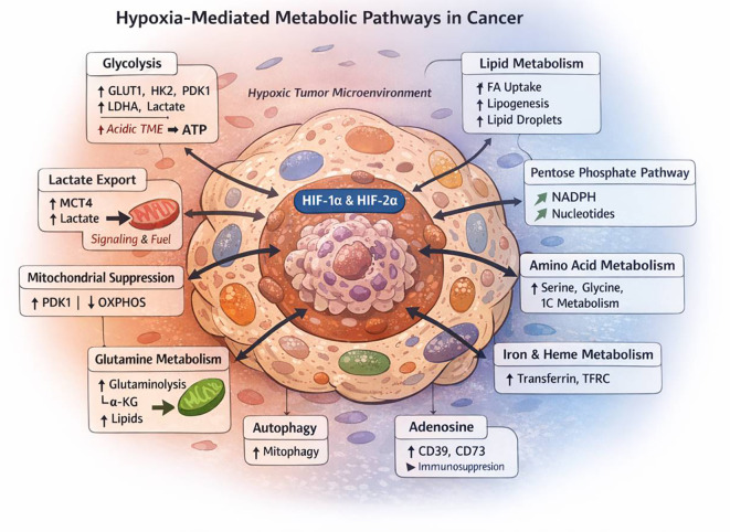
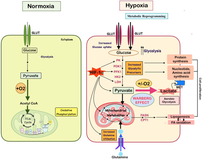
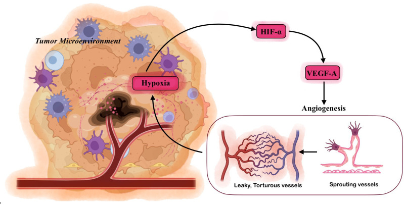
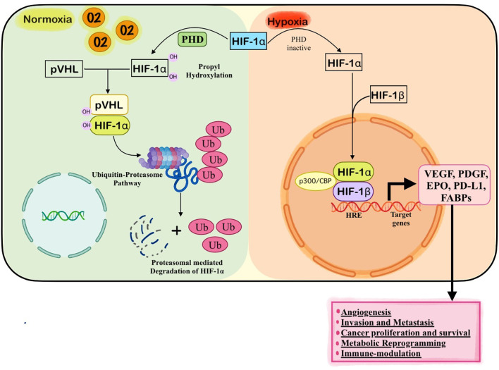
Hypoxia, a defining hallmark of solid tumors, arises from structurally and functionally abnormal vasculature, rapid cellular proliferation, and impaired perfusion, resulting in chronic and cycling oxygen deprivation within the tumor massThe hypoxic tumor microenvironment orchestrates extensive molecular reprogramming primarily through stabilization and activation of hypoxia-inducible factors (HIF-1α and HIF-2α), which regulate broad transcriptional networks governing metabolism, angiogenesis, stemness, invasion, and immune modulation. Under low oxygen tension, tumor cells shift toward aerobic glycolysis, enhance glutamine utilization, promote lipid synthesis and storage, suppress mitochondrial oxidative phosphorylation, and fine-tune redox balance through coordinated regulation of ROS-generating and antioxidant systems. These adaptations not only sustain proliferation and survival under metabolic stress but also facilitate epithelial–mesenchymal transition, extracellular matrix remodeling, and metastatic dissemination. Beyond malignant cells, hypoxia reprograms stromal compartments—including cancer-associated fibroblasts, endothelial cells, tumor-associated macrophages, and myeloid-derived suppressor cells—thereby establishing a metabolically cooperative, angiogenic, and profoundly immunosuppressive microenvironment. Hypoxia-induced acidosis, lactate accumulation, and HIF-driven cytokine signaling further impair cytotoxic T-cell and NK-cell activity, contributing to immune escape and resistance to radiotherapy, chemotherapy, and immunotherapy. Emerging evidence from single-cell multi-omics, spatial transcriptomics, metabolic imaging, and early-phase clinical trials targeting HIF signaling, angiogenic pathways, and metabolic enzymes has uncovered actionable vulnerabilities in hypoxia-driven malignancies. This review synthesizes the mechanistic foundations of hypoxia-induced metabolic reprogramming, its role in tumor progression and therapeutic resistance, and discusses innovative strategies aimed at exploiting hypoxia-associated metabolic dependencies to advance precision oncology.
Overview
The claims and statistics of this paper haven’t been checked yet.
Run the check — free, no account needed. It reads the open-access text and checks the sources it cites.
Integrity signals
Registry
Cites
Statistics
Figures
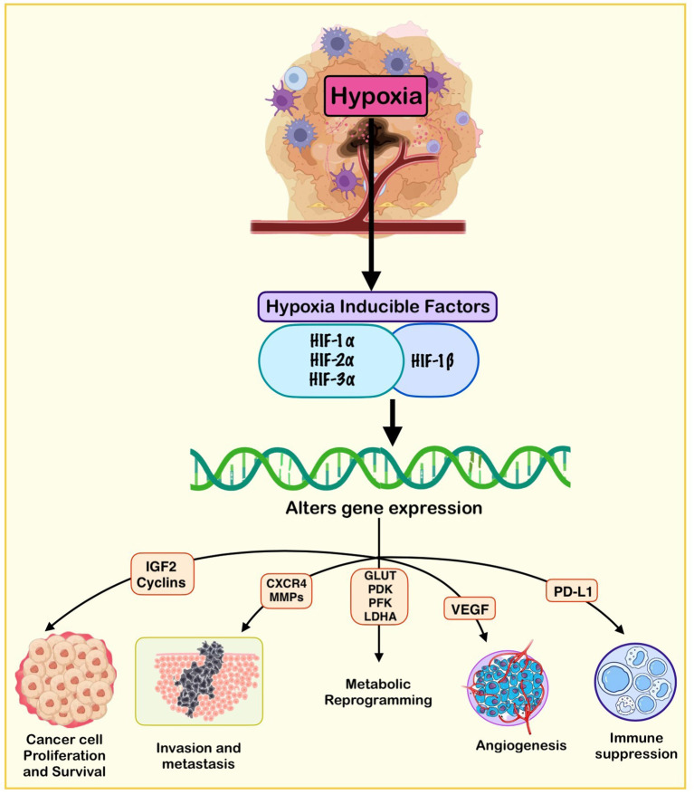
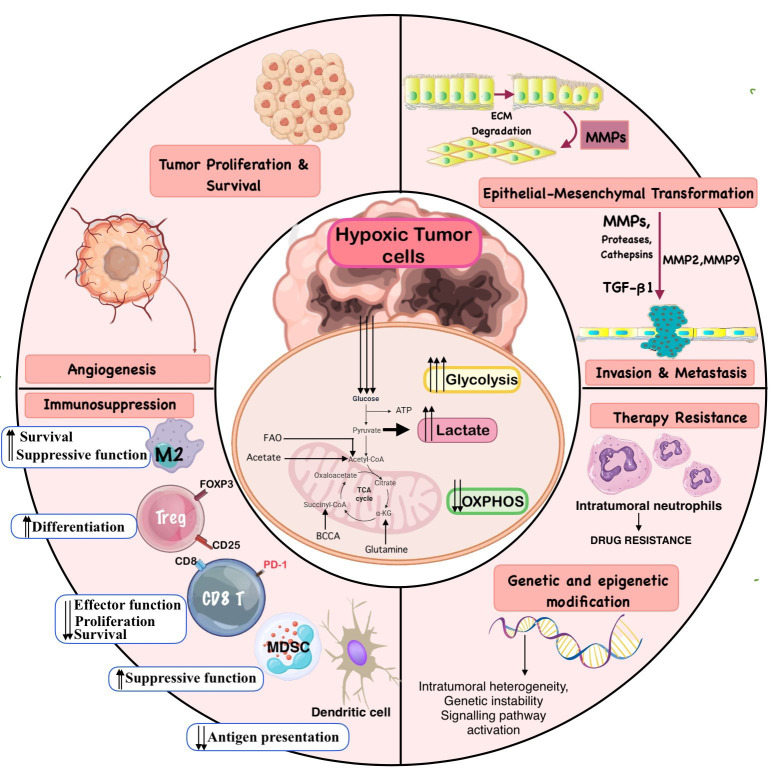
Verification
Claims the paper makes
The claims and statistics of this paper haven’t been checked yet.
Run the check — free, no account needed. It reads the open-access text and checks the sources it cites.
Claims other papers make about this one
Each is checked against this paper’s own text: does it say what the citing paper says it does?
The cited source says what the claim attributes to it.
In the citing paper
“As tumors grow beyond the reach of blood vessels, hypoxia leads to the stabilization of hypoxia-inducible factor 1α (HIF-1α), which then induces the expression of vascular endothelial growth factor (VEGF) and promotes angiogenesis [54].”
From Overcoming Resistance to Anti-EGFR Therapies: Mechanisms of Cetuximab and Panitumumab Resistance and Emerging Combination Strategies (2026) · cites this paper as [54]
Self-check
Percentages, sums and reported differences re-computed from the paper’s own numbers.
The self-check needs the paper’s full text.
Citations
Cites
Wang, G. L., Jiang, B. H., Rue, E. A., & Semenza, G. L. (1995) · Proceedings of the National Academy of Sciences · doi:10.1073/pnas.92.12.5510 · Cited by 6,212
The Warburg Effect: How Does it Benefit Cancer Cells?
Liberti, M. V., & Locasale, J. W. (2016) · Trends in Biochemical Sciences · doi:10.1016/j.tibs.2015.12.001 · Cited by 5,150
Kim, J.-W., Tchernyshyov, I., Semenza, G. L., & Dang, C. V. (2006) · Cell Metabolism · doi:10.1016/j.cmet.2006.02.002 · Cited by 3,905
Hypoxia-inducible factors in physiology and medicine
Semenza, G. L. (2012) · Cell · doi:10.1016/j.cell.2012.01.021 · Cited by 3,645
Functional Polarization of Tumour-Associated Macrophages by Tumour-Derived Lactic Acid
Colegio, O. R., Chu, N.-Q., Szabo, A. L., Chu, T., Rhebergen, A. M., Jairam, V., Cyrus, N., Brokowski, C. E., Eisenbarth, S. C., Phillips, G. M., et al. (2014) · Nature · doi:10.1038/nature13490 · Cited by 3,046
Mao, X., Xu, J., Wang, W., Liang, C., Hua, J., Liu, J., Zhang, B., Meng, Q., Yu, X., & Shi, S. (2021) · Molecular Cancer · doi:10.1186/s12943-021-01428-1 · Cited by 2,290
HIF-1 mediates adaptation to hypoxia by actively downregulating mitochondrial oxygen consumption
Papandreou, I., Cairns, R. A., Fontana, L., Lim, A. L., & Denko, N. C. (2006) · Cell Metabolism · doi:10.1016/j.cmet.2006.01.012 · Cited by 2,267
The Histological Structure of Some Human Lung Cancers and the Possible Implications for Radiotherapy
Thomlinson, R. H., & Gray, L. H. (1955) · British Journal of Cancer · doi:10.1038/bjc.1955.55 · Cited by 2,228
Noman, M. Z., Desantis, G., Janji, B., Hasmim, M., Karray, S., Dessen, P., Bronte, V., & Chouaib, S. (2014) · Journal of Experimental Medicine · doi:10.1084/jem.20131916 · Cited by 2,166
Tumor angiogenesis: causes, consequences, challenges and opportunities
Lugano, R., Ramachandran, M., & Dimberg, A. (2019) · Cellular and Molecular Life Sciences · doi:10.1007/s00018-019-03351-7 · Cited by 2,149
Research works printed but not linked: 8
- Calorini, 2012, Extracellular acidity as favouring factor of tumor progression and metastatic dissemination, Exp Oncol, 34, 79
- Gao, 2015, Acidic extracellular microenvironment promotes the invasion and cathepsin B secretion of PC-3 cells, Int J Clin Exp Med, 8
- Jarc, 2019, Lipid droplets and the management of cellular stress, Yale J Biol Med, 92
- Leek, 2002, Relation of hypoxia-inducible factor-2 alpha (HIF-2 alpha) expression in tumor-infiltrative macrophages to tumor angiogenesis and the oxidative thymidine phosphorylase pathway in Human breast cancer, Cancer Res, 62
- Boucher, 1990, Interstitial pressure gradients in tissue-isolated and subcutaneous tumors: implications for therapy, Cancer Res, 50
- Zhang, 2025, Systematic and comprehensive insights into HIF-1 stabilization under normoxic conditions: implications for cellular adaptation and therapeutic strategies in cancer, Cell Mol Biol Lett, 30, 10.1186/s11658-024-00682-7 · DOI
- Han, 2023, Natural compounds as lactate dehydrogenase inhibitors: potential therapeutics for lactate dehydrogenase inhibitors-related diseases, Front Pharmacol, 14, 10.3389/fphar.2023.1275000 · DOI
- Hay, 2003, Structure-activity relationships of 1, 2, 4-benzotriazine 1, 4-dioxides as hypoxia-selective analogues of tirapazamine, J Med Chem, 46, 10.1021/jm020367 · DOI
Cited by
Sorted by citations
Henrykowska, G., Bartusik-Aebisher, D., Dynarowicz, K., Ramesh, T. S., Smolak, B., & Aebisher, D. (2026) · Pharmaceuticals · doi:10.3390/ph19071041 · Cited by 1
“As tumors grow beyond the reach of blood vessels, hypoxia leads to the stabilization of hypoxia-inducible factor 1 α (HIF-1 α ), which then induces the expression of vascular endothelial growth factor (VEGF) and promotes angiogenesis [54].”
Glycolytic reprogramming and the EMT drive cervical cancer progression via the NQO1/HIF1α-SIX1 axis
Zhu, G., Piao, Z., Gao, Y., Cong, X., Wei, P., Han, A., Zhou, H., Lin, Z., & Jin, T. (2026) · iScience · doi:10.1016/j.isci.2026.117007 · Cited by 1
Hammouz, R. Y., Maciejek, K., & Bednarek, A. K. (2026) · International Journal of Molecular Sciences · doi:10.3390/ijms27156740
Chronobiology of Cancer: How Aging Fuels Oncogenesis at the Molecular Level
Singh, A., Misra, A., & Zaheer, S. (2026) · Aging and Cancer · doi:10.1002/aac2.70027
Self-citationLi, X., Yang, K., Liang, X., Qi, F., & Lv, Y. (2026) · Precision medicine and engineering. · doi:10.1016/j.preme.2026.100085
Shan, C., Guo, M., Feng, F., Wang, X., Mohang, L., Xu, W., Jia, K., Liu, Y., & Jia, Y. (2026) · The EPMA Journal · doi:10.1007/s13167-026-00465-4
Hypoxia-Driven Alterations in the Tumour Microenvironment of Oral Cancer
Reddy, S. S., Ravichandran, A., Reddy, A., Radhakrishnan, A., & Linda Christabel, S. (2026) · Onco · doi:10.3390/onco6030046
Lei, X., Song, B., Jin, D., Zhou, J., Guo, T., & Kong, J. (2026) · Frontiers in Physiology · doi:10.3389/fphys.2026.1848297
Design and Limitations of Tumor Microenvironment-Responsive Nanocarriers for Targeted Drug Delivery
Yang, X. (2026) · Theoretical and Natural Science · doi:10.54254/2753-8818/2027.mb37184
Histone Lactylation in Cancer: Linking Hypoxia-Driven Metabolism to Epigenetic Regulation
Ostapowicz, J., & Kulcenty, K. I. (2026) · Letters in Oncology Science · doi:10.21641/los.2026.23.3.306
Obeagu, E. I., & Edeoga, I. H. (2026) · Oncology Reviews · doi:10.3389/or.2026.1959034
Macrophage immunometabolism in glioblastoma: from microglial reprogramming to B cell dysfunction
Yu, E., Jones, G., & Lee-Chang, C. (2026) · Frontiers in Immunology · doi:10.3389/fimmu.2026.1947853
Saeed, A. F., Ashraf, M. N., & Iqbal, M. Z. (2026) · Frontiers in Immunology · doi:10.3389/fimmu.2026.1920004
Metadata
- Authors
- Sureka, N., Maheshwari, R., Agravat, A., Singhal, S., Zaman, S., Rishi, B., Siraj, F., Zaheer, S., & Misra, A.
- Affiliations
- Safdarjang Hospital (IN)
- Department of Pathology, Pandit Deendayal Upadhyay (PDU) Medical College
- Indian Council of Medical Research (IN)
- National Institute of Child Health (PK)
- Corresponding author
- Zaheer, Sufian
- Misra, Aroonima
- ORCID
- Venue
- Frontiers in Oncology
- Publisher
- Frontiers Media SA
- Published
- 2026-03-31
- Volume
- 16
- Pages
- 1771365-1771365
- ISSN
- 2234-943X (ISSN-L)
- PMC
- PMC13076347
- OpenAlex
- W7143706570
- Cited by
- 11
- References
- 313
- Citations by year
- 2026: 11
- FWCI
- 9.23
- Type
- journal article
- Language
- en
- Open access
- gold
- doi.org
- Licence
- Locations
- Frontiers in Oncology (publishedVersion, cc-by)
- PubMed Central (publishedVersion, cc-by)
- PubMed (publishedVersion)
- Topics
- Life Sciences › Biochemistry, Genetics and Molecular Biology › Cancer Research › Cancer, Hypoxia, and Metabolism
- Life Sciences › Biochemistry, Genetics and Molecular Biology › Molecular Biology › Angiogenesis and VEGF in Cancer
- Health Sciences › Medicine › Oncology › Cancer Cells and Metastasis
- Keywords
- tumor hypoxia, hypoxic tumor microenvironment, metabolic reprogramming, hypoxia-inducible factor, HIF-1α, HIF-2α, aerobic glycolysis, glutamine metabolism, lipid metabolism, redox homeostasis, reactive oxygen species, epithelial-mesenchymal transition, extracellular matrix remodeling, metastasis, cancer-associated fibroblasts, tumor-associated macrophages, myeloid-derived suppressor cells, tumor immunosuppression, immune evasion, treatment resistance, HIF-targeted therapy, precision oncology
About these checks
Registry, retraction, Cites and statistics results are computed, not judged, with one exception: a printed reference that no index lists is read once by a model before it counts as a citation error. Claims are extracted by a model and kept only when their quote appears verbatim in the text; each cited source is then checked, with a model’s help, for whether it says what the claim relies on. Those verdicts can be wrong — read the claim’s quote and the cited source before relying on one.
Statistics and arithmetic are recomputed, not judged.
Confidence bands are shown only for fields calibrated on our annotated test set. None is calibrated yet, so no band is shown.
What counts as a claim ›How we measure confidence ›How we check this ›
Are you an author, or do you see a problem with this page? Request a review
