

The DOI matches its Crossref record; 1 of the 1 references it cites were found in a registry or an index.
Mechanisms of isoniazid and rifampicin-induced liver injury and the effects of natural medicinal ingredients: A review
Abstract
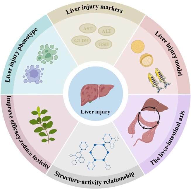
Isoniazid (INH) and rifampicin (RFP) are the first-line medications for tuberculosis treatment, and liver injury is the major adverse effect. Natural medicinal ingredients provide distinct benefits in alleviating patients’ symptoms, lowering the liver injury risk, delaying disease progression, and strengthening the body’s ability to heal. This paper summarises the recent research on the mechanisms of INH and RFP-induced liver injury and the effects of natural medicinal ingredients. It is believed that INH-induced liver injury may be attributed to oxidative stress, mitochondrial dysfunction, drug metabolic enzymes, protoporphyrin IX accumulation, endoplasmic reticulum stress, bile transport imbalance, and immune response. RFP-induced liver injury is mainly related to cholestasis, endoplasmic reticulum stress, and liver lipid accumulation. However, the combined effect of INH and RFP on liver injury risk is still uncertain. RFP can increase INH-induced hepatotoxicity by regulating the expression of drug-metabolizing enzymes and transporters. In contrast, INH can antagonize RFP-induced liver injury by reducing the total bilirubin level in the blood. Sagittaria sagittifolia polysaccharide, quercetin, gallic acid, and other natural medicinal ingredients play protective roles on INH and RFP-induced liver injury by enhancing the body’s antioxidant capacity, regulating metabolism, inhibiting cell apoptosis, and reducing the inflammatory response. There are still many gaps in the literature on INH and RFP-induced liver injury mechanisms and the effects of natural medicinal ingredients. Thus, further research should be carried out from the perspectives of liver injury phenotype, injury markers, in vitro and in vivo liver injury model construction, and liver-gut axis. This paper comprehensively reviewed the literature on mechanisms involved in INH and RFP-induced liver injury and the status of developing new drugs against INH and RFP-induced liver injury. In addition, this review also highlighted the uses and advantages of natural medicinal ingredients in treating drug-induced liver injury.
Overview
The claims and statistics of this paper haven’t been checked yet.
Run the check — free, no account needed. It reads the open-access text and checks the sources it cites.
Integrity signals
Registry
Cites
Statistics
Figures
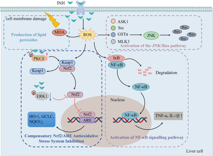
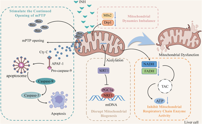
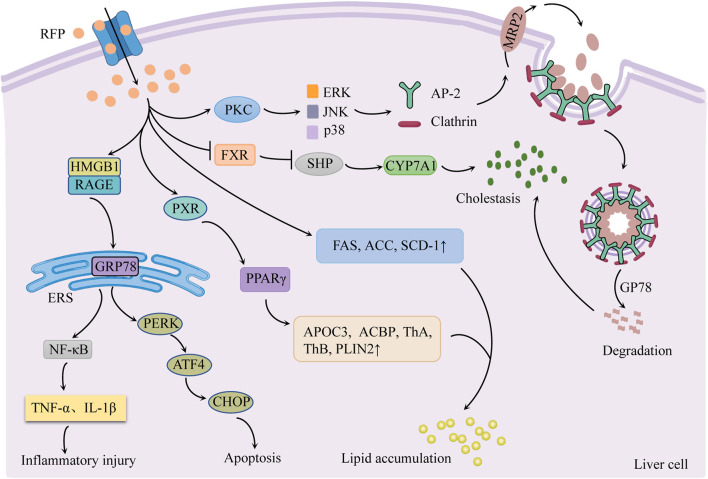
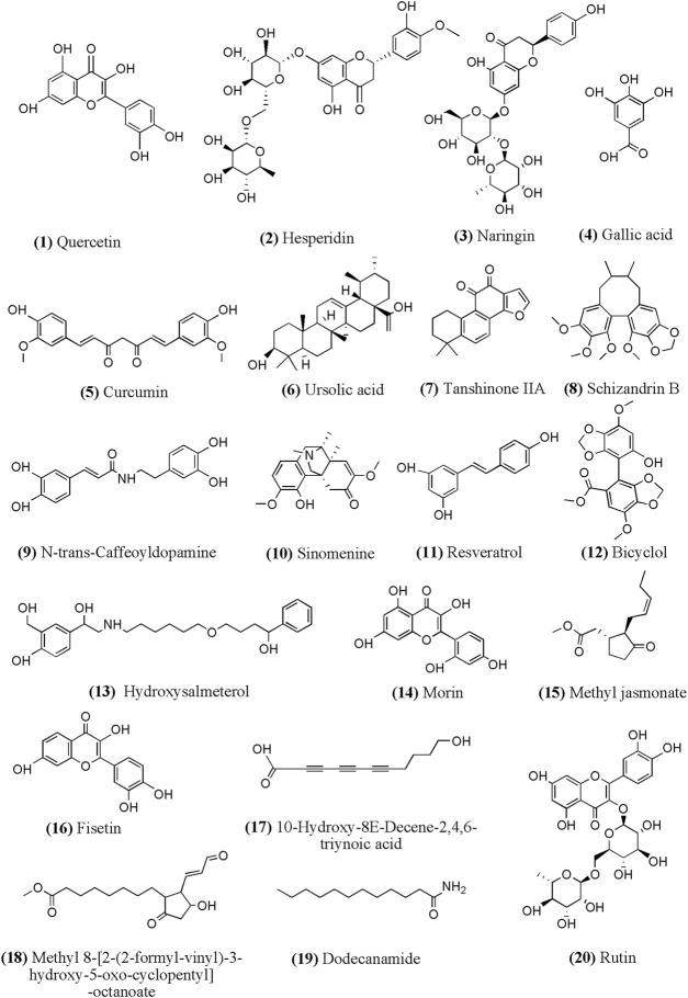
Verification
Claims the paper makes
The claims and statistics of this paper haven’t been checked yet.
Run the check — free, no account needed. It reads the open-access text and checks the sources it cites.
Claims other papers make about this one
Each is checked against this paper’s own text: does it say what the citing paper says it does?
In the citing paper
“Similarly, rifampin, a strong CYP inducer, has been demonstrated to increase the incidence of hepatotoxicity when administered together with isoniazid as an anti-TB treatment [36,37].”
From Chinese guideline for the diagnosis and treatment of drug-induced liver injury: an update (2024) · cites this paper as [36]
Self-check
Percentages, sums and reported differences re-computed from the paper’s own numbers.
The self-check needs the paper’s full text.
Citations
Cites
Incidence and Etiology of Drug-Induced Liver Injury in Mainland China
Shen, T., Liu, Y., Shang, J., Xie, Q., Li, J., Yan, M., Xu, J., Niu, J., Liu, J., Watkins, P. B., et al. (2019) · Gastroenterology · doi:10.1053/j.gastro.2019.02.002 · Cited by 615
Research works printed but not linked: 111
- Barretto, 2019, Gene expression profiling reveals that PXR activation inhibits hepatic PPARα activity and decreases FGF21 secretion in male C57Bl6/J mice, Int. J. Mol. Sci., 20, 3767, 10.3390/ijms20153767 · DOI
- Barua, 2021, Therapeutic potential of curcumin as an antimycobacterial agent, Biomolecules, 11, 1278, 10.3390/biom11091278 · DOI
- Biswas, 2020, Isoniazid and rifampicin produce hepatic fibrosis through an oxidative stress-dependent mechanism, Int. J. Hepatol., 2020, 6987295, 10.1155/2020/6987295 · DOI
- Boelsterli, 2014, Mechanisms of isoniazid-induced idiosyncratic liver injury: Emerging role of mitochondrial stress, J. Gastroenterol. Hepatol., 29, 678, 10.1111/jgh.12516 · DOI
- Brewer, 2020, Toxicoproteomic profiling of hPXR transgenic mice treated with rifampicin and isoniazid, Cells, 9, 1654, 10.3390/cells9071654 · DOI
- Brewer, 2019, The isoniazid metabolites hydrazine and pyridoxal isonicotinoyl hydrazone modulate heme biosynthesis, Toxicol. Sci., 168, 209, 10.1093/toxsci/kfy294 · DOI
- Chakaya, 2021, Global tuberculosis report 2020 - reflections on the global TB burden, treatment and prevention efforts, Int. J. Infect. Dis., 113, S7, 10.1016/j.ijid.2021.02.107 · DOI
- Chanhom, 2020, GSTM1 and GSTT1 genetic polymorphisms and their association with antituberculosis drug-induced liver injury, Biomed. Rep., 12, 153, 10.3892/br.2020.1275 · DOI
- Chen, 2016, Study of characteristics and mechanism of liver damaged in using simvastatin combined with HRZ, Anti Infect. Pharm., 13, 729, 10.13493/j.issn.1672-7878.2016.04-003 · DOI
- Chen, 2022, 4-Phenylbutyrate protects against rifampin-induced liver injury via regulating MRP2 ubiquitination through inhibiting endoplasmic reticulum stress, Bioengineered, 13, 2866, 10.1080/21655979.2021.2024970 · DOI
- Chen, 2016, The role of reactive oxygen species in isoniazid-induced DNA damage in L-02 cells and the protective effect of quercetin, Carcinog. Teratogenesis Mutagen., 28, 472, 10.3969/j.issn.1004-616x.2016.06.012 · DOI
- Chen, 2018, Dexmedetomidine ameliorates acute stress-induced kidney injury by attenuating oxidative stress and apoptosis through inhibition of the ROS/JNK signaling pathway, Oxid. Med. Cell. Longev., 2018, 4035310, 10.1155/2018/4035310 · DOI
- Chen, 2014, Down-regulation of PERK-ATF4-CHOP pathway by Astragaloside IV is associated with the inhibition of endoplasmic reticulum stress-induced podocyte apoptosis in diabetic rats, Cell. Physiol. biochem., 33, 1975, 10.1159/000362974 · DOI
- Chen, The role of ROS/JNK pathway in the inhibition of isoniazid-induced hepatocyte apoptosis by quercetin, J. Huzhou Univ., 43, 59
- Chen, Role of the Nrf2/ARE signaling pathway on quercetin-inhibition of INH-induced mitochondrial oxidative damage in hepatocytes, Carcinog. Teratogenesis Mutagen., 33, 208, 10.3969/j.issn.1004-616x.2021.03.009 · DOI
- Chen, 2019, Involvement of the ROS/Caspase-3 signaling pathway in isoniazidinduced apoptosis in L-02 cells and the protective effect of quercetin, Carcinog. Teratogenesis Mutagen., 31, 53, 10.3969/j.issn.1004-616x.2019.01.010 · DOI
- Cheng, 2020, Schizandrin B mitigates rifampicin-induced liver injury by inhibiting endoplasmic reticulum stress, Biol. Pharm. Bull., 43, 145, 10.1248/bpb.b19-00725 · DOI
- Church, 2019, Candidate biomarkers for the diagnosis and prognosis of drug-induced liver injury: An international collaborative effort, Hepatology, 69, 760, 10.1002/hep.29802 · DOI · Paper page
- Combrink, 2020, Metabolomics describes previously unknown toxicity mechanisms of isoniazid and rifampicin, Toxicol. Lett., 322, 104, 10.1016/j.toxlet.2020.01.018 · DOI
- Devarbhavi, 2013, Outcome and determinants of mortality in 269 patients with combination anti-tuberculosis drug-induced liver injury, J. Gastroenterol. Hepatol., 28, 161, 10.1111/j.1440-1746.2012.07279.x · DOI · Paper page
- Dong, 2014, Hepatoprotective effects of Yulangsan polysaccharide against isoniazid and rifampicin-induced liver injury in mice, J. Ethnopharmacol., 152, 201, 10.1016/j.jep.2014.01.001 · DOI
- Dou, 2021, Protective effect of Hippophae Fructus ursolic acid on NLRP3/caspase-1 in human hepatocyte injury induced by anti-tuberculosis drugs, Chin. Tradit. Pat. Med., 43, 1319, 10.3969/j.issn.1001-1528.2021.05.041 · DOI
- Fang, 2017, Protective effects of seaweed polysaccharide on isoniazid and rifampicin induced liver injury rats, J. Lanzhou Univ. Med. Sci., 43, 23, 10.13885/j.issn.1000-2812.2017.04.005 · DOI
- Garcia-Cortes, 2020, Drug induced liver injury: An update, Arch. Toxicol., 94, 3381, 10.1007/s00204-020-02885-1 · DOI
- Gong, Magnesium isoglycyrrhizinate attenuates anti-tuberculosis drug-induced liver injury by enhancing intestinal barrier function and inhibiting the LPS/TLRs/NF-κB signaling pathway in mice, Pharmaceuticals, 15, 1130, 10.3390/ph15091130 · DOI
- Gong, Comparative effectiveness of glycyrrhizic acid preparations aimed at preventing and treating anti-tuberculosis drug-induced liver injury: A network meta-analysis of 97 randomized controlled trials, Phytomedicine., 98, 153942, 10.1016/j.phymed.2022.153942 · DOI
- Guo, Research progress on drug-induced liver injury caused by isoniazid-induced mitochondrial damag, Chin. J. Clin. Pharmacol. Ther., 20, 356
- Guo, The inhibition of hepatic bile acids transporters Ntcp and Bsep is involved in the pathogenesis of isoniazid/rifampicin-induced hepatotoxicity, Toxicol. Mech. Methods, 25, 382, 10.3109/15376516.2015.1033074 · DOI
- Hassan, 2016, Role of inflammatory and oxidative stress, cytochrome P450 2E1, and bile acid disturbance in rat liver injury induced by isoniazid and lipopolysaccharide cotreatment, Antimicrob. Agents Chemother., 60, 5285, 10.1128/AAC.00854-16 · DOI
- He, 2017, Involvement of protoporphyrin IX accumulation in the pathogenesis of isoniazid/rifampicin-induced liver injury: The prevention of curcumin, Xenobiotica., 47, 154, 10.3109/00498254.2016.1160159 · DOI
- He, 2021, Application of three-dimensional models in prediction of drug-induced hepatotoxicity, J. Pharm. Res., 40, 678, 10.13506/j.cnki.jpr.2021.10.012 · DOI
- Hong, 2015, Current status of herbal medicines in chronic liver disease therapy: The biological effects, molecular targets and future prospects, Int. J. Mol. Sci., 16, 28705, 10.3390/ijms161226126 · DOI
- Huang, 2016, Rifampicin-induced hepatic lipid accumulation: Association with up-regulation of peroxisome proliferator-activated receptor γ in mouse liver, PloS one, 11, e0165787, 10.1371/journal.pone.0165787 · DOI
- Iranshahy, 2018, The role of nuclear factor erythroid 2-related factor 2 in hepatoprotective activity of natural products: A review, Food Chem. Toxicol., 120, 261, 10.1016/j.fct.2018.07.024 · DOI
- Jia, 2019, Mechanism of isoniazid-induced hepatotoxicity in zebrafish larvae: Activation of ROS-mediated ERS, apoptosis and the Nrf2 pathway, Chemosphere, 227, 541, 10.1016/j.chemosphere.2019.04.026 · DOI
- Ke, 2019, Metabolomic study on the protective effect of Sagittaria Sagittifolia polysaccharide on liver injury[D]
- Ke, 2018, Metabolomic study to determine the mechanism underlying the effects of Sagittaria sagittifolia polysaccharide on isoniazid- and rifampicin-induced hepatotoxicity in mice, Molecules, 23, 3087, 10.3390/molecules23123087 · DOI
- Kim, 2017, Mechanism investigation of rifampicin-induced liver injury using comparative toxicoproteomics in mice, Int. J. Mol. Sci., 18, 1417, 10.3390/ijms18071417 · DOI
- Lebeaupin, 2018, Endoplasmic reticulum stress signalling and the pathogenesis of non-alcoholic fatty liver disease, J. Hepatol., 69, 927, 10.1016/j.jhep.2018.06.008 · DOI
- Lee, 2013, Isoniazid-induced cell death is precipitated by underlying mitochondrial complex I dysfunction in mouse hepatocytes, Free Radic. Biol. Med., 65, 584, 10.1016/j.freeradbiomed.2013.07.038 · DOI
- Lee, 2019, Mitochondrial DNA variants in patients with liver injury due to anti-tuberculosis drugs, J. Clin. Med., 8, 1207, 10.3390/jcm8081207 · DOI
- Lee, 2020, BAX-dependent mitochondrial pathway mediates the crosstalk between ferroptosis and apoptosis, Apoptosis, 25, 625, 10.1007/s10495-020-01627-z · DOI
- Lei, 2021, Clinical perspectives of isoniazid-induced liver injury, Liver Res., 5, 45, 10.1016/j.livres.2021.02.001 · DOI
- Li, GSTM1 and GSTT1 genetic polymorphisms and risk of anti-tuberculosis drug-induced hepatotoxicity: An updated meta-analysis, Eur. J. Clin. Microbiol. Infect. Dis., 32, 859, 10.1007/s10096-013-1831-y · DOI
- Li, Human PXR modulates hepatotoxicity associated with rifampicin and isoniazid co-therapy, Nat. Med., 19, 418, 10.1038/nm.3104 · DOI
- Li, 2019, Mitochondrial damage and Drp1 overexpression in rifampicin- and isoniazid-induced liver injury cell model, J. Clin. Transl. Hepatol., 7, 40, 10.14218/JCTH.2018.00052 · DOI
- Liu H S, 2021, Effects of Sagittaria Sagittifolia polysaccharide on HepG2 cell hepatic damage HO-1 and GCLC induced by isoniazid/rifampicin, Chin. Archives Traditional Chin. Med., 36, 4977
- Liu, 2022, In vivo and in vitro experimental study on the protective effect of Nrf2 activated by Sagittaria Sagittifolia polysaccharides on liver injury caused by isoniazid and rifampicin in combination, China J. Traditional Chin. Med. Pharm., 37, 1112
- Liu X, 2021, The association between cytochrome P450 polymorphisms and anti-tuberculosis drug-induced liver injury: A systematic review and meta-analysis, Ann. Palliat. Med., 10, 6518, 10.21037/apm-21-1224 · DOI
- Liu, 2017, Protective effect of bicyclol on anti-tuberculosis drug induced liver injury in rats, Molecules, 22, 524, 10.3390/molecules22040524 · DOI
- Lu, 2020, The role of HMGB1/ERS signaling pathway in rifampin induced liver injury in mice and its possible mechanism[D]
- Lu, 2017, Advantages of in vitro 3D liver model in study of liver toxicity, Acta Pharm. Sin., 52, 1859, 10.16438/j.0513-4870.2017-0475 · DOI
- Mansouri, 2018, Mitochondrial dysfunction and signaling in chronic liver diseases, Gastroenterology, 155, 629, 10.1053/j.gastro.2018.06.083 · DOI
- Metushi, IgG3 is the dominant subtype of anti-isoniazid antibodies in patients with isoniazid-induced liver failure, Chem. Res. Toxicol., 27, 738, 10.1021/tx500108u · DOI
- Metushi, Detection of anti-isoniazid and anti-cytochrome P450 antibodies in patients with isoniazid-induced liver failure, Hepatology, 59, 1084, 10.1002/hep.26564 · DOI
- Metushi, Mild isoniazid-induced liver injury in humans is associated with an increase in Th17 cells and T cells producing IL-10, Chem. Res. Toxicol., 27, 683, 10.1021/tx500013z · DOI
- Mu, 2020, Hinokiflavone induces apoptosis via activating mitochondrial ROS/JNK/caspase pathway and inhibiting NF-κB activity in hepatocellular carcinoma, J. Cell. Mol. Med., 24, 8151, 10.1111/jcmm.15474 · DOI
- Nicoletti, 2014, Protective effects of resveratrol on hepatotoxicity induced by isoniazid and rifampicin via SIRT1 modulation, J. Nat. Prod., 77, 2190, 10.1021/np5003143 · DOI
- Pan, 2020, Lipid peroxidation aggravates anti-tuberculosis drug-induced liver injury: Evidence of ferroptosis induction, Biochem. Biophys. Res. Commun., 533, 1512, 10.1016/j.bbrc.2020.09.140 · DOI
- Qu, 2018, Dysregulation of BSEP and MRP2 may play an important role in isoniazid-induced liver injury via the SIRT1/FXR pathway in rats and HepG2 cells, Biol. Pharm. Bull., 41, 1211, 10.1248/bpb.b18-00028 · DOI
- Richardson, 2019, NAT2 variants and toxicity related to anti-tuberculosis agents: A systematic review and meta-analysis, Int. J. Tuberc. Lung Dis., 23, 293, 10.5588/ijtld.18.0324 · DOI
- Sachar, 2016, Chronic Treatment with isoniazid causes protoporphyrin IX accumulation in mouse liver, Chem. Res. Toxicol., 29, 1293, 10.1021/acs.chemrestox.6b00121 · DOI
- Sanjay, 2021, Gallic acid attenuates isoniazid and rifampicin-induced liver injury by improving hepatic redox homeostasis through influence on Nrf2 and NF-κB signalling cascades in Wistar Rats, J. Pharm. Pharmacol., 73, 473, 10.1093/jpp/rgaa048 · DOI
- Sanoh, 2019, Changes in bile acid concentrations after administration of ketoconazole or rifampicin to chimeric mice with humanized liver, Biol. Pharm. Bull., 42, 1366, 10.1248/bpb.b19-00249 · DOI
- Santos, 2019, Relationship of anti-tuberculosis drug-induced liver injury and genetic polymorphisms in CYP2E1 and GST, Braz. J. Infect. Dis., 23, 381, 10.1016/j.bjid.2019.09.003 · DOI
- Segovia-Zafra, 2021, Preclinical models of idiosyncratic drug-induced liver injury (iDILI): Moving towards prediction, Acta Pharm. Sin. B, 11, 3685, 10.1016/j.apsb.2021.11.013 · DOI · Paper page
- Shah, 2016, Isoniazid induced lupus presenting as oral mucosal ulcers with pancytopenia, J. Clin. Diagn. Res., 10, OD03, 10.7860/JCDR/2016/22543.8629 · DOI
- Shi, 2015, First-line anti-tuberculosis drug-induced liver injury in mice on the NF-κB expression in liver cells[D]
- Singh, 2022, A versatile flavonoid Quercetin: Study of its toxicity and differential gene expression in the liver of mice, Phytomedicine Plus, 2, 100148, 10.1016/j.phyplu.2021.100148 · DOI
- Su, 2021, Antituberculosis drugs (rifampicin and isoniazid) induce liver injury by regulating NLRP3 inflammasomes, Mediat. Inflamm., 2021, 8086253, 10.1155/2021/8086253 · DOI
- Suvichapanich, 2018, NAT2 ultra-slow acetylator and risk of anti-tuberculosis drug-induced liver injury: A genotype-based meta-analysis, Pharmacogenet. Genomics, 28, 167, 10.1097/FPC.0000000000000339 · DOI
- Udomsinprasert, 2022, Mitochondrial DNA content as a diagnostic marker for antituberculosis drug-induced liver injury, Int. J. Infect. Dis., 122, 1034, 10.1016/j.ijid.2022.07.071 · DOI
- Verma, 2015, Isoniazid prevents Nrf2 translocation by inhibiting ERK1 phosphorylation and induces oxidative stress and apoptosis, Redox Biol., 6, 80, 10.1016/j.redox.2015.06.020 · DOI
- Verma, 2018, Isoniazid induces apoptosis: Role of oxidative stress and inhibition of nuclear translocation of nuclear factor (erythroid-derived 2)-like 2 (Nrf2), Life Sci., 199, 23, 10.1016/j.lfs.2018.02.037 · DOI
- Wali, 2020, Crocus sativus L. extract containing polyphenols modulates oxidative stress and inflammatory response against anti-tuberculosis drugs-induced liver injury, Plants, 9, 167, 10.3390/plants9020167 · DOI
- Wang, 2020, Protective effect and mechanism of water extract from persimmon leaves on liver injury induced by isoniazid and rifampicin in mice, J. Guangxi Med. Univ., 37, 37, 10.16190/j.cnki.45-1211/r.2020.01.005 · DOI
- Wang, 2016, Naringenin protects against isoniazid- and rifampicin-induced apoptosis in hepatic injury, World J. Gastroenterol., 22, 9775, 10.3748/wjg.v22.i44.9775 · DOI
- Wang, 2020, PXR-Mediated idiosyncratic drug-induced liver injury: Mechanistic insights and targeting approaches, Expert Opin. Drug Metab. Toxicol., 16, 711, 10.1080/17425255.2020.1779701 · DOI
- Wang, Sagittaria sagittifolia polysaccharide protects against isoniazid- and rifampicin-induced hepatic injury via activation of nuclear factor E2-related factor 2 signaling in mice, J. Ethnopharmacol., 227, 237, 10.1016/j.jep.2018.09.002 · DOI
- Wang, Effects of Sagittaria sagittifolin polysaccharide on CYP2E1 and CYP3A4 in INH/RFP-induced hepatotoxicity in HepG2 cells, Chin. Archives Traditional Chin. Med., 36, 2230, 10.13193/j.issn.1673-7717.2018.09.046 · DOI
- Wang, 2018, Protective effect and mechanism of Sagittaria Sagittifolia polysaccharide on INH/RFP-induced hepatocyte HepG2 injury[D]
- Wang, 2016, Isoniazid metabolism and hepatotoxicity, Acta Pharm. Sin. B, 6, 384, 10.1016/j.apsb.2016.07.014 · DOI
- Wang, 2021, Protective effect and mechanism of Prunella vulgaris sulfate polysaccharide on liver injury induced by isoniazid mice, Chin. J. Clin. Gastroenterol., 33, 242, 10.3870/lcxh.j.issn.1005-541X.2021.04.04 · DOI
- Wang, 2021, Protective effect of total flavonoids extracted from Polygonum perfoliatum L. on the liver injury caused by anti-tuberculosis drugs in mice, Chin. J. Clin. Pharmacol., 37, 713, 10.13699/j.cnki.1001-6821.2021.06.017 · DOI
- Wang Y, 2021, Peroxiredoxin 3 inhibits acetaminophen-induced liver pyroptosis through the regulation of mitochondrial ROS., Front. Immunol., 12, 652782, 10.3389/fimmu.2021.652782 · DOI
- Wang, 2019, Association of PXR and CAR polymorphisms and antituberculosis drug-induced hepatotoxicity, Sci. Rep., 9, 2217, 10.1038/s41598-018-38452-z · DOI
- Wu, 2019, Modeling of isoniazid-induced liver injury in mice and the intervention effect of mitochondrial targeting drug MitoQ, Zhejiang Pract. Med. April., 24, 79, 10.16794/j.cnki.cn33-1207/r.2019.02.001 · DOI
- Wu, 2015, Protective effects of the bioactive natural product N-trans-Caffeoyldopamine on hepatotoxicity induced by isoniazid and rifampicin, Bioorg. Med. Chem. Lett., 25, 5424, 10.1016/j.bmcl.2015.09.003 · DOI
- Xi, 2017, The protective effect and mechanism of total flavonoids extracted from Polygonum perfoliatum L. in anti-tuberculosis drugs-induced liver injury in mice, Pharmacol. Clin. Chin. Materia Medica, 33, 51, 10.13412/j.cnki.zyyl.2017.05.014 · DOI
- Xiang, 2019, Study on the characteristics of mouse liver injury model induced by rifampicin and isoniazid, Chin. Pharmacol. Bull., 35, 586, 10.3969/j.issn.1001-1978.2019.04.028 · DOI
- Xu, 2020, Rifampicin induces clathrin-dependent endocytosis and ubiquitin-proteasome degradation of MRP2 via oxidative stress-activated PKC-ERK/JNK/p38 and PI3K signaling pathways in HepG2 cells, Acta Pharmacol. Sin., 41, 56, 10.1038/s41401-019-0266-0 · DOI
- Xu, 2013, Comparison of the exposure of glycyrrhizin and its metabolites and the pseudoaldosteronism after intravenous administration of alpha- and beta-glycyrrhizin in rat, Drug Res., 63, 620, 10.1055/s-0033-1349837 · DOI
- Xu, 2016, Effects of rifampicin on hepatotoxicity and bile acid metabolism genes in mice, Chin. Pharmacol. Bull., 32, 841, 10.3969/j.issn.1001-1978.2016.06.020 · DOI
- Yang, 2019, Association of genetic polymorphisms of CYP2E1, NAT2, GST and SLCO1B1 with the risk of anti-tuberculosis drug-induced liver injury: A systematic review and meta-analysis, BMJ open, 9, e027940, 10.1136/bmjopen-2018-027940 · DOI
- Yang, 2017, Study on drug-induced liver injury caused by isoniazid, Hebei Med. J., 39, 1653, 10.3969/j.issn.1002-7386.2017.11.014 · DOI
- Yang, 2020, Tanshinone IIA prevents rifampicin-induced liver injury by regulating BSEP/NTCP expression via epigenetic activation of NRF2, Liver Int., 40, 141, 10.1111/liv.14262 · DOI
- Yu, 2021, Protective effect of sinomenine on liver injury induced by isoniazid and rifampicin in mice, Zhejiang Med. J., 43, 828, 10.12056/j.issn.1006-2785.2021.43.8.2020-3770 · DOI
- Zahra, 2020, Study on genotyping polymorphism and sequencing of N-Acetyltransferase 2 (NAT2) among Al-Ahsa population, Biomed. Res. Int., 2020, 8765347, 10.1155/2020/8765347 · DOI
- Zhang, 2020, Pyridoxal isonicotinoyl hydrazone inhibition of FXR is involved in the pathogenesis of isoniazid-induced liver injury, Toxicol. Appl. Pharmacol., 402, 115134, 10.1016/j.taap.2020.115134 · DOI
- Zhang, 2016, Hesperidin alleviates oxidative stress and upregulates the multidrug resistance protein 2 in isoniazid and rifampicin-induced liver injury in rats, J. Biochem. Mol. Toxicol., 30, 342, 10.1002/jbt.21799 · DOI
- Zhang, 2019, Genetic polymorphisms in PXR and NF-κB1 influence susceptibility to anti-tuberculosis drug-induced liver injury, PloS one, 14, e0222033, 10.1371/journal.pone.0222033 · DOI
- Zhang, 2020, Research progress on the mechanism of first-line antituberculosis drugs-induced liver injury, Clin. Medicat. Journa, 18, 21, 10.3969/j.issn.1672-3384.2020.07.006 · DOI
- Zhang, 2017, Impairment of mitochondrial biogenesis and dynamics involved in isoniazid-induced apoptosis of HepG2 cells was alleviated by p38 MAPK pathway, Front. Pharmacol., 8, 753, 10.3389/fphar.2017.00753 · DOI
- Zhang, 2016, Rifampicin-induced injury in L02 cells is alleviated by 4-PBA via inhibition of the PERK-ATF4-CHOP pathway, Toxicol. Vitro, 36, 186, 10.1016/j.tiv.2016.07.017 · DOI
- Zhang, Hepatotoxicity induced by isoniazid-lipopolysaccharide through endoplasmic reticulum stress, autophagy, and apoptosis pathways in zebrafish, Antimicrob. Agents Chemother., 63, 016399, 10.1128/AAC.01639-18 · DOI
- Zhang, 2020, Quercetin attenuates NLRP3 inflammasome activation and apoptosis to protect INH-induced liver injury via regulating SIRT1 pathway, Int. Immunopharmacol., 85, 106634, 10.1016/j.intimp.2020.106634 · DOI
- Zhang, Quercetin protected against isoniazide-induced HepG2 cell apoptosis by activating the SIRT1/ERK pathway, J. Biochem. Mol. Toxicol., 33, e22369, 10.1002/jbt.22369 · DOI
- Zhao, 2016, Correlation between liver and spleen theory and liver injury induced by intestinal endotoxemia, Chin. J. Integr. Traditional West. Med. Liver Dis., 26, 91, 10.3969/j.issn.1003-0264.2016.02.010 · DOI
- Zheng, 2021, Mitochondrial stress response in drug-induced liver injury, Mol. Biol. Rep., 48, 6949, 10.1007/s11033-021-06674-6 · DOI
- Zhou, 2017, The mechanism of moderate inflammation induced by LPS enhances the liver injury caused by isoniazid in zebrafish[D]
- Zhu, 2020, Cell type-specific roles of CD38 in the interactions of isoniazid with NAD+ in the liver, Drug Metab. Dispos., 48, 1372, 10.1124/dmd.120.000139 · DOI
Cited by
Sorted by citations
Chinese guideline for the diagnosis and treatment of drug-induced liver injury: an update
Mao, Y., Ma, S., Liu, C., Liu, X., Su, M., Li, D., Li, Y., Chen, G., Chen, J., Chen, J., et al. (2024) · Hepatology International · doi:10.1007/s12072-023-10633-7 · Cited by 62
Metadata
- Authors
- Zhuang, X., Li, L., Liu, T., Zhang, R., Yang, P., Wang, X., & Dai, L.
- Venue
- Frontiers in Pharmacology
- Publisher
- Frontiers Media SA
- Published
- 2022-10-10
- Volume
- 13
- ISSN
- 1663-9812
- PMC
- PMC9589499
- Cited by
- 88
- References
- 1
- Funders
- National Natural Science Foundation of China (NO.82104506)
About these checks
Registry, retraction, Cites and statistics results are computed, not judged, with one exception: a printed reference that no index lists is read once by a model before it counts as a citation error. Claims are extracted by a model and kept only when their quote appears verbatim in the text; each cited source is then checked, with a model’s help, for whether it says what the claim relies on. Those verdicts can be wrong — read the claim’s quote and the cited source before relying on one.
Statistics and arithmetic are recomputed, not judged.
Confidence bands are shown only for fields calibrated on our annotated test set. None is calibrated yet, so no band is shown.
What counts as a claim ›How we measure confidence ›How we check this ›
Are you an author, or do you see a problem with this page? Request a review
