

The DOI matches its Crossref record; 48 of the 48 references it cites were found in a registry or an index.
Kv7-specific activators hyperpolarize resting membrane potential and modulate human iPSC-derived sensory neuron excitability
Open accessAbstract
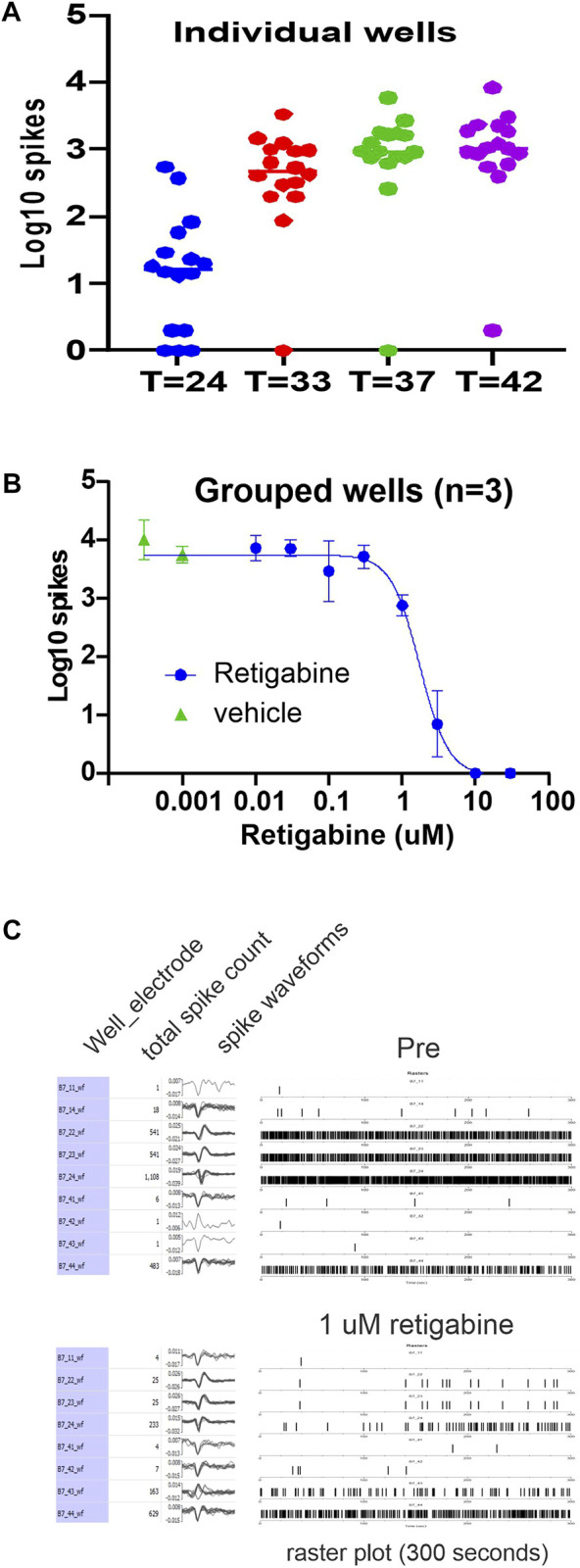
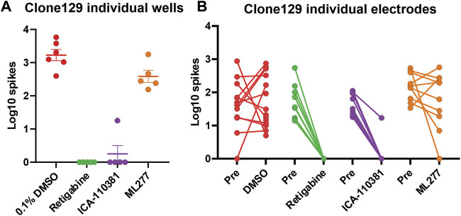
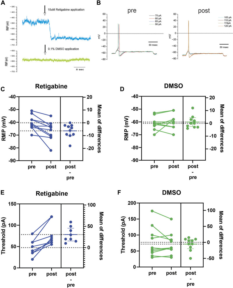
Chronic pain is highly prevalent and remains a significant unmet global medical need. As part of a search for modulatory genes that confer pain resilience, we have studied two family cohorts where one individual reported much less pain than other family members that share the same pathogenic gain-of-function Nav1.7 mutation that confers hyperexcitability on pain-signaling dorsal root ganglion (DRG) neurons. In each of these kindreds, the pain-resilient individual carried a gain-of-function variant in Kv7.2 or Kv7.3, two potassium channels that stabilize membrane potential and reduce excitability. Our observation in this molecular genetic study that these gain-of-function Kv7.2 and 7.3 variants reduce DRG neuron excitability suggests that agents that activate or open Kv7 channels should attenuate sensory neuron firing. In the present study, we assess the effects on sensory neuron excitability of three Kv7 modulators—retigabine (Kv7.2 thru Kv7.5 activator), ICA-110381 (Kv7.2/Kv7.3 specific activator), and as a comparator ML277 (Kv7.1 specific activator)—in a “human-pain-in-a-dish” model (human iPSC-derived sensory neurons, iPSC-SN). Multi-electrode-array (MEA) recordings demonstrated inhibition of firing with retigabine and ICA-110381 (but not with ML277), with the concentration-response curve indicating that retigabine can achieve a 50% reduction of firing with sub-micromolar concentrations. Current-clamp recording demonstrated that retigabine hyperpolarized iPSC-SN resting potential and increased threshold. This study implicates Kv7.2/Kv7.3 channels as effective modulators of sensory neuron excitability, and suggest that compounds that specifically target Kv7.2/Kv7.3 currents in sensory neurons, including human sensory neurons, might provide an effective approach toward pain relief.
Overview
The claims and statistics of this paper haven’t been checked yet.
Free: it reads the open-access text and checks the first 5 claims that cite a source we can check. Create a free account to check every claim.
Integrity signals
Registry
Cites
Statistics
Figures

Verification
Claims the paper makes
The claims and statistics of this paper haven’t been checked yet.
Free: it reads the open-access text and checks the first 5 claims that cite a source we can check. Create a free account to check every claim.
Claims other papers make about this one
Each is checked against this paper’s own text: does it say what the citing paper says it does?
The source backs part of the claim, not all of it — or backs it for a different population, condition or comparison.
In the citing paper
“Within prefrontal and limbic circuits, Kv7 activity normalizes neuronal gain and suppresses stress-evoked firing escalation that impairs working memory and mood regulation [241,250,251].”
From Synaptic Plasticity—Intrinsic Excitability and Antidepressant Discovery (2026) · cites this paper as [241]
Experimental — accuracy not yet measured
Self-check
Percentages, sums and reported differences re-computed from the paper’s own numbers.
The self-check needs the paper’s full text.
Citations
Cites
KCNQ2 and KCNQ3 Potassium Channel Subunits: Molecular Correlates of the M-Channel
Wang, H. (1998) · Science · doi:10.1126/science.282.5395.1890 · Cited by 1,294
Krebs, E. E., Gravely, A., Nugent, S., Jensen, A. C., DeRonne, B., Goldsmith, E. S., Kroenke, K., Bair, M. J., & Noorbaloochi, S. (2018) · JAMA · doi:10.1001/jama.2018.0899 · Cited by 983 · cited 1×
“In a long (12 months) randomized clinical trial in veterans with chronic pain, opioids were not better than non-steroidal anti-inflammatory drugs (Krebs et al., 2018).”
BackgroundBrown, D. A., & Passmore, G. M. (2009) · British Journal of Pharmacology · doi:10.1111/j.1476-5381.2009.00111.x · Cited by 676 · cited 2×
“3 channel subunits co-assemble to form a heteromeric channel responsible for producing potassium M-current conductance that regulates subthreshold excitability of sensory neurons (Wang et al., 1998; Brown and Passmore, 2009).”
BackgroundMethodChambers, S. M., Qi, Y., Mica, Y., Lee, G., Zhang, X.-J., Niu, L., Bilsland, J., Cao, L., Stevens, E., Whiting, P., et al. (2012) · Nature Biotechnology · doi:10.1038/nbt.2249 · Cited by 602
Gain-of-function mutation in Nav1.7 in familial erythromelalgia induces bursting of sensory neurons
Dib-Hajj, S. D., Rush, A. M., Cummins, T. R., Hisama, F. M., Novella, S., Tyrrell, L., Marshall, L., & Waxman, S. G. (2005) · Brain · doi:10.1093/brain/awh514 · Cited by 490
Self-citationHumphreys, K., Shover, C. L., Andrews, C. M., Bohnert, A. S. B., Brandeau, M. L., Caulkins, J. P., Chen, J. H., Cuéllar, M.-F., Hurd, Y. L., Juurlink, D. N., et al. (2022) · The Lancet · doi:10.1016/s0140-6736(21)02252-2 · Cited by 421
Tatulian, L., Delmas, P., Abogadie, F. C., & Brown, D. A. (2001) · The Journal of Neuroscience · doi:10.1523/jneurosci.21-15-05535.2001 · Cited by 406
KCNQ/M Currents in Sensory Neurons: Significance for Pain Therapy
Passmore, G. M., Selyanko, A. A., Mistry, M., Al-Qatari, M., Marsh, S. J., Matthews, E. A., Dickenson, A. H., Brown, T. A., Burbidge, S. A., Main, M., et al. (2003) · The Journal of Neuroscience · doi:10.1523/jneurosci.23-18-07227.2003 · Cited by 366
Gunthorpe, M. J., Large, C. H., & Sankar, R. (2012) · Epilepsia · doi:10.1111/j.1528-1167.2011.03365.x · Cited by 319
Severe pain in veterans: the effect of age and sex, and comparisons with the general population
Nahin, R. L. (2017) · The Journal of Pain · doi:10.1016/j.jpain.2016.10.021 · Cited by 293 · cited 1×
“Chronic pain is highly prevalent and remains a significant unmet global medical need because current treatments are not effective (Nahin, 2017).”
Background
Cited by
Beyond the hype: a comprehensive exploration of CBD’s biological impacts and mechanisms of action
Swenson, K. S. (2025) · Journal of Cannabis Research · doi:10.1186/s42238-025-00274-y · Cited by 23
Targeting sodium channels — challenges for acute pain and the leap to chronic pain
Alsaloum, M., Vasylyev, D. V., & Waxman, S. G. (2025) · Nature Reviews Neurology · doi:10.1038/s41582-025-01127-1 · Cited by 15
Self-citationCarotenuto, L., Keminer, O., Carleo, G., Zaliani, A., Leo, A., Citraro, R., De Sarro, G., Dirkx, N., Kaji, M., Weckhuysen, S., et al. (2025) · British Journal of Pharmacology · doi:10.1111/bph.70119 · Cited by 10
Zhang, B., Shi, X., Liu, X., Liu, Y., Li, X., Wang, Q., Huang, D., Zhao, W., Cui, J., Cao, Y., et al. (2024) · Journal of Pharmaceutical Analysis · doi:10.1016/j.jpha.2024.101132 · Cited by 10
Jones, D. T., Zurek, N. A., & Alles, S. R. A. (2026) · Journal of Neuroscience · doi:10.1523/jneurosci.0926-25.2025 · Cited by 8
Metadata
- Authors
- Estacion, M., Liu, S., Cheng, X., Dib-Hajj, S., & Waxman, S. G.
- Affiliations
- Yale University (US)
- Corresponding author
- Waxman, Stephen G.
- Venue
- Frontiers in Pharmacology
- Publisher
- Frontiers Media SA
- Published
- 2023-02-27
- Volume
- 14
- Pages
- 1138556-1138556
- ISSN
- 1663-9812 (ISSN-L)
- PMC
- PMC10008904
- OpenAlex
- W4322506649
- Cited by
- 11
- References
- 48
- Citations by year
- 2026: 5 · 2025: 5 · 2024: 1
- FWCI
- 0.98
- Type
- journal article
- Language
- en
- Open access
- gold
- doi.org
- Licence
- Locations
- Frontiers in Pharmacology (publishedVersion, cc-by)
- PubMed Central (publishedVersion, cc-by)
- PubMed (publishedVersion)
- DOAJ (DOAJ: Directory of Open Access Journals) (submittedVersion, cc-by-sa)
- Funders
- U.S. Department of Veterans Affairs
- Topics
- Life Sciences › Biochemistry, Genetics and Molecular Biology › Molecular Biology › Ion channel regulation and function
- Health Sciences › Medicine › Physiology (medical) › Pain Mechanisms and Treatments
- Life Sciences › Neuroscience › Sensory Systems › Ion Channels and Receptors
- Keywords
- Kv7.2, Kv7.3, Kv7 channels, retigabine, sensory neuron excitability, dorsal root ganglion neurons, resting membrane potential, hyperpolarization, chronic pain, pain resilience, potassium channel modulators
About these checks
Registry, retraction, Cites and statistics results are computed, not judged, with one exception: a printed reference that no index lists is read once by a model before it counts as a citation error. Claims are extracted by a model and kept only when their quote appears verbatim in the text; each cited source is then checked, with a model’s help, for whether it says what the claim relies on. Those verdicts can be wrong — read the claim’s quote and the cited source before relying on one.
Statistics and arithmetic are recomputed, not judged.
Confidence bands are shown only for fields calibrated on our annotated test set. None is calibrated yet, so no band is shown.
What counts as a claim ›How we measure confidence ›How we check this ›
Are you an author, or do you see a problem with this page? Request a review
How to cite
Cite CoChat for the checks on this page, and the paper for its own findings.
- This page
- CoChat. (2026). Citation check: Kv7-specific activators hyperpolarize resting membrane potential and modulate human iPSC-derived sensory neuron excitability. https://app.cochat.ai/papers/10.3389/fphar.2023.1138556
- The paper
- Estacion, M., Liu, S., Cheng, X., Dib-Hajj, S., & Waxman, S. G. (2023). Kv7-specific activators hyperpolarize resting membrane potential and modulate human iPSC-derived sensory neuron excitability. Frontiers in Pharmacology. https://doi.org/10.3389/fphar.2023.1138556
Data sources
Bibliographic data: Semantic Scholar, OpenAlex, Crossref.
Contains information from the Semantic Scholar Academic Graph Datasets, made available under the ODC Attribution License (ODC-BY 1.0).
Semantic Scholar’s data is described in: Kinney, R., et al. (2023). The Semantic Scholar Open Data Platform. arXiv:2301.10140.
