

The DOI matches its Crossref record; 2 of the 2 references it cites were found in a registry or an index.
Evaluating the Pathogenic Potential of IgE Targeting Cross-Reactive Carbohydrate Determinants in Dogs
Abstract
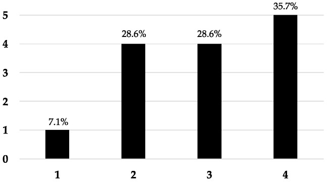
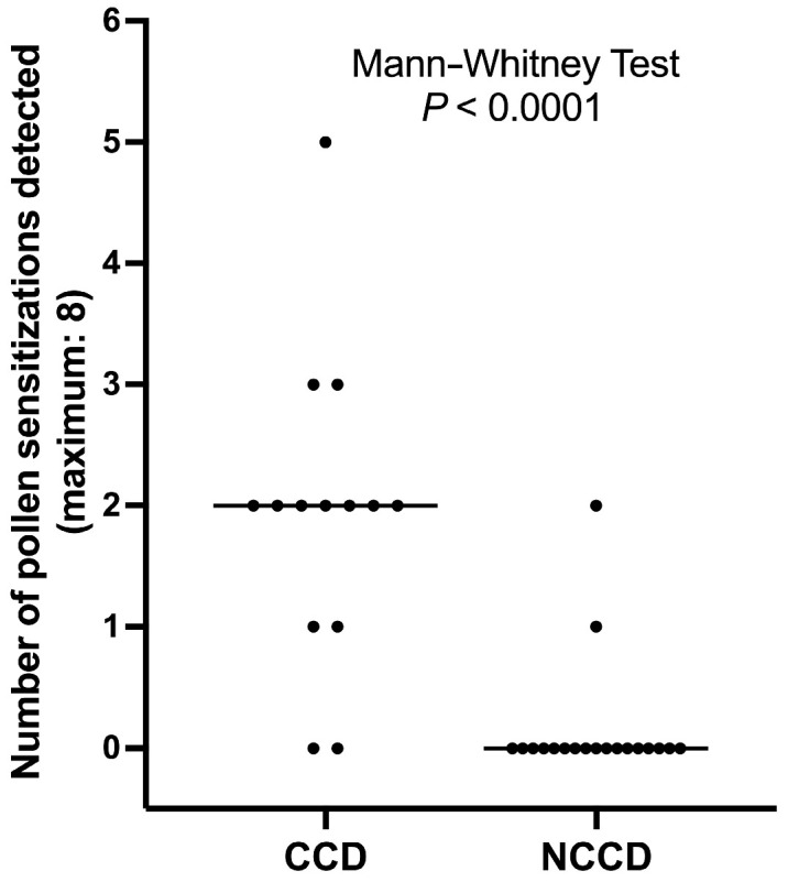
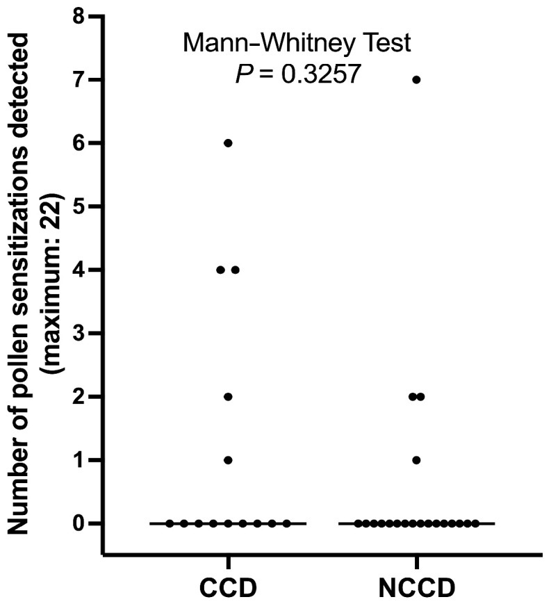
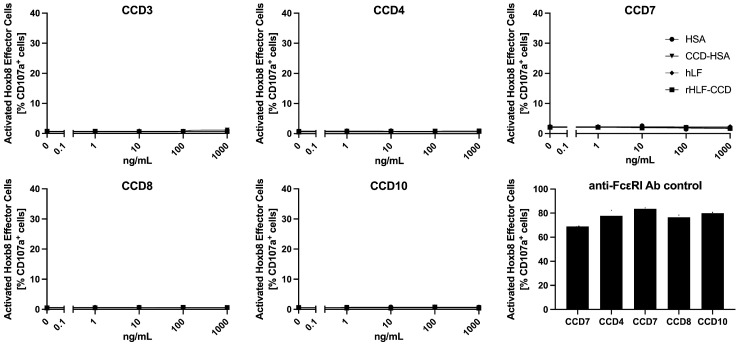
Cross-reactive carbohydrate determinants (CCDs) are complex N-glycans shared among allergens of plant, insect venom, and nematode origin. In allergic humans, IgE anti-CCD often develop and cause discrepancies between serological and skin tests. Overall, CCD-IgE are believed to be of low pathogenic relevance. IgE-targeting CCDs are also detected in companion animals, but their pathogenic potential and biological relevance are unknown. Herein, we first establish that, in 34 dogs with atopic dermatitis, the presence of serum anti-CCD IgE was detected in 14 pets (41.2%). In dogs, as in humans, IgE-targeting CCDs are heterogeneous, as they differentially recognized four distinct CCD-expressing proteins. The presence of CCD-IgE was associated with a higher and more frequent recognition of plant extracts in serological but not intradermal tests. Two different CCD-expressing proteins did not elicit immediate reactions when injected intradermally in dogs with detectable serum anti-CCD IgE. Similarly, two different CCD-expressing proteins did not induce the activation of mast cells passively transferred with canine anti-CCD IgE. Altogether, these results suggest that in dogs, as in humans, anti-CCD IgE are likely to have little pathogenic potential and blocking them in allergen-specific IgE serological tests is warranted to avoid false-positive results to plant extracts.
Overview
The claims and statistics of this paper haven’t been checked yet.
Run the check — free, no account needed. It reads the open-access text and checks the sources it cites.
Integrity signals
Registry
Cites
Statistics
Figures
Verification
Claims the paper makes
The claims and statistics of this paper haven’t been checked yet.
Run the check — free, no account needed. It reads the open-access text and checks the sources it cites.
Claims other papers make about this one
Each is checked against this paper’s own text: does it say what the citing paper says it does?
In the citing paper
“Anti-CCD IgE antibodies may affect in vitro allergy test results and complicate their interpretation.”
From Parasites and allergies: a complex bidirectional relationship from evolutionary origins to modern therapeutics (2026) · cites this paper as Olivry et al. 2024
In the citing paper
“However, in many cases, CCD-specific IgE is of limited clinical relevance and does not correlate with symptoms”
From Parasites and allergies: a complex bidirectional relationship from evolutionary origins to modern therapeutics (2026) · cites this paper as Olivry et al. 2024
In the citing paper
“In addition, epitopes such as galactose-β-1,4-fucose have been identified in N-glycans shared by A. suum and D. pteronyssinus.”
From Parasites and allergies: a complex bidirectional relationship from evolutionary origins to modern therapeutics (2026) · cites this paper as Olivry et al. 2024
Self-check
Percentages, sums and reported differences re-computed from the paper’s own numbers.
The self-check needs the paper’s full text.
Citations
Cites
The Role of Protein Glycosylation in Allergy
Altmann, F. (2007) · International Archives of Allergy and Immunology · doi:10.1159/000096114 · Cited by 393 · cited 2×
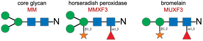
“…rst, abbreviated as MUXF3, only has two terminal The “classical” plant CCDs have two crucial additions to the basic glycan core: a xylose with a β 1,2 linkage to the most proximal mannose and a fucose attached with a α 1,3 linkage to the proximal N-acetylglucosamine (Figure 1) [2,3]. mannose and…”
Mari, A., Iacovacci, P., Afferni, C., Barletta, B., Tinghino, R., Di Felice, G., & Pini, C. (1999) · Journal of Allergy and Clinical Immunology · doi:10.1016/s0091-6749(99)70171-5 · Cited by 217
Research works printed but not linked: 46
- Commins, 2015, Carbohydrates as Allergens, Curr. Allergy Asthma Rep., 15, 492, 10.1007/s11882-014-0492-y · DOI
- Hilger, 2021, Carbohydrate Epitopes Currently Recognized as Targets for IgE Antibodies, Allergy, 76, 2383, 10.1111/all.14802 · DOI
- Ishihara, 1979, Complete Structure of the Carbohydrate Moiety of Stem Bromelain. An Application of the Almond Glycopeptidase for Structural Studies of Glycopeptides, J. Biol. Chem., 254, 10715, 10.1016/S0021-9258(19)86580-8 · DOI
- Bardor, 2003, Immunoreactivity in Mammals of Two Typical Plant Glyco-Epitopes, Core Alpha(1,3)-Fucose and Core Xylose, Glycobiology, 13, 427, 10.1093/glycob/cwg024 · DOI
- Aalberse, 1981, Immunoglobulin E Antibodies That Crossreact with Vegetable Foods, Pollen, and Hymenoptera Venom, J. Allergy Clin. Immunol., 68, 356, 10.1016/0091-6749(81)90133-0 · DOI
- Homann, 2017, Glycans and Glycan-Specific IgE in Clinical and Molecular Allergology: Sensitization, Diagnostics, and Clinical Symptoms, J. Allergy Clin. Immunol., 140, 356, 10.1016/j.jaci.2017.04.019 · DOI
- Tretter, 1993, Fucose Alpha 1,3-Linked to the Core Region of Glycoprotein N-Glycans Creates an Important Epitope for IgE from Honeybee Venom Allergic Individuals, Int. Arch. Allergy Immunol., 102, 259, 10.1159/000236534 · DOI
- Hemmer, 2004, Specificity of IgG and IgE Antibodies against Plant and Insect Glycoprotein Glycans Determined with Artificial Glycoforms of Human Transferrin, Glycobiology, 14, 457, 10.1093/glycob/cwh058 · DOI
- Levy, 2018, A Preliminary Study of Serum IgE against Cross-Reactive Carbohydrate Determinants (CCD) in Client-Owned Atopic Dogs, Vet. Dermatol., 29, 243-e90, 10.1111/vde.12529 · DOI
- Bexley, 2018, Inhibition of Canine Serum IgE Binding to Cross-Reactive Carbohydrate Determinants in Environmental Allergens (Abstract), Vet. Dermatol., 29, 357
- Piccione, 2019, Serum IgE against Cross-Reactive Carbohydrate Determinants (CCD) in Healthy and Atopic Dogs, Vet. Dermatol., 30, 507-e153, 10.1111/vde.12799 · DOI
- Gedon, 2019, Agreement of Serum Allergen Test Results with Unblocked and Blocked IgE against Cross-Reactive Carbohydrate Determinants (CCD) and Intradermal Test Results in Atopic Dogs, Vet. Dermatol., 30, 195-e61, 10.1111/vde.12742 · DOI
- Lee, 2020, Detection and Inhibition of IgE for Cross-Reactive Carbohydrate Determinants Evident in an Enzyme-Linked Immunosorbent Assay for Detection of Allergen-Specific IgE in the Sera of Dogs and Cats, Vet. Dermatol., 31, 439-e116, 10.1111/vde.12904 · DOI
- Mohammaddavoodi, 2021, Prevalence of Immunoglobulin E against Cross-Reactive Carbohydrate Determinants and the Impact of a Blocker in Canine Sera, Tierarztl. Praxis. Ausg. K Kleintiere/Heimtiere, 49, 272, 10.1055/a-1442-3928 · DOI
- Canning, 2021, Multivariable Analysis of the Influence of Cross-Reactive Carbohydrate Determinant Inhibition and Other Factors on Intradermal and Serological Allergen Test Results: A Prospective, Multicentre Study, Vet. Dermatol., 32, 347-e96, 10.1111/vde.12974 · DOI
- Kasper, 2023, Cross-Reactive Carbohydrate Determinants in Atopic and Healthy Dogs and Their Influence on Allergy Test Specificity, Vet. Rec., 193, e3308, 10.1002/vetr.3308 · DOI
- Mohammaddavoodi, 2019, Prevalence of Immunoglobulin E (IgE) against Cross-Reactive Carbohydrate Determinants (CCD) and Impact of CCD Blocking in Serum from Cats (Abstract), Vet. Dermatol., 31, 34
- Enck, 2021, Detection and Inhibition of IgE Antibodies Reactive with Cross-Reactive Carbohydrate Determinants in an ELISA for Allergen-Specific IgE in Horses, Vet. Dermatol., 32, 685-e184, 10.1111/vde.12963 · DOI
- Aalberse, 1997, Poor Biologic Activity of Cross-Reactive IgE Directed to Carbohydrate Determinants of Glycoproteins, J. Allergy Clin. Immunol., 100, 327, 10.1016/S0091-6749(97)70245-8 · DOI
- Mari, 2002, IgE to Cross-Reactive Carbohydrate Determinants: Analysis of the Distribution and Appraisal of the in Vivo and in Vitro Reactivity, Int. Arch. Allergy Immunol., 129, 286, 10.1159/000067591 · DOI
- Mari, 2008, Evaluation by Double-Blind Placebo-Controlled Oral Challenge of the Clinical Relevance of IgE Antibodies against Plant Glycans, Allergy, 63, 891, 10.1111/j.1398-9995.2008.01703.x · DOI
- Ebo, 2004, Sensitization to Cross-Reactive Carbohydrate Determinants and the Ubiquitous Protein Profilin: Mimickers of Allergy, Clin. Exp. Allergy, 34, 137, 10.1111/j.1365-2222.2004.01837.x · DOI
- Bolhaar, 2005, A Mutant of the Major Apple Allergen, Mal d 1, Demonstrating Hypo-Allergenicity in the Target Organ by Double-Blind Placebo-Controlled Food Challenge, Clin. Exp. Allergy, 35, 1638, 10.1111/j.1365-2222.2005.02390.x · DOI
- Batanero, 1999, IgE-Binding and Histamine-Release Capabilities of the Main Carbohydrate Component Isolated from the Major Allergen of Olive Tree Pollen, Ole e 1, J. Allergy Clin. Immunol., 103, 147, 10.1016/S0091-6749(99)70538-5 · DOI
- Wicklein, 2004, Carbohydrate Moieties Can Induce Mediator Release: A Detailed Characterization of Two Major Timothy Grass Pollen Allergens, Biol. Chem., 385, 397, 10.1515/BC.2004.044 · DOI
- Foetisch, 2003, Biological Activity of IgE Specific for Cross-Reactive Carbohydrate Determinants, J. Allergy Clin. Immunol., 111, 889, 10.1067/mai.2003.173 · DOI
- Iacovacci, 2002, Comparison between the Native Glycosylated and the Recombinant Cup A1 Allergen: Role of Carbohydrates in the Histamine Release from Basophils, Clin. Exp. Allergy, 32, 1620, 10.1046/j.1365-2222.2002.01516.x · DOI
- Vieths, 2001, N- and O-Linked Oligosaccharides of Allergenic Glycoproteins, Glycoconj. J., 18, 373, 10.1023/A:1014860030380 · DOI · Paper page
- Jin, 2008, Affinity of IgE and IgG against Cross-Reactive Carbohydrate Determinants on Plant and Insect Glycoproteins, J. Allergy Clin. Immunol., 121, 185, 10.1016/j.jaci.2007.07.047 · DOI
- Keswani, 2024, Neutralizing IgG4 Antibodies Are a Biomarker of Sustained Efficacy after Peanut Oral Immunotherapy, J. Allergy Clin. Immunol., 153, 1611, 10.1016/j.jaci.2024.02.017 · DOI
- Furiness, K.N., El Ansari, Y.S., Oettgen, H.C., and Kanagaratham, C. (2024). Allergen-Specific IgA and IgG Antibodies as Inhibitors of Mast Cell Function in Food Allergy. Front. Allergy, 5. · DOI
- Holzweber, 2013, Inhibition of IgE Binding to Cross-Reactive Carbohydrate Determinants Enhances Diagnostic Selectivity, Allergy, 68, 1269, 10.1111/all.12229 · DOI
- Olivry, T., Fontao, A.M., Aumayr, M., Ivanovova, N.P., Mitterer, G., and Harwanegg, C. (2024). Validation of a Multiplex Molecular Macroarray for the Determination of Allergen-Specific IgE Sensitizations in Dogs. Vet. Sci., 11. · DOI
- Hensel, P., Santoro, D., Favrot, C., Hill, P., and Griffin, C. (2015). Canine Atopic Dermatitis: Detailed Guidelines for Diagnosis and Allergen Identification. BMC Vet. Res., 11. · DOI
- Fujiyama, 2004, N-Linked Glycan Structures of Human Lactoferrin Produced by Transgenic Rice, Biosci. Biotechnol. Biochem., 68, 2565, 10.1271/bbb.68.2565 · DOI
- Hammerberg, 1997, Auto IgG Anti-IgE and IgG x IgE Immune Complex Presence and Effects on ELISA-Based Quantitation of IgE in Canine Atopic Dermatitis, Demodectic Acariasis and Helminthiasis, Vet. Immunol. Immunopathol., 60, 33, 10.1016/S0165-2427(97)00119-0 · DOI
- Olivry, 2013, Evidence-Based Guidelines for Anti-Allergic Drug Withdrawal Times before Allergen-Specific Intradermal and IgE Serological Tests in Dogs, Vet. Dermatol., 24, 225-e49, 10.1111/vde.12016 · DOI
- Hubbard, 2011, Comparison of Subjective and Objective Intradermal Allergy Test Scoring Methods in Dogs with Atopic Dermatitis, J. Am. Anim. Hosp. Assoc., 47, 399, 10.5326/JAAHA-MS-5638 · DOI
- Brigger, 2022, A Novel Functional Mast Cell Assay for the Detection of Allergies, J. Allergy Clin. Immunol., 149, 1018, 10.1016/j.jaci.2021.08.006 · DOI
- Bachmeier-Zbären, N., Celik, A., van Brummelen, R., Roos, N., Steinmann, M., Hoang, J.A., Yin, X., Ditlof, C.M., Duan, L., and Upton, J.E.M. (2024). Clinical Utility Analysis of the Hoxb8 Mast Cell Activation Test for the Diagnosis of Peanut Allergy. Allergy, in press. · DOI
- Stedman, 2001, Measurement of Canine IgE Using the Alpha Chain of the Human High Affinity IgE Receptor, Vet. Immunol. Immunopathol., 78, 349, 10.1016/S0165-2427(01)00242-2 · DOI
- Akkerdaas, 2000, Beta(1,2)-Xylose and Alpha(1,3)-Fucose Residues Have a Strong Contribution in IgE Binding to Plant Glycoallergens, J. Biol. Chem., 275, 11451, 10.1074/jbc.275.15.11451 · DOI
- McCall, 2001, Characterization and Cloning of a Major High Molecular Weight House Dust Mite Allergen (Der f 15) for Dogs, Vet. Immunol. Immunopathol., 78, 231, 10.1016/S0165-2427(00)00258-0 · DOI
- Weber, 2003, Identification, Characterization, and Cloning of a Complementary DNA Encoding a 60-Kd House Dust Mite Allergen (Der f 18) for Human Beings and Dogs, J. Allergy Clin. Immunol., 112, 79, 10.1067/mai.2003.1602 · DOI
- Olivry, 2017, The Novel High Molecular Weight Dermatophagoides Farinae Protein Zen-1 Is a Major Allergen in North American and European Mite Allergic Dogs with Atopic Dermatitis, Vet. Dermatol., 28, 177-e38, 10.1111/vde.12393 · DOI
- Mallmann, 2021, Clinical Relevance of Intradermal Test Results in Atopic Dogs, Tierarztl. Prax. Ausg. K. Kleintiere/Heimtiere, 49, 349, 10.1055/a-1584-4965 · DOI
Cited by
Sorted by citations
Packi, K., Świerczewski, D., Klimczak, S., Ochab, D., Gałęziewska, J., Gromek, P., Kruczkowska, W., Płuciennik, E., Kałuzińska-Kołat, Ż., & Kołat, D. (2026) · Antonie van Leeuwenhoek · doi:10.1007/s10482-026-02425-4
Metadata
- Authors
- Olivry, T., Mas Fontao, A., Widorn, L., & Mueller, R. S.
- Venue
- Animals
- Publisher
- MDPI AG
- Published
- 2024-11-14
- Volume
- 14
- Issue
- 22
- Pages
- 3275
- ISSN
- 2076-2615
- PMC
- PMC11591198
- Cited by
- 6
- References
- 2
- Language
- en
- Funders
- Nextmune AB
About these checks
Registry, retraction, Cites and statistics results are computed, not judged, with one exception: a printed reference that no index lists is read once by a model before it counts as a citation error. Claims are extracted by a model and kept only when their quote appears verbatim in the text; each cited source is then checked, with a model’s help, for whether it says what the claim relies on. Those verdicts can be wrong — read the claim’s quote and the cited source before relying on one.
Statistics and arithmetic are recomputed, not judged.
Confidence bands are shown only for fields calibrated on our annotated test set. None is calibrated yet, so no band is shown.
What counts as a claim ›How we measure confidence ›How we check this ›
Are you an author, or do you see a problem with this page? Request a review
