

The DOI matches its Crossref record; 1 of the 1 references it cites were found in a registry or an index.
Staphylococcal Biofilm on the Surface of Catheters: Electron Microscopy Evaluation of the Inhibition of Biofilm Growth by RNAIII Inhibiting Peptide
Abstract
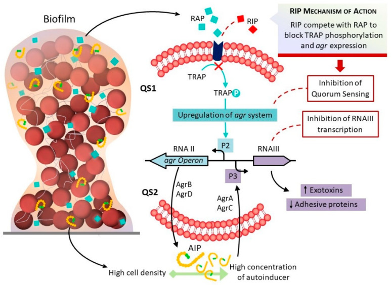
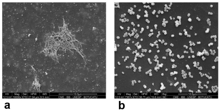
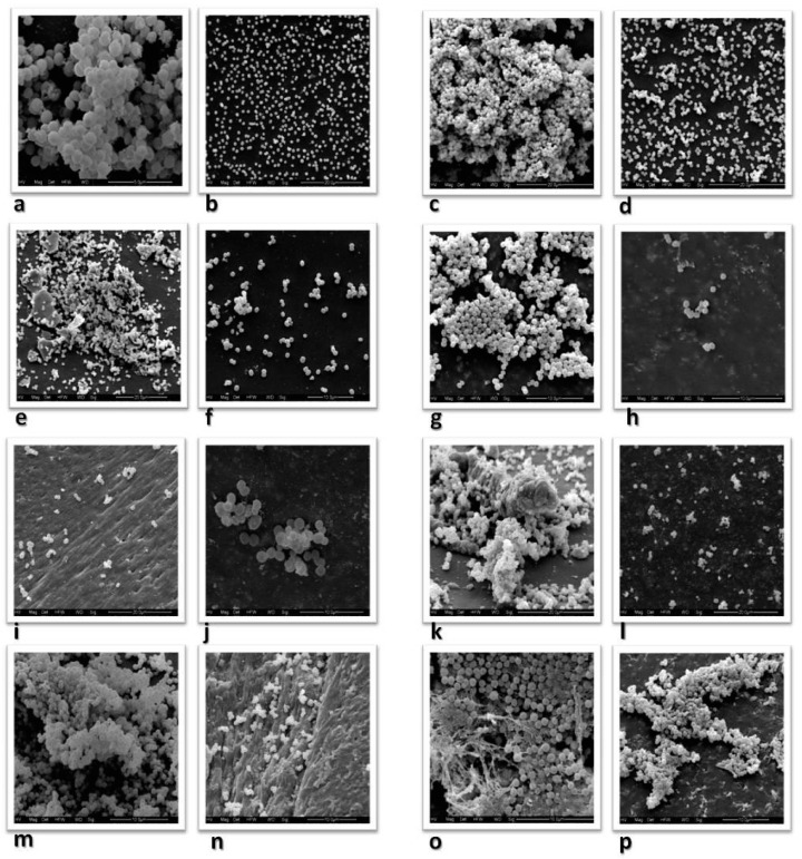
Staphylococcus aureus and coagulase-negative staphylococci (CoNS) have become the main causative agents of medical device-related infections due to their biofilm-forming capability, which protects them from the host’s immune system and from the action of antimicrobials. This study evaluated the ability of RNA III inhibiting peptide (RIP) to inhibit biofilm formation in 10 strains isolated from clinical materials, including one S. aureus strain, two S. epidermidis, two S. haemolyticus, two S. lugdunensis, and one isolate each of the following species: S. warneri, S. hominis, and S. saprophyticus. The isolates were selected from a total of 200 strains evaluated regarding phenotypic biofilm production and the presence and expression of the ica operon. The isolates were cultured in trypticase soy broth with 2% glucose in 96-well polystyrene plates containing catheter segments in the presence and absence of RIP. The catheter segments were observed by scanning electron microscopy. The results showed inhibition of biofilm formation in the presence of RIP in all CoNS isolates; however, RIP did not interfere with biofilm formation by S. aureus. RIP is a promising tool that might be used in the future for the prevention of biofilm-related infections caused by CoNS.
Overview
The claims and statistics of this paper haven’t been checked yet.
Run the check — free, no account needed. It reads the open-access text and checks the sources it cites.
Integrity signals
Registry
Cites
Statistics
Figures
Verification
Claims the paper makes
The claims and statistics of this paper haven’t been checked yet.
Run the check — free, no account needed. It reads the open-access text and checks the sources it cites.
Claims other papers make about this one
Each is checked against this paper’s own text: does it say what the citing paper says it does?
In the citing paper
“Similarly, reverse transcription-polymerase chain reaction evaluates biofilm-related gene expression but does not offer insights into the spatial organization of the EPS matrix and biofilm morphology.45-47”
From SEMTWIST Quantification of Biofilm Infection in Human Chronic Wound Using Scanning Electron Microscopy and Machine Learning (2025) · cites this paper as [45]
Self-check
Percentages, sums and reported differences re-computed from the paper’s own numbers.
The self-check needs the paper’s full text.
Citations
Cites
Marrie, T. J., & Costerton, J. W. (1984) · Journal of Clinical Microbiology · doi:10.1128/jcm.19.5.687-693.1984 · Cited by 327
Research works printed but not linked: 30
- Morales, 2004, Biofilm: The microbial “bunker” for intravascular catheter-related infection, Support. Care Cancer, 12, 701, 10.1007/s00520-004-0630-5 · DOI
- Claesson, 2005, Adherence of Staphylococcus epidermidis to extracellular matrix proteins and effects of fibrinogen-bound bacteria on oxidase activity and apoptosis in neutrophils, APMS, 113, 361
- Gotz, 2002, Staphylococcus and biofilms, Mol. Microbiol., 43, 1367, 10.1046/j.1365-2958.2002.02827.x · DOI
- Pozzi, 2007, Association between methicillin susceptibility and biofilm regulation in Staphylococcus aureus isolates from device-related infections, J. Clin. Microbiol., 45, 1379, 10.1128/JCM.02280-06 · DOI
- Donlan, 2002, Biofilm: Microbial life on surfaces, Emerg. Infect. Dis., 8, 881, 10.3201/eid0809.020063 · DOI · Paper page
- Cafiso, 2004, Presence of the ica operon in clinical isolates of Staphylococcus epidermidis and its role in biofilm production, Clin. Microbiol. Infect., 10, 1081, 10.1111/j.1469-0691.2004.01024.x · DOI
- Mack, 1996, The intercellular adhesin involved in biofilm accumulation of Staphylococcus epidermidis is a linear b-1,6-linked glucosaminoglycan: Purification and structural analysis, J. Bacteriol., 178, 175, 10.1128/jb.178.1.175-183.1996 · DOI · Paper page
- Kropec, 2002, Immunochemical properties of the staphylococcal poly-N-acetylglucosamine surface polysaccharide, Infect. Immun., 70, 4433, 10.1128/IAI.70.8.4433-4440.2002 · DOI
- Heilmann, 1996, Molecular basis of intercellular adhesion in the biofilm-forming Staphylococcus epidermidis, Mol. Microbiol., 20, 1083, 10.1111/j.1365-2958.1996.tb02548.x · DOI
- Cucarella, 2001, Bap, a Staphylococcus aureus surface protein involved in biofilm formation, J. Bacteriol., 183, 2888, 10.1128/JB.183.9.2888-2896.2001 · DOI
- Vadyvaloo, 2005, Molecular genetics of Staphylococcus epidermidis on indwelling medical devices, Int. J. Artif. Organs, 28, 1069, 10.1177/039139880502801104 · DOI
- Balaban, 2003, Use the quorum-sensing inhibitor RNAIII-inhibiting peptide to prevent biofilm formation in vivo by drug-resistant Staphylococcus epidermidis, J. Infect. Dis., 187, 625, 10.1086/345879 · DOI
- Balaban, 2007, Treatment of Staphylococcus aureus biofilm infection by the quorum sensing inhibitor RIP, Antimicrob. Agents Chemother., 51, 2226, 10.1128/AAC.01097-06 · DOI
- Simonetti, 2008, RNAIII-inhibiting peptide enhances healing of wounds infected with methicillin-resistant Staphylococcus aureus, Antimicrob. Agents Chemother., 52, 2205, 10.1128/AAC.01340-07 · DOI
- Shaw, 2007, Inactivation of traP has no effect on the agr quorum-sensing system or virulence of Staphylococcus aureus, Infect. Immun., 75, 4519, 10.1128/IAI.00491-07 · DOI
- Adhikari, 2007, A Nonsense mutation in agrA accounts for the defect in agr expression and the avirulence of Staphylococcus aureus 8325-4 traP:kan, Infect. Immun., 75, 4534, 10.1128/IAI.00679-07 · DOI
- Balaban, 2001, Regulation of Staphylococcus aureus pathogenesis via target of RNAIII-activating protein (TRAP), J. Biol. Chem., 276, 2658, 10.1074/jbc.M005446200 · DOI
- Korem, 2005, Transcriptional profiling (microarray analysis) of TRAP, a master regulator of staphylococcal virulence, Infect. Immun., 73, 6220, 10.1128/IAI.73.10.6220-6228.2005 · DOI
- Balaban, 2005, Prevention of staphylococcal biofilm-associated infections by the quorum sensing inhibitor RIP, Clin. Orthop. Relat. Res., 437, 48, 10.1097/01.blo.0000175889.82865.67 · DOI
- Fredheim, 2009, Biofilm formation by Staphylococcus haemolyticus, J. Clin. Microbiol., 47, 1172, 10.1128/JCM.01891-08 · DOI
- Kini, 2010, Unusual presentation of endocarditis caused by Staphylococcus warneri, Open Microbiol. J., 4, 103, 10.2174/1874285801004010103 · DOI
- Frank, 2007, Poly-N-acetylglucosamine is not a major component of the extracellular matrix in biofilms formed by icaADBC-positive Staphylococcus lugdunensis isolates, Infect. Immun., 75, 4728, 10.1128/IAI.00640-07 · DOI
- Sun, 2012, Growth properties of Staphylococcus aureus in biofilm formed on polystyrene plate, Afr. J. Microbiol. Res., 6, 3284
- Koneman, E.W., Allen, S.D., Janda, W.M., Winn, W.C., and Schreckenberger, P.C. (1997). Koneman’s Color Atlas and Textbook of Diagnostic Microbiology, JB Lippincott. [6th ed.].
- Couto, 2001, Identification of clinical staphylococcal isolates from humans by Internal Transcribed Spacer PCR, J. Clin. Microbiol., 39, 3099, 10.1128/JCM.39.9.3099-3103.2001 · DOI
- Christensen, 1985, Adherence of coagulase-negative Staphylococci to plastic tissue culture plates: A quantitative model for the adherence of Staphylococci to medical devices, J. Clin. Microbiol., 22, 996, 10.1128/jcm.22.6.996-1006.1985 · DOI
- Oliveira, 2010, Comparison of methods for the detection of biofilm production in coagulase-negative staphylococci, BMC Res. Notes, 3, 1, 10.1186/1756-0500-3-260 · DOI
- Arciola, 2001, Presence of icaA and icaD genes and slime production in a collection of staphylococcal strains from catheter-associated infections, J. Clin. Microbiol., 39, 2151, 10.1128/JCM.39.6.2151-2156.2001 · DOI
- Arciola, 2005, A multiplex PCR method for the detection of all five individual genes of ica locus in Staphylococcus epidermidis. A survey on 400 clinical isolates from prosthesis-associated infections, J. Biomed. Mater. Res. A, 75A, 408, 10.1002/jbm.a.30445 · DOI
- Rohde, 2007, Polysaccharide intercellular adhesin or protein factors in biofilm accumulation of Staphylococcus epidermidis and Staphylococcus aureus isolated from prosthetic hip and knee joint infections, Biomaterials, 28, 1711, 10.1016/j.biomaterials.2006.11.046 · DOI
Cited by
Sorted by citations
Singh, S., Muniz De Oliveira, F., Wang, C., Kumar, M., Xuan, Y., DeMazumder, D., Sen, C. K., & Roy, S. (2025) · Advances in Wound Care · doi:10.1089/wound.2024.0291 · Cited by 6
Metadata
- Authors
- de Oliveira, A., Pinheiro-Hubinger, L., Pereira, V. C., Riboli, D. F. M., Martins, K. B., Romero, L. C., & Cunha, M. D. L. R. D. S. D.
- Venue
- Antibiotics
- Publisher
- MDPI AG
- Published
- 2021-07-20
- Volume
- 10
- Issue
- 7
- Pages
- 879
- ISSN
- 2079-6382
- PMC
- PMC8300745
- Cited by
- 10
- References
- 1
- Language
- en
- Funders
- FAPESP (2011/07285-5)
- São Paulo State Department of Education (1)
About these checks
Registry, retraction, Cites and statistics results are computed, not judged, with one exception: a printed reference that no index lists is read once by a model before it counts as a citation error. Claims are extracted by a model and kept only when their quote appears verbatim in the text; each cited source is then checked, with a model’s help, for whether it says what the claim relies on. Those verdicts can be wrong — read the claim’s quote and the cited source before relying on one.
Statistics and arithmetic are recomputed, not judged.
Confidence bands are shown only for fields calibrated on our annotated test set. None is calibrated yet, so no band is shown.
What counts as a claim ›How we measure confidence ›How we check this ›
Are you an author, or do you see a problem with this page? Request a review
