

The DOI matches its Crossref record; 37 of the 37 references it cites were found in a registry or an index.
A Predicted Helix-Turn-Helix Core Is Critical for Bacteriophage Kil Peptide to Disrupt Escherichia coli Cell Division
Open accessAbstract
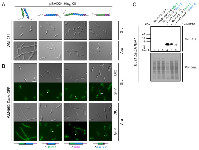
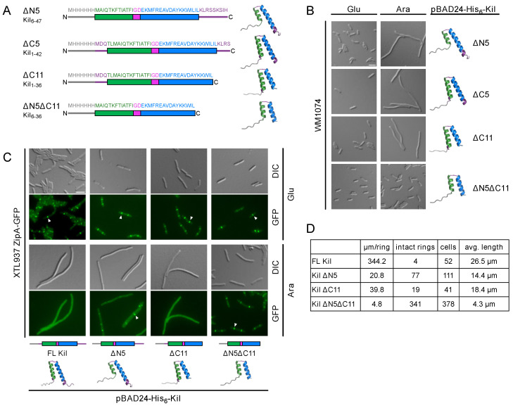
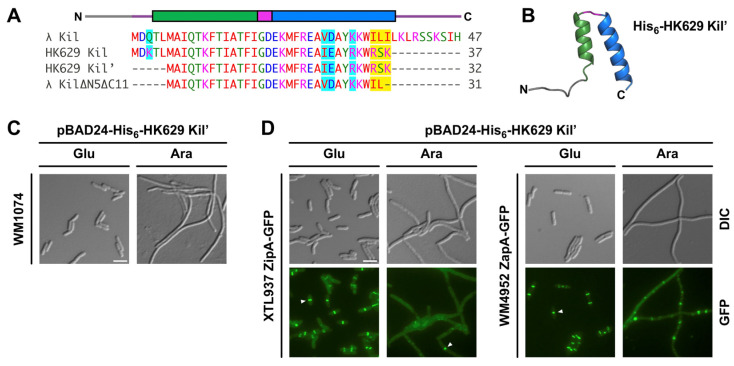
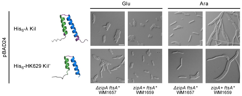
Background/objectives: FtsZ, a eukaryotic tubulin homolog and an essential component of the bacterial divisome, is the target of numerous antimicrobial compounds as well as proteins and peptides, most of which inhibit FtsZ polymerization dynamics. We previously showed that the Kil peptide from bacteriophage λ inhibits Escherichia coli cell division by disrupting FtsZ ring assembly, and this inhibition requires the presence of the essential FtsZ membrane anchor protein ZipA. Methods: To investigate Kil’s molecular mechanism further, we employed deletions, truncations, and molecular modeling to identify the minimal residues necessary for its activity. Results: Modeling suggested that Kil’s core segment folds into a helix-turn-helix (HTH) structure. Deleting either the C-terminal 11 residues or the N-terminal 5 residues of Kil still allowed the inhibition of E. coli cell division, but removing both termini nearly abolished this activity, indicating that a minimal region within the Kil HTH core is essential for its structure and function. Another Kil-like peptide from a closely related enterobacterial phage also disrupted FtsZ ring assembly and required ZipA for this activity. Consistent with its broader activity against FtsZ, λ Kil was able to efficiently inhibit cell division of a uropathogenic E. coli (UPEC) strain. Conclusions: Understanding the structure and function of Kil and similar peptides can potentially reveal additional ways to target FtsZ for antimicrobial therapies and elucidate how FtsZ functions in bacterial cell division.
Overview
The claims and statistics of this paper haven’t been checked yet.
Run the check — free, no account needed. It reads the open-access text and checks the sources it cites.
Integrity signals
Registry
Cites
Statistics
Figures
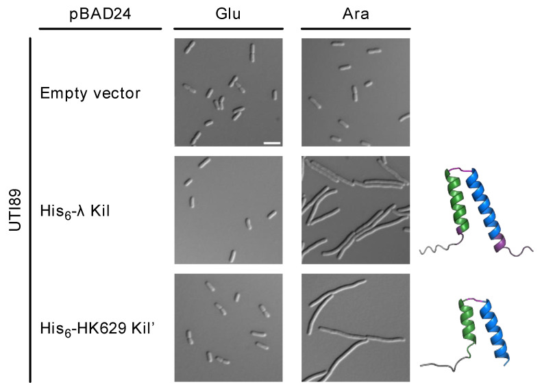
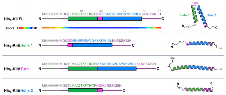
Verification
Claims the paper makes
The claims and statistics of this paper haven’t been checked yet.
Run the check — free, no account needed. It reads the open-access text and checks the sources it cites.
Claims other papers make about this one
Each is checked against this paper’s own text: does it say what the citing paper says it does?
The check wasn’t confident either way, so it gives no verdict — never disputed.
In the citing paper
“The second, the phage HK629, is also considered not dangerous for humans or animals. It is not inherently present in all E. coli strains, and it can integrate into the bacterial genome under certain conditions [140]. The third, the phage fiAA91, is not dangerous for humans or animals as well, showing similar features as HK629 [141].”
From Selection, Comparative Genomics, and Potential Probiotic Features of Escherichia coli 5C, a pks-Negative Strain Isolated from Healthy Infant Donor Feces (2026) · cites this paper as [140]
Experimental — accuracy not yet measured
Self-check
Percentages, sums and reported differences re-computed from the paper’s own numbers.
The self-check needs the paper’s full text.
Citations
Cites
Global burden of bacterial antimicrobial resistance in 2019: a systematic analysis
Murray, C. J. L., Ikuta, K. S., Sharara, F., Swetschinski, L., Robles Aguilar, G., Gray, A., Han, C., Bisignano, C., Rao, P., Wool, E., et al. (2022) · The Lancet · doi:10.1016/s0140-6736(21)02724-0 · Cited by 16,316
Accurate structure prediction of biomolecular interactions with AlphaFold 3
Abramson, J., Adler, J., Dunger, J., Evans, R., Green, T., Pritzel, A., Ronneberger, O., Willmore, L., Ballard, A. J., Bambrick, J., et al. (2024) · Nature · doi:10.1038/s41586-024-07487-w · Cited by 15,861
Guzman, L., Belin, D., Carson, M. J., & Beckwith, J. (1995) · Journal of Bacteriology · doi:10.1128/jb.177.14.4121-4130.1995 · Cited by 4,942
Urinary tract infections: epidemiology, mechanisms of infection and treatment options
Flores‐Mireles, A. L., Walker, J. N., Caparon, M. G., & Hultgren, S. J. (2015) · Nature Reviews Microbiology · doi:10.1038/nrmicro3432 · Cited by 4,343
FtsZ ring structure associated with division in Escherichia coli
Bi, E., & Lutkenhaus, J. (1991) · Nature · doi:10.1038/354161a0 · Cited by 1,471
Errington, J., Daniel, R. A., & Scheffers, D. (2003) · Microbiology and Molecular Biology Reviews · doi:10.1128/mmbr.67.1.52-65.2003 · Cited by 613
The highly diverse antiphage defence systems of bacteria
Georjon, H., & Bernheim, A. G. (2023) · Nature Reviews Microbiology · doi:10.1038/s41579-023-00934-x · Cited by 516
Thermosensitive Mutants of E. coli Affected in the Processes of DNA Synthesis and Cellular DivisionPreprint
Hirota, Y., Ryter, A., & Jacob, F. (1968) · Cold Spring Harbor Symposia on Quantitative Biology · doi:10.1101/sqb.1968.033.01.077 · Cited by 484
Diverse paths to midcell: assembly of the bacterial cell division machinery.
Goehring, N. W., & Beckwith, J. (2005) · Current Biology · doi:10.1016/j.cub.2005.06.038 · Cited by 440
Unique and overlapping roles for ZipA and FtsA in septal ring assembly in Escherichia coli
Pichoff, S., & Lutkenhaus, J. (2002) · The EMBO Journal · doi:10.1093/emboj/21.4.685 · Cited by 407
ZipA-Induced Bundling of FtsZ Polymers Mediated by an Interaction between C-Terminal Domains
Hale, C. A., Rhee, A. C., & de Boer, P. D. (2000) · Journal of Bacteriology · doi:10.1128/jb.182.18.5153-5166.2000 · Cited by 232
Geissler, B., Elraheb, D., & Margolin, W. (2003) · Proceedings of the National Academy of Sciences · doi:10.1073/pnas.0635003100 · Cited by 204
Self-citationThe crystal structure of ZapA and its modulation of FtsZ polymerisation.
Low, H. H., Moncrieffe, M., & Löwe, J. (2004) · Journal of Molecular Biology · doi:10.1016/j.jmb.2004.05.031 · Cited by 168
At the Heart of Bacterial Cytokinesis: The Z Ring
Du, S., & Lutkenhaus, J. (2019) · Trends in Microbiology · doi:10.1016/j.tim.2019.04.011 · Cited by 162
Structural and genetic analyses reveal the protein SepF as a new membrane anchor for the Z ring
Duman, R., Ishikawa, S., Celik, I., Strahl, H., Ogasawara, N., Troc, P., Löwe, J., & Hamoen, L. W. (2013) · Proceedings of the National Academy of Sciences · doi:10.1073/pnas.1313978110 · Cited by 150
Tsang, M., & Bernhardt, T. G. (2014) · Molecular Microbiology · doi:10.1111/mmi.12905 · Cited by 149
Insights into the assembly and regulation of the bacterial divisome
Cameron, T. A., & Margolin, W. (2023) · Nature Reviews Microbiology · doi:10.1038/s41579-023-00942-x · Cited by 143
Self-citationEzrA prevents aberrant cell division by modulating assembly of the cytoskeletal protein FtsZ
Haeusser, D. P., Schwartz, R. L., Smith, A. M., Oates, M. E., & Levin, P. A. (2004) · Molecular Microbiology · doi:10.1111/j.1365-2958.2004.04016.x · Cited by 129
Geissler, B., & Margolin, W. (2005) · Molecular Microbiology · doi:10.1111/j.1365-2958.2005.04858.x · Cited by 120
Self-citationGalli, E., & Gerdes, K. (2010) · Molecular Microbiology · doi:10.1111/j.1365-2958.2010.07183.x · Cited by 110
Bacterial cell division as a target for new antibiotics
Saß, P., & Brötz‐Oesterhelt, H. (2013) · Current Opinion in Microbiology · doi:10.1016/j.mib.2013.07.006 · Cited by 90
Haeusser, D. P., Hoashi, M., Weaver, A., Brown, N., Pan, J., Sawitzke, J., Thomason, L., Court, D., & Margolin, W. (2014) · PLoS Genetics · doi:10.1371/journal.pgen.1004217 · Cited by 88
Self-citationConter, A., Bouché, J., & Dassain, M. (1996) · Journal of Bacteriology · doi:10.1128/jb.178.17.5100-5104.1996 · Cited by 65
Gene product 0.4 increases bacteriophage T7 competitiveness by inhibiting host cell division
Kiro, R., Molshanski-Mor, S., Yosef, I., et al. (2013) · Proceedings of the National Academy of Sciences · doi:10.1073/pnas.1314096110 · Cited by 65
Recent progress of bacterial FtsZ inhibitors with a focus on peptides
Han, H., Wang, Z., Li, T., Teng, D., Mao, R., Hao, Y., Yang, N., Wang, X., & Wang, J. (2020) · FEBS Journal · doi:10.1111/febs.15489 · Cited by 62
Du, S., Park, K., & Lutkenhaus, J. (2014) · Molecular Microbiology · doi:10.1111/mmi.12854 · Cited by 50
Heller, D. M., Tavag, M., & Hochschild, A. (2017) · PLoS Genetics · doi:10.1371/journal.pgen.1007007 · Cited by 44
Ragunathan, P. T., & Vanderpool, C. K. (2019) · Journal of Bacteriology · doi:10.1128/jb.00475-19 · Cited by 41
Schoenemann, K. M., Krupka, M., Rowlett, V. W., et al. (2018) · Molecular Microbiology · doi:10.1111/mmi.14069 · Cited by 40
Bacterial divisome protein FtsA forms curved antiparallel double filaments when binding to FtsN
Nierhaus, T., McLaughlin, S. H., Bürmann, F., et al. (2022) · Nature Microbiology · doi:10.1038/s41564-022-01206-9 · Cited by 40
Hernández-Rocamora, V. M., Alfonso, C., Margolin, W., Zorrilla, S., & Rivas, G. (2015) · Journal of Biological Chemistry · doi:10.1074/jbc.m115.653329 · Cited by 35
Self-citationControl of septum thickness by the curvature of SepF polymers
Wenzel, M., Gulsoy, I. N. C., Gao, Y., et al. (2020) · Proceedings of the National Academy of Sciences · doi:10.1073/pnas.2002635118 · Cited by 31
Hernández‐Rocamora, V. M., García-Montañés, C., Rivas, G., & Llorca, Ó. (2012) · Journal of Structural Biology · doi:10.1016/j.jsb.2012.08.013 · Cited by 22
Anchors: A way for FtsZ filaments to stay membrane bound
Naha, A., Haeusser, D. P., & Margolin, W. (2023) · Molecular Microbiology · doi:10.1111/mmi.15067 · Cited by 22
Self-citationModels versus pathogens: how conserved is the FtsZ in bacteria?
Battaje, R. R., Piyush, R., Pratap, V., & Panda, D. (2023) · Bioscience Reports · doi:10.1042/bsr20221664 · Cited by 21
Recent advances in studies on FtsZ inhibitors
Wang, Y., Liu, L.-T., Hou, B., Yao, C.-M., Wang, X.-F., & Lu, B. (2024) · Biochemical Pharmacology · doi:10.1016/j.bcp.2024.116551 · Cited by 18
Investigating the effect of bacteriophages on bacterial FtsZ localisation
Dhanoa, G. K., Kushnir, I., Qimron, U., et al. (2022) · Frontiers in Cellular and Infection Microbiology · doi:10.3389/fcimb.2022.863712 · Cited by 10
Cited by
Sorted by citations
Di Pierro, F., Sagheddu, V., Galletti, S., Casaroli, A., Labrini, E., Soldi, S., Cazzaniga, M., Bertuccioli, A., Matera, M., Cavecchia, I., et al. (2026) · Probiotics and Antimicrobial Proteins · doi:10.1007/s12602-025-10522-5 · Cited by 5
“It is not inherently present in all E. coli strains, and it can integrate into the bacterial genome under certain conditions [140].”
Metadata
- Authors
- Naha, A., Cameron, T. A., & Margolin, W.
- Affiliations
- The University of Texas Health Science Center at Houston (US)
- Corresponding author
- Margolin, William
- Venue
- Antibiotics
- Publisher
- MDPI AG
- Published
- 2025-01-08
- Volume
- 14
- Issue
- 1
- Pages
- 52
- ISSN
- 2079-6382 (ISSN-L)
- PMC
- PMC11762379
- OpenAlex
- W4406173551
- Cited by
- 3
- References
- 37
- Citations by year
- 2026: 1 · 2025: 2
- FWCI
- 1.75
- Type
- journal article
- Language
- en
- Open access
- gold
- doi.org
- Licence
- Locations
- Antibiotics (publishedVersion, cc-by)
- PubMed Central (publishedVersion, cc-by)
- PubMed (publishedVersion)
- DOAJ (DOAJ: Directory of Open Access Journals) (submittedVersion, cc-by-sa)
- Funders
- he National Institutes of Health (NIH) (AI171856)
- Topics
- Life Sciences › Biochemistry, Genetics and Molecular Biology › Genetics › Bacterial Genetics and Biotechnology
- Life Sciences › Immunology and Microbiology › Virology › Bacteriophages and microbial interactions
- Life Sciences › Immunology and Microbiology › Microbiology › Antimicrobial Peptides and Activities
- Keywords
- FtsZ, bacterial cell division inhibition, divisome, ZipA, antimicrobial peptides, Escherichia coli, uropathogenic Escherichia coli
About these checks
Registry, retraction, Cites and statistics results are computed, not judged, with one exception: a printed reference that no index lists is read once by a model before it counts as a citation error. Claims are extracted by a model and kept only when their quote appears verbatim in the text; each cited source is then checked, with a model’s help, for whether it says what the claim relies on. Those verdicts can be wrong — read the claim’s quote and the cited source before relying on one.
Statistics and arithmetic are recomputed, not judged.
Confidence bands are shown only for fields calibrated on our annotated test set. None is calibrated yet, so no band is shown.
What counts as a claim ›How we measure confidence ›How we check this ›
Are you an author, or do you see a problem with this page? Request a review
