

The DOI matches its Crossref record; 2 of the 2 references it cites were found in a registry or an index.
Knockdown SENP1 Suppressed the Angiogenic Potential of Mesenchymal Stem Cells by Impacting CXCR4-Regulated MRTF-A SUMOylation and CCN1 Expression
Open accessAbstract
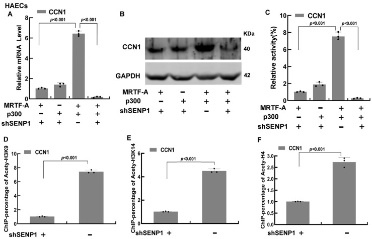
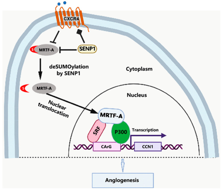
The angiogenic potential of mesenchymal stem cells (MSCs) is critical for adult vascular regeneration and repair, which is regulated by various growth factors and cytokines. In the current study, we report that knockdown SUMO-specific peptidase 1 (SENP1) stimulated the SUMOylation of MRTF-A and prevented its translocation into the nucleus, leading to downregulation of the cytokine and angiogenic factor CCN1, which significantly impacted MSC-mediated angiogenesis and cell migration. Further studies showed that SENP1 knockdown also suppressed the expression of a chemokine receptor CXCR4, and overexpression of CXCR4 could partially abrogate MRTF-A SUMOylation and reestablish the CCN1 level. Mutation analysis confirmed that SUMOylation occurred on three lysine residues (Lys-499, Lys-576, and Lys-624) of MRTF-A. In addition, SENP1 knockdown abolished the synergistic co-activation of CCN1 between MRTF-A and histone acetyltransferase p300 by suppressing acetylation on histone3K9, histone3K14, and histone4. These results revealed an important signaling pathway to regulate MSC differentiation and angiogenesis by MRTF-A SUMOylation involving cytokine/chemokine activities mediated by CCN1 and CXCR4, which may potentially impact a variety of cellular processes such as revascularization, wound healing, and progression of cancer.
Overview
The claims and statistics of this paper haven’t been checked yet.
Run the check — free, no account needed. It reads the open-access text and checks the sources it cites.
Integrity signals
Registry
Cites
Statistics
Figures
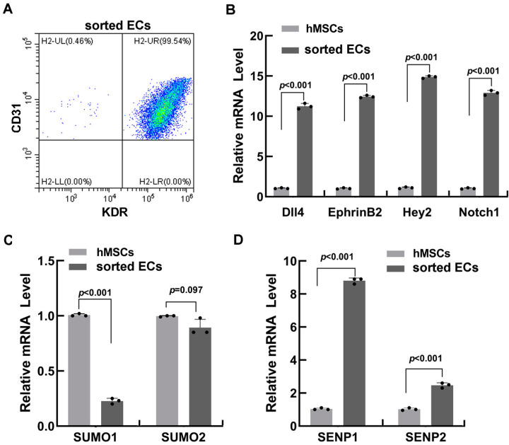
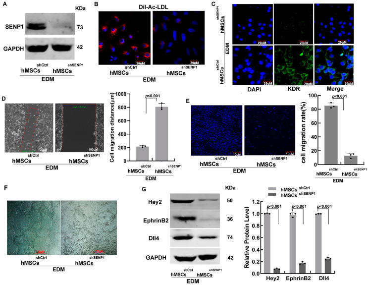
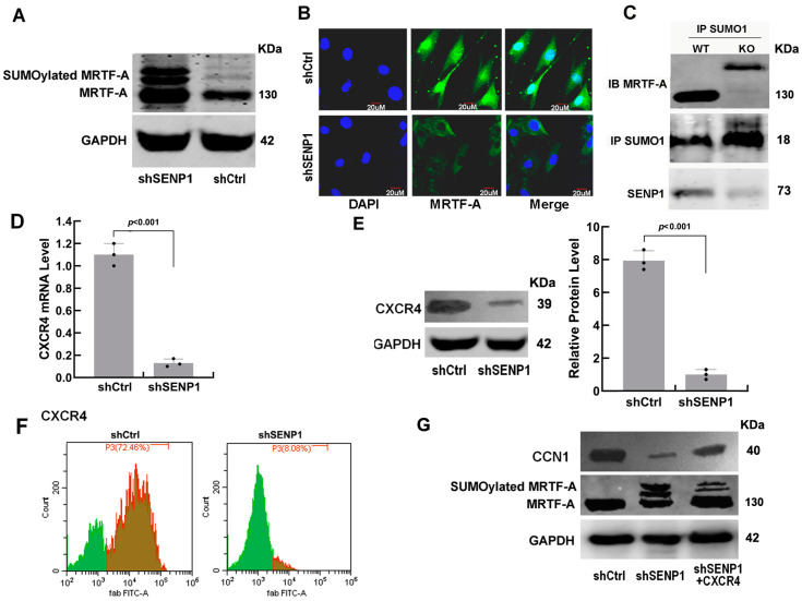
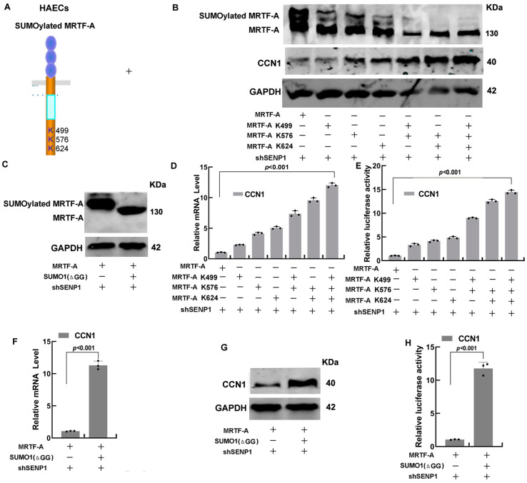
Verification
Claims the paper makes
The claims and statistics of this paper haven’t been checked yet.
Run the check — free, no account needed. It reads the open-access text and checks the sources it cites.
Claims other papers make about this one
Each is checked against this paper’s own text: does it say what the citing paper says it does?
The source backs part of the claim, not all of it — or backs it for a different population, condition or comparison.
In the citing paper
“Interestingly, lower bands (< 150 kDa) for MRTFA protein present in fibrotic livers are related to desumoylation that activates MRTFA (Fig. 1D and Fig. S1C) [50].”
From Mechanical stiffness orchestrates distinct regulation of MRTFs and YAP / TAZ transcriptional cofactors in hepatocytes (2026) · cites this paper as [50]
Experimental — accuracy not yet measured
Self-check
Percentages, sums and reported differences re-computed from the paper’s own numbers.
The self-check needs the paper’s full text.
Citations
Cites
Immunoregulatory mechanisms of mesenchymal stem and stromal cells in inflammatory diseases
Shi, Y., Wang, Y., Li, Q., Liu, K., Hou, J., Shao, C., & Wang, Y. (2018) · Nature Reviews Nephrology · doi:10.1038/s41581-018-0023-5 · Cited by 1,223
Hanna, M., Liu, H., Amir, J., Sun, Y., Morris, S. W., Siddiqui, M. A. Q., Lau, L. F., & Chaqour, B. (2009) · Journal of Biological Chemistry · doi:10.1074/jbc.m109.019059 · Cited by 106
Research works printed but not linked: 47
- Bagno, 2018, Mesenchymal Stem Cell-Based Therapy for Cardiovascular Disease: Progress and Challenges, Mol. Ther., 26, 1610, 10.1016/j.ymthe.2018.05.009 · DOI
- Kwon, 2014, Multiple Paracrine Factors Secreted by Mesenchymal Stem Cells Contribute to Angiogenesis, Vascul. Pharmacol., 63, 19, 10.1016/j.vph.2014.06.004 · DOI
- Watt, 2013, The Angiogenic Properties of Mesenchymal Stem/Stromal Cells and Their Therapeutic Potential, Br. Med. Bull., 108, 25, 10.1093/bmb/ldt031 · DOI
- Dulak, 2013, Therapeutic Angiogenesis for Revascularization in Peripheral Artery Disease, Gene, 525, 220, 10.1016/j.gene.2013.03.097 · DOI
- Zhang, 2016, Knockdown of DNMT1 and DNMT3a Promotes the Angiogenesis of Human Mesenchymal Stem Cells Leading to Arterial Specific Differentiation, Stem Cells, 34, 1273, 10.1002/stem.2288 · DOI
- Kim, 2018, Regulation of RhoA GTPase and Various Transcription Factors in the RhoA Pathway, J. Cell Physiol., 233, 6381, 10.1002/jcp.26487 · DOI · Paper page
- Esnault, 2014, Rho-Actin Signaling to the MRTF Coactivators Dominates the Immediate Transcriptional Response to Serum in Fibroblasts, Genes Dev., 28, 943, 10.1101/gad.239327.114 · DOI
- Miralles, 2003, Actin Dynamics Control SRF Activity by Regulation of Its Coactivator MAL, Cell, 113, 329, 10.1016/S0092-8674(03)00278-2 · DOI · Paper page
- Medjkane, 2009, Myocardin-Related Transcription Factors and SRF Are Required for Cytoskeletal Dynamics and Experimental Metastasis, Nat. Cell Biol., 11, 257, 10.1038/ncb1833 · DOI
- Vartiainen, 2007, Nuclear Actin Regulates Dynamic Subcellular Localization and Activity of the SRF Cofactor MAL, Science, 316, 1749, 10.1126/science.1141084 · DOI
- Baarlink, 2013, Nuclear Actin Network Assembly by Formins Regulates the SRF Coactivator MAL, Science, 340, 864, 10.1126/science.1235038 · DOI
- Muehlich, 2008, Serum-Induced Phosphorylation of the Serum Response Factor Coactivator MKL1 by the Extracellular Signal-Regulated Kinase 1/2 Pathway Inhibits Its Nuclear Localization, Mol. Cell Biol., 28, 6302, 10.1128/MCB.00427-08 · DOI
- Nakagawa, 2005, Transcriptional Activity of Megakaryoblastic Leukemia 1 (MKL1) Is Repressed by SUMO Modification, Genes Cells, 10, 835, 10.1111/j.1365-2443.2005.00880.x · DOI
- Qin, 2021, SUMOylation Wrestles with the Occurrence and Development of Breast Cancer, Front. Oncol., 11, 659661, 10.3389/fonc.2021.659661 · DOI
- Kunz, 2018, SUMO-Specific Proteases and Isopeptidases of the SENP Family at a Glance, J. Cell Sci., 131, jcs211904, 10.1242/jcs.211904 · DOI
- Zhu, 2017, SUMOylation Negatively Regulates Angiogenesis by Targeting Endothelial NOTCH Signaling, Circ. Res., 121, 636, 10.1161/CIRCRESAHA.117.310696 · DOI
- Wang, 2020, SUMOylation of Vascular Endothelial Growth Factor Receptor 2 Inhibits the Proliferation, Migration, and Angiogenesis Signaling Pathway in Non-Small Cell Lung Cancer, Anti-Cancer Drugs, 31, 492, 10.1097/CAD.0000000000000896 · DOI
- MacDonald, I.J., Huang, C.-C., Liu, S.-C., Lin, Y.-Y., and Tang, C.-H. (2021). Targeting CCN Proteins in Rheumatoid Arthritis and Osteoarthritis. Int. J. Mol. Sci., 22. · DOI
- Li, 2015, Emerging Role of CCN Family Proteins in Tumorigenesis and Cancer Metastasis (Review), Int. J. Mol. Med., 36, 1451, 10.3892/ijmm.2015.2390 · DOI
- Chen, 2017, CCN1 Promotes VEGF Production in Osteoblasts and Induces Endothelial Progenitor Cell Angiogenesis by Inhibiting miR-126 Expression in Rheumatoid Arthritis: CCN1 Increases VEGF Expression in Osteoblasts, J. Bone Miner. Res., 32, 34, 10.1002/jbmr.2926 · DOI
- Shi, 2020, The Role of the CXCL12/CXCR4/CXCR7 Chemokine Axis in Cancer, Front. Pharmacol., 11, 574667, 10.3389/fphar.2020.574667 · DOI
- Lu, 2022, The Role of CXC Chemokines in Cardiovascular Diseases, Front. Pharmacol., 12, 765768, 10.3389/fphar.2021.765768 · DOI
- Zagzag, 2006, Hypoxia-Inducible Factor 1 and VEGF Upregulate CXCR4 in Glioblastoma: Implications for Angiogenesis and Glioma Cell Invasion, Lab. Investig., 86, 1221, 10.1038/labinvest.3700482 · DOI
- Niklason, 2018, Arterial Venous Differentiation for Vascular Bioengineering, Annu. Rev. Biomed. Eng., 20, 431, 10.1146/annurev-bioeng-062117-121231 · DOI
- Aranguren, 2007, In Vitro and In Vivo Arterial Differentiation of Human Multipotent Adult Progenitor Cells, Blood, 109, 2634, 10.1182/blood-2006-06-030411 · DOI
- Song, 2016, The MRTF-A/B Function as Oncogenes in Pancreatic Cancer, Oncol. Rep., 35, 127, 10.3892/or.2015.4329 · DOI
- Chaqour, 2020, Caught between a “Rho” and a Hard Place: Are CCN1/CYR61 and CCN2/CTGF the Arbiters of Microvascular Stiffness?, J. Cell Commun. Signal., 14, 21, 10.1007/s12079-019-00529-3 · DOI
- He, 2015, Transcriptional Factors P300 and MRTF-A Synergistically Enhance the Expression of Migration-Related Genes in MCF-7 Breast Cancer Cells, Biochem. Biophys. Res. Commun., 467, 813, 10.1016/j.bbrc.2015.10.060 · DOI
- Meng, 2018, MRTF-A Mediates the Activation of COL1A1 Expression Stimulated by Multiple Signaling Pathways in Human Breast Cancer Cells, Biomed. Pharmacother., 104, 718, 10.1016/j.biopha.2018.05.092 · DOI
- Weinl, 2013, Endothelial SRF/MRTF Ablation Causes Vascular Disease Phenotypes in Murine Retinae, J. Clin. Investig., 123, 2193, 10.1172/JCI64201 · DOI
- Dorn, 2018, Interplay of Cell-Cell Contacts and RhoA/MRTF-A Signaling Regulates Cardiomyocyte Identity, EMBO J., 37, e98133, 10.15252/embj.201798133 · DOI
- Wang, 2020, Heat Shock Protein 27 Enhances SUMOylation of Heat Shock Protein B8 to Accelerate the Progression of Breast Cancer, Am. J. Pathol., 190, 2464, 10.1016/j.ajpath.2020.04.012 · DOI
- Ashikari, 2017, Androgen Induces G3BP2 and SUMO-Mediated P53 Nuclear Export in Prostate Cancer, Oncogene, 36, 6272, 10.1038/onc.2017.225 · DOI
- Pandareesh, 2021, Prostate Carcinogenesis: Insights in Relation to Epigenetics and Inflammation, Endocr. Metab. Immune Disord. Drug Targets, 21, 253, 10.2174/1871530320666200719020709 · DOI
- Zhang, X., Wang, H., Wang, H., Xiao, F., Seth, P., Xu, W., Jia, Q., Wu, C., Yang, Y., and Wang, L. (2017). SUMO-Specific Cysteine Protease 1 Promotes Epithelial Mesenchymal Transition of Prostate Cancer Cells via Regulating SMAD4 DeSUMOylation. Int. J. Mol. Sci., 18. · DOI
- Yeh, 2010, SUMO Losing Balance: SUMO Proteases Disrupt SUMO Homeostasis to Facilitate Cancer Development and Progression, Genes Cancer, 1, 748, 10.1177/1947601910382555 · DOI
- Mo, 2002, CYR61 (CCN1) Is Essential for Placental Development and Vascular Integrity, Mol. Cell Biol., 22, 8709, 10.1128/MCB.22.24.8709-8720.2002 · DOI · Paper page
- Brigstock, 2002, Regulation of Angiogenesis and Endothelial Cell Function by Connective Tissue Growth Factor (CTGF) and Cysteine-Rich 61 (CYR61), Angiogenesis, 5, 153, 10.1023/A:1023823803510 · DOI
- Lee, 2017, Interplay between CCN1 and Wnt5a in Endothelial Cells and Pericytes Determines the Angiogenic Outcome in a Model of Ischemic Retinopathy, Sci. Rep., 7, 1405, 10.1038/s41598-017-01585-8 · DOI
- Lee, 2019, CCN1-Yes-Associated Protein Feedback Loop Regulates Physiological and Pathological Angiogenesis, Mol. Cell Biol., 39, e00107-19, 10.1128/MCB.00107-19 · DOI · Paper page
- Liu, 2023, Mitochondrial SENP2 Regulates the Assembly of SDH Complex under Metabolic Stress, Cell Rep., 42, 112041, 10.1016/j.celrep.2023.112041 · DOI
- Chen, 2021, Inhibition of SENP2-Mediated Akt DeSUMOylation Promotes Cardiac Regeneration via Activating Akt Pathway, Clin. Sci., 135, 811, 10.1042/CS20201408 · DOI
- Yu, 2016, Myocardin-Related Transcription Factor A and Yes-Associated Protein Exert Dual Control in G Protein-Coupled Receptor- and RhoA-Mediated Transcriptional Regulation and Cell Proliferation, Mol. Cell Biol., 36, 39, 10.1128/MCB.00772-15 · DOI
- Hinkel, 2014, MRTF-A Controls Vessel Growth and Maturation by Increasing the Expression of CCN1 and CCN2, Nat. Commun., 5, 3970, 10.1038/ncomms4970 · DOI
- Morein, 2020, Beyond Cell Motility: The Expanding Roles of Chemokines and Their Receptors in Malignancy, Front. Immunol., 11, 952, 10.3389/fimmu.2020.00952 · DOI
- Jiang, 2022, Modifying Strategies for SDF-1/CXCR4 Interaction during Mesenchymal Stem Cell Transplantation, Gen. Thorac. Cardiovasc. Surg., 70, 1, 10.1007/s11748-021-01696-0 · DOI
- Liu, 2011, Critical Role of SDF-1α-Induced Progenitor Cell Recruitment and Macrophage VEGF Production in the Experimental Corneal Neovascularization, Mol. Vis., 17, 2129
Cited by
Sorted by citations
Torres‐Ortiz, B. S., Pérez‐Calixto, D., Carretero‐Ortega, J., Guzman‐López, A. G., Pinto‐Dueñas, D. C., Hernández‐Juárez, M., Jiménez‐Escobar, A., Rodríguez‐Hernández, A., Díaz‐Bello, B., Serna‐Márquez, N., et al. (2026) · The FEBS Journal · doi:10.1111/febs.70570
Metadata
- Authors
- Zhang, R., Liu, Q., Lyu, C., Gao, X., & Ma, W.
- Affiliations
- Nankai University (CN)
- Qilu University of Technology (CN)
- Tianjin University of Science and Technology (CN)
- Corresponding author
- Liu, Qingxi
- Ma, Wenjian
- Venue
- Biomedicines
- Publisher
- MDPI AG
- Published
- 2023-03-15
- Volume
- 11
- Issue
- 3
- Pages
- 914
- ISSN
- 2227-9059 (ISSN-L)
- PMC
- PMC10046070
- OpenAlex
- W4327595726
- Cited by
- 3
- References
- 2
- Citations by year
- 2025: 1 · 2024: 2
- FWCI
- 0.52
- Type
- journal article
- Language
- en
- Open access
- gold
- doi.org
- Licence
- Locations
- Biomedicines (publishedVersion, cc-by)
- PubMed Central (publishedVersion, cc-by)
- PubMed (publishedVersion)
- DOAJ (DOAJ: Directory of Open Access Journals) (submittedVersion, cc-by-sa)
- Funders
- National Key R&D Program of China (2018YFA0901702, TJYXZDXK-056B, 2019GSF107088, ZR2020MC077, ZR202111220001)
- Tianjin Key Medical Discipline (Specialty) Construction Project (2018YFA0901702, TJYXZDXK-056B, 2019GSF107088, ZR2020MC077, ZR202111220001)
- Shandong Key R&D Program (2018YFA0901702, TJYXZDXK-056B, 2019GSF107088, ZR2020MC077, ZR202111220001)
- National Science Foundation of Shandong (2018YFA0901702, TJYXZDXK-056B, 2019GSF107088, ZR2020MC077, ZR202111220001)
- Topics
- Life Sciences › Biochemistry, Genetics and Molecular Biology › Cell Biology › Mesenchymal stem cell research
- Life Sciences › Biochemistry, Genetics and Molecular Biology › Molecular Biology › Connective Tissue Growth Factor Research
- Life Sciences › Biochemistry, Genetics and Molecular Biology › Molecular Biology › Angiogenesis and VEGF in Cancer
- Keywords
- SENP1, MRTF-A, SUMOylation, CYR61, CXCR4, mesenchymal stem cells, angiogenesis, cell migration, P300, histone acetylation, nuclear translocation, lysine residues, vascular regeneration
About these checks
Registry, retraction, Cites and statistics results are computed, not judged, with one exception: a printed reference that no index lists is read once by a model before it counts as a citation error. Claims are extracted by a model and kept only when their quote appears verbatim in the text; each cited source is then checked, with a model’s help, for whether it says what the claim relies on. Those verdicts can be wrong — read the claim’s quote and the cited source before relying on one.
Statistics and arithmetic are recomputed, not judged.
Confidence bands are shown only for fields calibrated on our annotated test set. None is calibrated yet, so no band is shown.
What counts as a claim ›How we measure confidence ›How we check this ›
Are you an author, or do you see a problem with this page? Request a review
