

The DOI matches its Crossref record.
Recent Advancements of LSPR Fiber‐Optic Biosensing: Combination Methods, Structure, and Prospects
Open accessAbstract
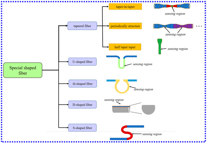
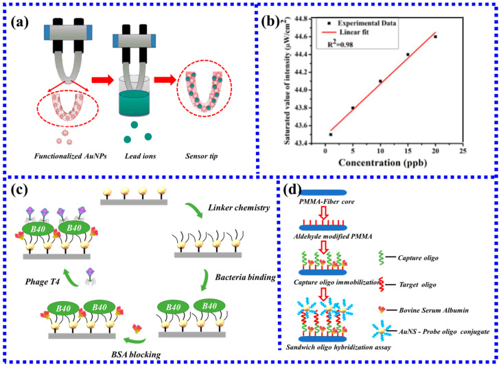
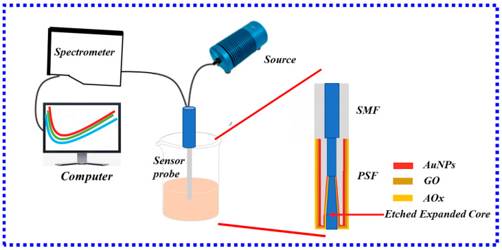
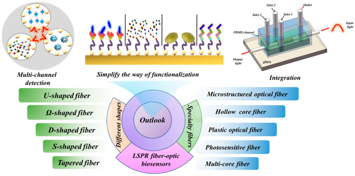
Fiber-optic biosensors based on localized surface plasmon resonance (LSPR) have the advantages of great biocompatibility, label-free, strong stability, and real-time monitoring of various analytes. LSPR fiber-optic biosensors have attracted extensive research attention in the fields of environmental science, clinical medicine, disease diagnosis, and food safety. The latest development of LSPR fiber-optic biosensors in recent years has focused on the detection of clinical disease markers and the detection of various toxic substances in the environment and the progress of new sensitization mechanisms in LSPR fiber-optic sensors. Therefore, this paper reviews the LSPR fiber-optic sensors from the aspects of working principle, structure, and application fields in biosensors. According to the structure, the sensor can be divided into three categories: traditional ordinary optical fiber, special shape optical fiber, and specialty optical fiber. The advantages and disadvantages of existing and future LSPR fiber-optic biosensors are discussed in detail. Additionally, the prospect of future development of fiber-optic biosensors based on LSPR is addressed.
Overview
The claims and statistics of this paper haven’t been checked yet.
Run the check — free, no account needed. It reads the open-access text and checks the sources it cites.
Integrity signals
Registry
Cites
Statistics
Figures
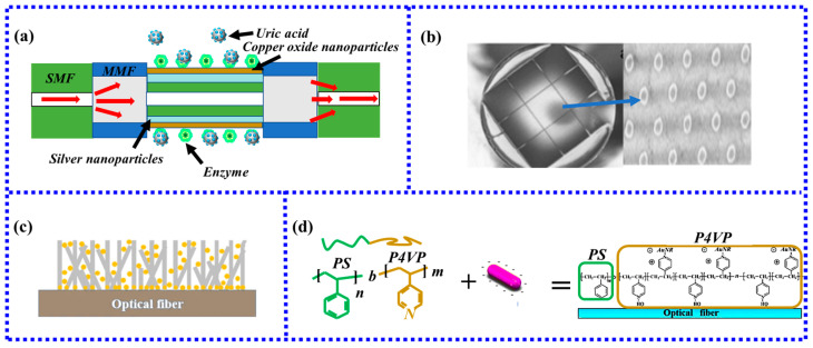
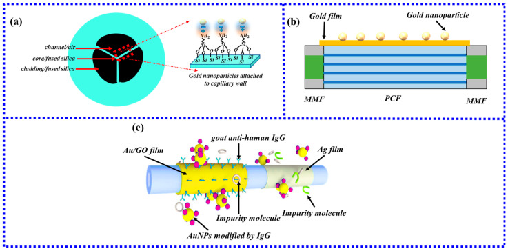
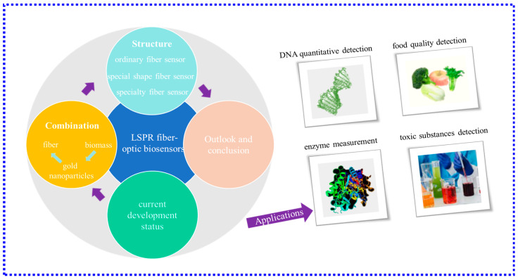
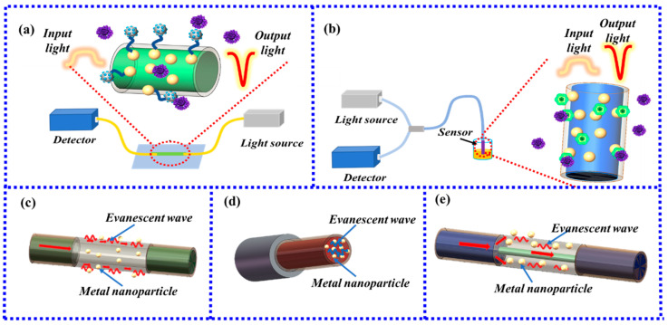
Verification
Claims the paper makes
The claims and statistics of this paper haven’t been checked yet.
Run the check — free, no account needed. It reads the open-access text and checks the sources it cites.
Claims other papers make about this one
Each is checked against this paper’s own text: does it say what the citing paper says it does?
The cited source says what the claim attributes to it.
In the citing paper
“LSPR biosensing detects biomolecular interactions by monitoring shifts in this band.”
From Current PAT Landscape in the Downstream Processing of Biopharmaceuticals (2025) · cites this paper as [95]
Self-check
Percentages, sums and reported differences re-computed from the paper’s own numbers.
The self-check needs the paper’s full text.
Citations
Cites
No references could be resolved yet.
Research works printed but not linked: 115
- Chang, T.-C., Sun, A.Y., Huang, Y.-C., Wang, C.-H., Wang, S.-C., and Chau, L.-K. (2022). Integration of Power-Free and Self-Contained Microfluidic Chip with Fiber Optic Particle Plasmon Resonance Aptasensor for Rapid Detection of SARS-CoV-2 Nucleocapsid Protein. Biosensors, 12. · DOI
- Li, 2022, In-situ DNA detection with an interferometric-type optical sensor based on tapered exposed core microstructured optical fiber, Sens. Actuators B Chem., 351, 130942, 10.1016/j.snb.2021.130942 · DOI
- Sypabekova, 2020, Reflector-less nanoparticles doped optical fiber biosensor for the detection of Case thrombin, Biosens. Bioelectron., 165, 112365, 10.1016/j.bios.2020.112365 · DOI
- Arjmand, 2020, Quantitative Analysis of Methyl-Parathion Pesticide in Presence of Enzyme Substrate Using Tapered Fiber Optic Biosensor, IEEE Sens. J., 20, 5243, 10.1109/JSEN.2020.2968895 · DOI
- Mahani, 2021, LSPR biosensing for the early-stage prostate cancer detection using hydrogen bonds between PSA and antibody: Molecular dynamic and experimental study, J. Mol. Liq., 324, 114736, 10.1016/j.molliq.2020.114736 · DOI
- Fang, 2020, Nanoporous layer fiber biosensing platform for real time culture- and separation-free detecting bacterial pathogens and measuring their susceptibility to antibiotics, Sens. Actuators B-Chem., 325, 128748, 10.1016/j.snb.2020.128748 · DOI
- Han, 2016, Practical, highly sensitive, and regenerable evanescent-wave biosensor for detection of Hg2+ and Pb2+ in water, Biosens. Bioelectron., 80, 265, 10.1016/j.bios.2016.01.070 · DOI
- Sharma, 2019, A highly selective LSPR biosensor for the detection of taurine realized on optical fiber substrate and gold nanoparticles, Opt. Fiber Technol., 52, 101962, 10.1016/j.yofte.2019.101962 · DOI
- Wang, 2018, An interferometric optical fiber biosensor with high sensitivity for IgG/anti-IgG immunosensing, Opt. Commun., 426, 388, 10.1016/j.optcom.2018.05.058 · DOI
- Li, 2022, Measurement. Optical fiber DNA biosensor with temperature monitoring based on double microcavities Fabry-Perot interference and Vernier combined effect, IEEE Trans. Instrum. Meas., 72, 7001208
- Guo, 2020, Hyperboloid-Drum Microdisk Laser Biosensors for Ultrasensitive Detection of Human IgG, Small, 16, 202000239, 10.1002/smll.202000239 · DOI
- Wang, 2019, Highly Sensitive SPR Biosensor Based on Graphene Oxide and Staphylococcal Protein A Co-Modified TFBG for Human IgG Detection, IEEE Trans. Instrum. Meas., 68, 3350, 10.1109/TIM.2018.2875961 · DOI
- Li, 2022, Plug-in optical fiber SPR biosensor for lung cancer gene detection with temperature and pH compensation, Sens. Actuators B Chem., 359, 131596, 10.1016/j.snb.2022.131596 · DOI
- Semwal, V., and Gupta, B.D. (2019, January 23–27). Localized Surface Plasmon Resonance Based Tapered Fiber Optic Ethanol Sensor. Proceedings of the Conference on Lasers and Electro-Optics Europe/European Quantum Electronics Conference (CLEO/Europe-EQEC), Munich, Germany. · DOI
- You, 2020, Laser-induced noble metal nanoparticle-graphene composites enabled flexible biosensor for pathogen detection, Biosens. Bioelectron., 150, 111896, 10.1016/j.bios.2019.111896 · DOI
- Agnarsson, 2009, Evanescent-wave fluorescence microscopy using symmetric planar waveguides, Opt. Express, 17, 5075, 10.1364/OE.17.005075 · DOI
- Zhang, 2017, Probing the binding affinity of plasma proteins adsorbed on Au nanoparticles, Nanoscale, 9, 4787, 10.1039/C7NR01523B · DOI
- Velusamy, 2016, Bio-Inspired Green Nanoparticles: Synthesis, Mechanism, and Antibacterial Application, Toxicol. Res., 32, 95, 10.5487/TR.2016.32.2.095 · DOI
- Luo, 2018, A novel immunosensor based on excessively tilted fiber grating coated with gold nanospheres improves the detection limit of Newcastle disease virus, Biosens. Bioelectron., 100, 169, 10.1016/j.bios.2017.08.064 · DOI
- Paul, 2017, LSPR based Ultra-sensitive low cost U-bent optical fiber for volatile liquid sensing, Sens. Actuators B-Chem., 250, 198, 10.1016/j.snb.2017.04.171 · DOI
- Xu, 2021, A portable optical fiber biosensor for the detection of zearalenone based on the localized surface plasmon resonance, Sens. Actuators B-Chem., 336, 129752, 10.1016/j.snb.2021.129752 · DOI
- Zhou, X., Gong, P., Ai, Y., Hou, Y., and Li, X. (2023). Miniature optical fiber DNA hybridization sensor with temperature compensation using a gold nanofilm-coated optical fiber. IEEE Sens. J. · DOI
- Chen, S., Zhang, C., Wang, J., Li, N., Song, Y., Wu, H., and Liu, Y. (2023). A Fiber Bragg Grating Sensor Based on Cladding Mode Resonance for Label-Free Biosensing. Biosensors, 13. · DOI
- Abakah, B. (2023). Non-Destructive Imaging of Phytosulfokine Trafficking in Plants Using Fiber-Optic Fluorescence Microscopy. [Master’s Thesis, East Tennessee State University].
- Liu, 2023, Microstructured Optical Fiber-Enhanced Light–Matter Interaction Enables Highly Sensitive Exosome-Based Liquid Biopsy of Breast Cancer, Anal. Chem., 95, 1095
- Deng, 2022, Practical tapered optical fiber system for in-situ label-free sensing of various antigens, Opt. Eng., 61, 074102, 10.1117/1.OE.61.7.074102 · DOI
- Zhao, 2022, Aptamer-based chemiluminescent optical fiber immunosensor with enhanced signal amplification for ultrasensitive detection of tumor biomarkers, Biosens. Bioelectron., 214, 114505, 10.1016/j.bios.2022.114505 · DOI
- Nag, 2020, Beta-lactam antibiotics induced bacteriolysis on LSPR sensors for assessment of antimicrobial resistance and quantification of antibiotics, Sens. Actuators B-Chem., 311, 127945, 10.1016/j.snb.2020.127945 · DOI
- Ding, 2016, Mesoporous nanospheres functionalized optical microfiber biosensor for low concentration neurotransmitter detection, Opt. Express, 24, 27152, 10.1364/OE.24.027152 · DOI
- Yuan, 2020, Au nanoparticles as label-free competitive reporters for sensitivity enhanced fiber-optic SPR heparin sensor, Biosens. Bioelectron., 154, 112039, 10.1016/j.bios.2020.112039 · DOI
- Sung, 2013, Colorimetric Detection of Co2+ Ion Using Silver Nanoparticles with Spherical, Plate, and Rod Shapes, Langmuir, 29, 8978, 10.1021/la401408f · DOI
- Zhao, 2021, Maintaining the localized surface plasmon resonance of copper nanoparticles by defective TiO2 thin films, Appl. Phys. A-Mater. Sci. Process., 127, 930, 10.1007/s00339-021-05073-9 · DOI
- Liu, 2021, MXene (Ti3C2Tx)-Ag nanocomplex as efficient and quantitative SERS biosensor platform by in-situ PDDA electrostatic self-assembly synthesis strategy, Sens. Actuators B Chem., 333, 129581, 10.1016/j.snb.2021.129581 · DOI
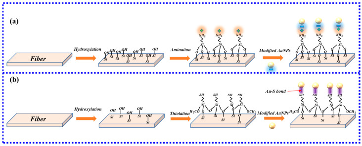
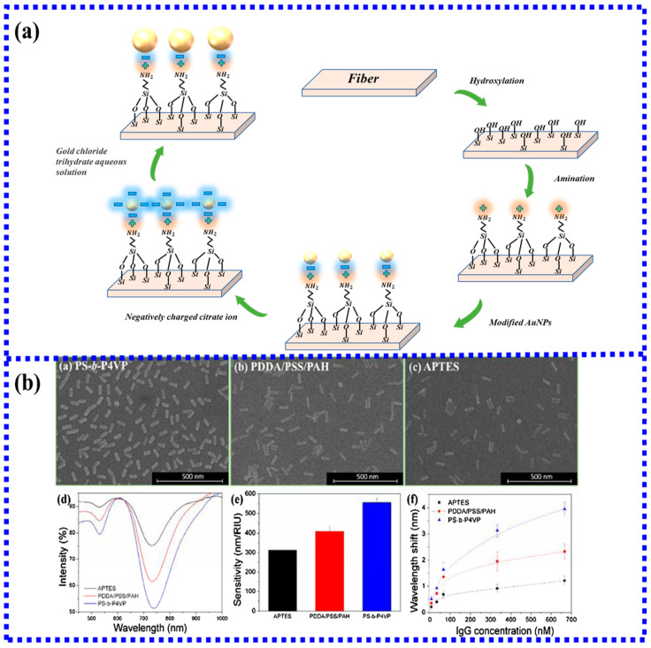
- Shao, 2010, Optical Fiber LSPR Biosensor Prepared by Gold Nanoparticle Assembly on Polyelectrolyte Multilayer, Sensors, 10, 3585, 10.3390/s100403585 · DOI
- Zhang, 2012, Fiber Surface Modification Technology for Fiber-Optic Localized Surface Plasmon Resonance Biosensors, Sensors, 12, 2729, 10.3390/s120302729 · DOI
- Khatri, A., Punjabi, N., Ghosh, D., Maji, S., and Mukherji, S. (2014, January 13–16). Detection of α-Synuclein, Marker for Parkinson’s disease using Localized Surface Plasmon Resonance Fiber Optic Sensor. Proceedings of the International Conference on Fibre Optics and Photonics, Kharagpur, India. M4A. 45. · DOI
- Tseng, Y.-T., Li, W.-Y., Yu, Y.-W., Chiang, C.-Y., Liu, S.-Q., Chau, L.-K., Lai, N.-S., and Chou, C.-C. (2020). Fiber Optic Particle Plasmon Resonance Biosensor for Label-Free Detection of Nucleic Acids and Its Application to HLA-B27 mRNA Detection in Patients with Ankylosing Spondylitis. Sensors, 20. · DOI
- 2021, Gold Nanoparticles Modified a Multimode Clad-Free Fiber for Ultrasensitive Detection of Bovine Serum Albumin, J. Nanomater., 2021, 5530709
- Dhara, 2019, Optical Fiber-Based Heavy Metal Detection Using the Localized Surface Plasmon Resonance Technique, IEEE Sens. J., 19, 8720, 10.1109/JSEN.2019.2921701 · DOI
- Yang, 2020, Development of Glucose Sensor Using Gold Nanoparticles and Glucose-Oxidase Functionalized Tapered Fiber Structure, Plasmonics, 15, 841, 10.1007/s11468-019-01104-7 · DOI
- Kumar, 2019, LSPR-based cholesterol biosensor using a tapered optical fiber structure, Biomed. Opt. Express, 10, 2150, 10.1364/BOE.10.002150 · DOI
- Hu, 2018, Avoiding Thiol Compound Interference: A Nanoplatform Based on High-Fidelity Au-Se Bonds for Biological Applications, Angew. Chem.-Int. Ed., 57, 5306, 10.1002/anie.201712921 · DOI
- Piwonski, 2005, Investigation of 3-mercaptopropyltrimethoxysilane self-assembled monolayers on Au(111) surface, Appl. Surf. Sci., 242, 147, 10.1016/j.apsusc.2004.08.009 · DOI
- Jia, S., Bian, C., Sun, J., and Xia, S. (2018, January 22–26). Gold nanospheres-coated lspr fiber sensor with high ri sensitivity by a rapid fabricating method. Proceedings of the 2018 IEEE 13th Annual International Conference on Nano/Micro Engineered and Molecular Systems (NEMS), Singapore. · DOI
- Goicoechea, J., Rivero, P.J., Sada, S., and Arregui, F.J. (2019). Self-Referenced Optical Fiber Sensor for Hydrogen Peroxide Detection Based on LSPR of Metallic Nanoparticles in Layer-by-Layer Films. Sensors, 19. · DOI
- Lu, M., Peng, W., Lin, M., Wang, F., and Zhang, Y. (2021). Gold Nanoparticle-Enhanced Detection of DNA Hybridization by a Block Copolymer-Templating Fiber-Optic Localized Surface Plasmon Resonance Biosensor. Nanomaterials, 11. · DOI
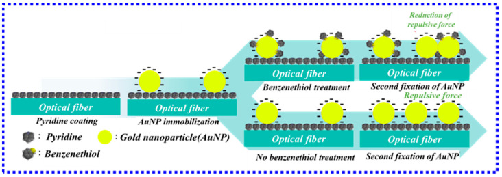
- Kim, 2019, Improved stability of gold nanoparticles on the optical fiber and their application to refractive index sensor based on localized surface plasmon resonance, Opt. Laser Technol., 114, 171, 10.1016/j.optlastec.2019.02.002 · DOI
- Lu, 2021, Comparative study of block copolymer-templated localized surface plasmon resonance optical fiber biosensors: CTAB or citrate-stabilized gold nanorods, Sens. Actuators B Chem., 329, 129094, 10.1016/j.snb.2020.129094 · DOI
- Kim, 2018, Real-time detection of prostate-specific antigens using a highly reliable fiber-optic localized surface plasmon resonance sensor combined with micro fluidic channel, Sens. Actuators B-Chem., 273, 891, 10.1016/j.snb.2018.07.007 · DOI
- Kim, 2019, Localized surface plasmon resonance biosensor using nanopatterned gold particles on the surface of an optical fiber, Sens. Actuators B-Chem., 280, 183, 10.1016/j.snb.2018.10.059 · DOI
- Dhawan, 2009, Fabrication of Nanodot Plasmonic Waveguide Structures Using FIB Milling and Electron Beam-Induced Deposition, Scanning, 31, 139, 10.1002/sca.20152 · DOI
- Hong, 2020, Solvent-free nanofabrication based on ice-assisted electron-beam lithography, Nano Lett., 20, 8841, 10.1021/acs.nanolett.0c03809 · DOI
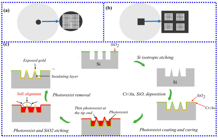
- Kim, 2018, Fabrication and measurement of microtip-array-based LSPR sensor using bundle fiber, Sens. Actuators A-Phys., 271, 146, 10.1016/j.sna.2018.01.021 · DOI
- Bresin, M., Nehru, N., and Hastings, J.T. (2013, January 5–6). Focused electron-beam induced deposition of plasmonic nanostructures from aqueous solutions. Proceedings of the Advanced Fabrication Technologies for Micro/Nano Optics and Photonics VI, San Francisco, CA, USA. · DOI
- Dhawan, 2007, Theoretical simulation and focused ion beam fabrication of gold nanostructures for surface-enhanced Raman scattering (SERS), Nanobiotechnology, 3, 164, 10.1007/s12030-008-9017-x · DOI
- Hoeflich, 2017, Direct Electron Beam Writing of Silver-Based Nanostructures, ACS Appl. Mater. Interfaces, 9, 24071, 10.1021/acsami.7b04353 · DOI
- Wang, 2004, Electron-beam induced initial growth of platinum films using Pt(PF3)(4), J. Vac. Sci. Technol. B, 22, 1803, 10.1116/1.1761266 · DOI
- Chien, 2021, Analysis of carbon content in direct-write plasmonic Au structures by nanomechanical scanning absorption microscopy, J. Appl. Phys., 129, 063105, 10.1063/5.0035234 · DOI
- Flatae, 2017, Plasmonic Gold Nanocones in the Near-Infrared for Quantum Nano-Optics, Adv. Opt. Mater., 5, 1700586, 10.1002/adom.201700586 · DOI
- Vaiano, 2023, Characterization of Lab-on-Fiber-based dosimeters in ultra-high dose radiation fields, Opt. Laser Technol., 161, 109177, 10.1016/j.optlastec.2023.109177 · DOI
- Keiser, 2014, Review of diverse optical fibers used in biomedical research and clinical practice, J. Biomed. Opt., 19, 080902, 10.1117/1.JBO.19.8.080902 · DOI
- Khanikar, 2021, A review on infiltrated or liquid core fiber optic SPR sensors, Photonics Nanostruct. Fundam. Appl., 46, 100945, 10.1016/j.photonics.2021.100945 · DOI
- Agrawal, 2020, Development of Uric Acid Sensor Using Copper Oxide and Silver Nanoparticles Immobilized SMSMS Fiber Structure-Based Probe, IEEE Trans. Instrum. Meas., 69, 9097, 10.1109/TIM.2020.2998876 · DOI
- Sanders, 2014, An enhanced LSPR fiber-optic nanoprobe for ultrasensitive detection of protein biomarkers, Biosens. Bioelectron., 61, 95, 10.1016/j.bios.2014.05.009 · DOI
- Malara, 2018, Resonant enhancement of plasmonic nanostructured fiber optic sensors, Sens. Actuators B Chem., 273, 1587, 10.1016/j.snb.2018.07.030 · DOI
- Kim, 2019, Fiber optic sensor based on ZnO nanowires decorated by Au nanoparticles for improved plasmonic biosensor, Sci. Rep., 9, 15605, 10.1038/s41598-019-52056-1 · DOI
- Kim, 2021, Fabrication and measurement of fiber optic localized surface plasmon resonance sensor based on gold nanoparticle dimer, Spectrochim. Acta Part A-Mol. Biomol. Spectrosc., 261, 120034, 10.1016/j.saa.2021.120034 · DOI
- Flauraud, 2017, In-Plane Plasmonic Antenna Arrays with Surface Nanogaps for Giant Fluorescence Enhancement, Nano Lett., 17, 1703, 10.1021/acs.nanolett.6b04978 · DOI
- Yang, 2020, Highly sensitive and selective sensor probe using glucose oxidase/gold nanoparticles/graphene oxide functionalized tapered optical fiber structure for detection of glucose, Optik, 208, 164536, 10.1016/j.ijleo.2020.164536 · DOI
- Dalila, 2019, Current and future envision on developing biosensors aided by 2D molybdenum disulfide (MoS2) productions, Biosens. Bioelectron., 132, 248, 10.1016/j.bios.2019.03.005 · DOI
- Esfahani Monfared, Y. (2020). Overview of Recent Advances in the Design of Plasmonic Fiber-Optic Biosensors. Biosensors, 10. · DOI
- Semwal, 2019, Experimental studies on the sensitivity of the propagating and localized surface plasmon resonance-based tapered fiber optic refractive index sensors, Appl. Opt., 58, 4149, 10.1364/AO.58.004149 · DOI
- Wang, 2012, Evanescent-Light Deposition of Graphene Onto Tapered Fibers for Passive Q-Switch and Mode-Locker, IEEE Photonics J., 4, 1295, 10.1109/JPHOT.2012.2208736 · DOI
- Wang, 2021, Taper-in-taper fiber structure-based LSPR sensor for alanine aminotransferase detection, Opt. Express, 29, 43793, 10.1364/OE.447202 · DOI
- Zhu, 2020, A novel periodically tapered structure-based gold nanoparticles and graphene oxide—Immobilized optical fiber sensor to detect ascorbic acid, Opt. Laser Technol., 127, 106156, 10.1016/j.optlastec.2020.106156 · DOI
- Wen, H.-Y., Hsu, H.-C., Tsai, Y.-T., Feng, W.-K., Lin, C.-L., and Chiang, C.-C.J.P. (2021). U-shaped optical fiber probes coated with electrically doped GQDs for humidity measurements. Polymers, 13. · DOI
- Sadani, 2019, LSPR based optical fiber sensor with chitosan capped gold nanoparticles on BSA for trace detection of Hg (II) in water, soil and food samples, Biosens. Bioelectron., 134, 90, 10.1016/j.bios.2019.03.046 · DOI
- Halkare, 2019, Bacteria functionalized gold nanoparticle matrix based fiber-optic sensor for monitoring heavy metal pollution in water, Sens. Actuators B-Chem., 281, 643, 10.1016/j.snb.2018.10.119 · DOI
- Arcas, A.d.S., Dutra, F.d.S., Allil, R.C.S.B., and Werneck, M.M. (2018). Surface Plasmon Resonance and Bending Loss-Based U-Shaped Plastic Optical Fiber Biosensors. Sensors, 18. · DOI
- Luo, 2021, Development of a rapid and ultra-sensitive cytosensor: Ω-shaped fiber optic LSPR integrated with suitable AuNPs coverage, Sens. Actuators B Chem., 336, 129706, 10.1016/j.snb.2021.129706 · DOI
- Xu, 2018, Omega-Shaped Fiber-Optic Probe-Based Localized Surface Plasmon Resonance Biosensor for Real-Time Detection of Salmonella Typhimurium, Anal. Chem., 90, 13640, 10.1021/acs.analchem.8b03905 · DOI
- Li, 2022, Sandwich method-based sensitivity enhancement of Ω-shaped fiber optic LSPR for time-flexible bacterial detection, Biosens. Bioelectron., 201, 113911, 10.1016/j.bios.2021.113911 · DOI
- He, 2021, Hybridized nanolayer modified Omega-shaped fiber-optic synergistically enhances localized surface plasma resonance for ultrasensitive cytosensor and efficient photothermal therapy, Biosens. Bioelectron., 194, 113599, 10.1016/j.bios.2021.113599 · DOI
- Chauhan, S.K., Tharion, J., Punjabi, N., Sharma, D.K., and Mukherji, S. (2014, January 27–31). A comparison of S-shaped and U-shaped optical fiber sensors. Proceedings of the Optical Sensors, Barcelona, Spain. · DOI
- Chauhan, 2016, Evanescent wave absorption based S-shaped fiber-optic biosensor for immunosensing applications, Procedia Eng., 168, 117, 10.1016/j.proeng.2016.11.161 · DOI
- Dimpi, 2022, Clad modified varying geometries of fiber optic LSPR sensors towards detection of hazardous volatile liquids and their comparative analysis, Environ. Technol. Innov., 25, 102112, 10.1016/j.eti.2021.102112 · DOI
- Gong, 2019, Experimental and theoretical investigation for surface plasmon resonance biosensor based on graphene/Au film/D-POF, Opt. Express, 27, 3483, 10.1364/OE.27.003483 · DOI
- Virk, 2022, D-shape optical fiber probe dimension optimization for LSPR based bio-sensor, Opt. Fiber Technol., 71, 102930, 10.1016/j.yofte.2022.102930 · DOI
- Liu, 2016, Evanescent wave analysis and experimental realization of refractive index sensor based on D-shaped plastic optical fiber, Optik, 127, 690, 10.1016/j.ijleo.2015.10.129 · DOI
- Cennamo, 2015, Sensitive detection of 2,4,6-trinitrotoluene by tridimensional monitoring of molecularly imprinted polymer with optical fiber and five-branched gold nanostars, Sens. Actuators B-Chem., 208, 291, 10.1016/j.snb.2014.10.079 · DOI
- Pathak, A.K., Rahman, B.M.A., Singh, V.K., and Kumari, S. (2019). Sensitivity Enhancement of a Concave Shaped Optical Fiber Refractive Index Sensor Covered with Multiple Au Nanowires. Sensors, 19. · DOI
- Feng, 2022, LSPR optical fiber sensor based on 3D gold nanoparticles with monolayer graphene as a spacer, Opt. Express, 30, 10187, 10.1364/OE.453806 · DOI
- Luo, 2019, Ultrasensitive U-shaped fiber optic LSPR cytosensing for label-free and in situ evaluation of cell surface N-glycan expression, Sens. Actuators B-Chem., 284, 582, 10.1016/j.snb.2019.01.015 · DOI
- Cennamo, 2013, Localized surface plasmon resonance with five-branched gold nanostars in a plastic optical fiber for bio-chemical sensor implementation, Sensors, 13, 14676, 10.3390/s131114676 · DOI
- Liu, Y., Zhang, N., Li, P., Yu, L., Chen, S., Zhang, Y., Jing, Z., and Peng, W.J.N. (2019). Low-cost localized surface plasmon resonance biosensing platform with a response enhancement for protein detection. Nanomaterials, 9. · DOI
- Chen, 2020, Microcapillary-based integrated LSPR device for refractive index detection and biosensing, J. Light. Technol., 38, 2485, 10.1109/JLT.2020.2969016 · DOI
- Li, 2021, 2D material assisted SMF-MCF-MMF-SMF based LSPR sensor for creatinine detection, Opt. Express, 29, 38150, 10.1364/OE.445555 · DOI
- Kumar, 2021, MoS2 functionalized multicore fiber probes for selective detection of shigella bacteria based on localized plasmon, J. Light. Technol., 39, 4069, 10.1109/JLT.2020.3036610 · DOI
- Singh, 2020, Etched multicore fiber sensor using copper oxide and gold nanoparticles decorated graphene oxide structure for cancer cells detection, Biosens. Bioelectron., 168, 112557, 10.1016/j.bios.2020.112557 · DOI
- Zhu, 2021, Localized Plasmon-Based Multicore Fiber Biosensor for Acetylcholine Detection, IEEE Trans. Instrum. Meas., 71, 1
- Li, 2021, A review of specialty fiber biosensors based on interferometer configuration, J. Biophotonics, 14, e202100068, 10.1002/jbio.202100068 · DOI
- Mollah, 2021, Novel Single Hole Exposed-Suspended Core Localized Surface Plasmon Resonance Sensor, IEEE Sens. J., 21, 2813
- Csaki, 2010, Nanoparticle Layer Deposition for Plasmonic Tuning of Microstructured Optical Fibers, Small, 6, 2584, 10.1002/smll.201001071 · DOI
- Wang, 2018, Sensitivity-Enhanced Optical Fiber Biosensor Based on Coupling Effect Between SPR and LSPR, IEEE Sens. J., 18, 8303, 10.1109/JSEN.2018.2866169 · DOI
- Wang, 2020, A dual channel self-compensation optical fiber biosensor based on coupling of surface plasmon polariton, Opt. Laser Technol., 124, 106002, 10.1016/j.optlastec.2019.106002 · DOI
- Kuriki, 2002, Plastic optical fiber lasers and amplifiers containing lanthanide complexes, Chem. Rev., 102, 2347, 10.1021/cr010309g · DOI
- Boruah, 2018, Localized surface plasmon resonance based U-shaped optical fiber probe for the detection of Pb2+ in aqueous medium, Sens. Actuators B-Chem., 276, 89, 10.1016/j.snb.2018.08.086 · DOI
- Halkare, P., Punjabi, N., Wangchuk, J., Kondabagil, K., and Mukherji, S. (2015, January 16–17). LSPR based fiber optic sensor for detection of E. coli using bacteriophage T4. Proceedings of the 2015 Workshop on Recent Advances in Photonics (WRAP), Bangalore, India. · DOI
- Gowri, A., and Sai, V. (2017, January 24–27). U-bent plastic optical fiber based plasmonic biosensor for nucleic acid detection. Proceedings of the Optical Sensors 2017, Prague, Czech Republic.
- Kumar, 2021, Highly Sensitive, Selective and Portable Sensor Probe Using Germanium-Doped Photosensitive Optical Fiber for Ascorbic Acid Detection, IEEE Sens. J., 21, 62
- Wang, 2022, 2-D Nanomaterials Assisted LSPR MPM Optical Fiber Sensor Probe for Cardiac Troponin I Detection, IEEE Trans. Instrum. Meas., 71, 1, 10.1109/TIM.2022.3216413 · DOI
- Kumar, 2019, LSPR-Based Cholesterol Biosensor Using Hollow Core Fiber Structure, IEEE Sens. J., 19, 7399, 10.1109/JSEN.2019.2916818 · DOI
- Vaiano, 2016, Lab on Fiber Technology for biological sensing applications, Laser Photonics Rev., 10, 922, 10.1002/lpor.201600111 · DOI
- Ricciardi, 2015, Lab-on-fiber technology: A new vision for chemical and biological sensing, Analyst, 140, 8068, 10.1039/C5AN01241D · DOI
- Liu, Q., Liu, Y., Yuan, H., Wang, J., Guang, J., and Peng, W. (2018, January 26–29). Smartphone based LSPR biosensor. Proceedings of the Asia Communications and Photonics Conference, Hangzhou, China. Su2A. 148. · DOI
Cited by
Sorted by citations
Current PAT Landscape in the Downstream Processing of Biopharmaceuticals
Sathiyapriyan, P., Mukherjee, S., Vogel, T., Essen, L., Boerema, D., Vey, M., & Kalina, U. (2025) · Analytical Science Advances · doi:10.1002/ansa.70013 · Cited by 45
Duarah, R., Torné‐Morató, H., Zhang, G., Amin, Y., Pabast, M., Sharma, N., He, S., Das, M. R., & Pompa, P. P. (2026) · Advanced Healthcare Materials · doi:10.1002/adhm.202504419 · Cited by 24
Metadata
- Authors
- Zhang, H., Zhou, X., Li, X., Gong, P., Zhang, Y., & Zhao, Y.
- Affiliations
- Northeastern University (CN)
- State Key Laboratory of Synthetical Automation for Process Industries (CN)
- Corresponding author
- Li, Xuegang
- Zhao, Yong
- Venue
- Biosensors
- Publisher
- MDPI AG
- Published
- 2023-03-20
- Volume
- 13
- Issue
- 3
- Pages
- 405
- ISSN
- 2079-6374 (ISSN-L)
- PMC
- PMC10046874
- OpenAlex
- W4328124534
- Cited by
- 99
- References
- 0
- Citations by year
- 2026: 29 · 2025: 34 · 2024: 27 · 2023: 9
- FWCI
- 1.51
- Type
- journal article
- Language
- en
- Open access
- gold
- doi.org
- Licence
- Locations
- Biosensors (publishedVersion, cc-by)
- PubMed Central (publishedVersion, cc-by)
- PubMed (publishedVersion)
- DOAJ (DOAJ: Directory of Open Access Journals) (submittedVersion, cc-by-sa)
- Funders
- National Natural Science Foundation of China (61903073, 6193300, 2013ZCX09, F2020501040)
- State Key Laboratory of Synthetical Automation for Process Industries (61903073, 6193300, 2013ZCX09, F2020501040)
- Hebei Natural Science Foundation (61903073, 6193300, 2013ZCX09, F2020501040)
- Topics
- Physical Sciences › Engineering › Electrical and Electronic Engineering › Advanced Fiber Optic Sensors
- Physical Sciences › Physics and Astronomy › Atomic and Molecular Physics, and Optics › Plasmonic and Surface Plasmon Research
- Physical Sciences › Materials Science › Electronic, Optical and Magnetic Materials › Gold and Silver Nanoparticles Synthesis and Applications
- Keywords
- localized surface plasmon resonance, optical fiber biosensor, label-free detection, food safety, specialty optical fibers
- MeSH terms
- Fiber Optic Technology, Biosensing Techniques, Biomarkers, Surface Plasmon Resonance, Optical Fibers
About these checks
Registry, retraction, Cites and statistics results are computed, not judged, with one exception: a printed reference that no index lists is read once by a model before it counts as a citation error. Claims are extracted by a model and kept only when their quote appears verbatim in the text; each cited source is then checked, with a model’s help, for whether it says what the claim relies on. Those verdicts can be wrong — read the claim’s quote and the cited source before relying on one.
Statistics and arithmetic are recomputed, not judged.
Confidence bands are shown only for fields calibrated on our annotated test set. None is calibrated yet, so no band is shown.
What counts as a claim ›How we measure confidence ›How we check this ›
Are you an author, or do you see a problem with this page? Request a review
