

The DOI matches its Crossref record; 411 of the 414 references it cites were found in a registry or an index.
Microglia: Agents of the CNS Pro-Inflammatory Response
Open accessAbstract
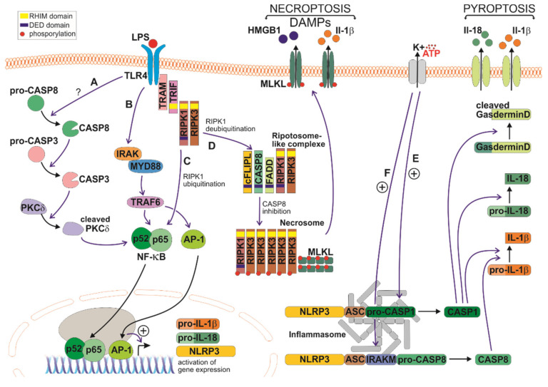
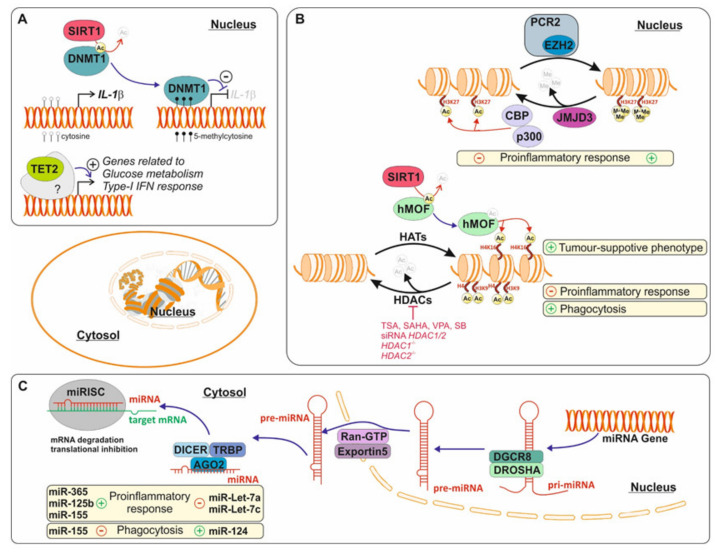
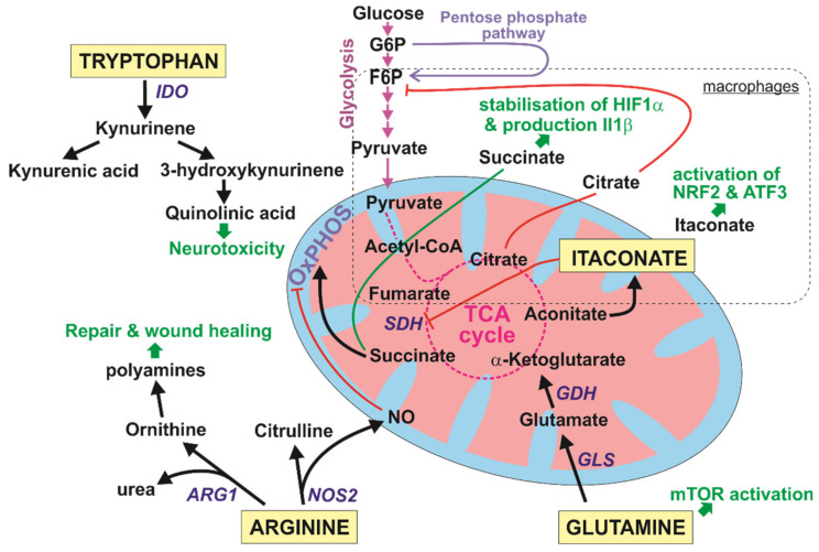
The pro-inflammatory immune response driven by microglia is a key contributor to the pathogenesis of several neurodegenerative diseases. Though the research of microglia spans over a century, the last two decades have increased our understanding exponentially. Here, we discuss the phenotypic transformation from homeostatic microglia towards reactive microglia, initiated by specific ligand binding to pattern recognition receptors including toll-like receptor-4 (TLR4) or triggering receptors expressed on myeloid cells-2 (TREM2), as well as pro-inflammatory signaling pathways triggered such as the caspase-mediated immune response. Additionally, new research disciplines such as epigenetics and immunometabolism have provided us with a more holistic view of how changes in DNA methylation, microRNAs, and the metabolome may influence the pro-inflammatory response. This review aimed to discuss our current knowledge of pro-inflammatory microglia from different angles, including recent research highlights such as the role of exosomes in spreading neuroinflammation and emerging techniques in microglia research including positron emission tomography (PET) scanning and the use of human microglia generated from induced pluripotent stem cells (iPSCs). Finally, we also discuss current thoughts on the impact of pro-inflammatory microglia in neurodegenerative diseases.
Overview
The claims and statistics of this paper haven’t been checked yet.
Run the check — free, no account needed. It reads the open-access text and checks the sources it cites.
Integrity signals
Registry
Cites
Statistics
Figures
Verification
Claims the paper makes
The claims and statistics of this paper haven’t been checked yet.
Run the check — free, no account needed. It reads the open-access text and checks the sources it cites.
Claims other papers make about this one
Each is checked against this paper’s own text: does it say what the citing paper says it does?
The source backs part of the claim, not all of it — or backs it for a different population, condition or comparison.
In the citing paper
“Activated microglia migrate to the site of damage via a process called chemotaxis and release chemokines (e.g., CCL2 and CXCL1), pro- and anti-inflammatory cytokines (e.g., IL-12, IL-10, TNF, TGF-β), and a variety of other inflammatory mediators [341].”
From Revisiting the Pathogenesis of X-Linked Adrenoleukodystrophy (2025) · cites this paper as [341]
Experimental — accuracy not yet measured
Self-check
Percentages, sums and reported differences re-computed from the paper’s own numbers.
The self-check needs the paper’s full text.
Citations
Cites
Takahashi, K., & Yamanaka, S. (2006) · Cell · doi:10.1016/j.cell.2006.07.024 · Cited by 26,673
Induction of Pluripotent Stem Cells from Adult Human Fibroblasts by Defined Factors
Takahashi, K., Tanabe, K., Ohnuki, M., Narita, M., Ichisaka, T., Tomoda, K., & Yamanaka, S. (2007) · Cell · doi:10.1016/j.cell.2007.11.019 · Cited by 20,221
Pathogen Recognition and Innate Immunity
Akira, S., Uematsu, S., & Takeuchi, O. (2006) · Cell · doi:10.1016/j.cell.2006.02.015 · Cited by 11,976
Amyloid oligomers exacerbate tau pathology in a mouse model of tauopathy
Selenica, M.-L. B., Brownlow, M., Jimenez, J. P., Lee, D. C., Pena, G., Dickey, C. A., Gordon, M. N., & Morgan, D. (2013) · Neurodegenerative Diseases · doi:10.1159/000337230 · Cited by 10,359
Corder, E. H., Saunders, A. M., Strittmatter, W. J., Schmechel, D. E., Gaskell, P. C., Small, G. W., Roses, A. D., Haines, J. L., & Pericak-Vance, M. A. (1993) · Science · doi:10.1126/science.8346443 · Cited by 9,429
Polazzi, E., Mengoni, I., Caprini, M., Peña‐Altamira, E., Kurtys, E., & Monti, B. (2012) · Neurosignals · doi:10.1159/000337115 · Cited by 9,023
Pattern Recognition Receptors and Inflammation
Takeuchi, O., & Akira, S. (2010) · Cell · doi:10.1016/j.cell.2010.01.022 · Cited by 8,895
NF-κB signaling in inflammation
Liu, T., Zhang, L., Joo, D., & Sun, S.-C. (2017) · Signal Transduction and Targeted Therapy · doi:10.1038/sigtrans.2017.23 · Cited by 8,791
Establishment in culture of pluripotential cells from mouse embryos
Evans, M. J., & Kaufman, M. H. (1981) · Nature · doi:10.1038/292154a0 · Cited by 8,623
Mosmann, T. R., Cherwinski, H. M., Bond, M. W., Giedlin, M. A., & Coffman, R. L. (1986) · The Journal of Immunology · doi:10.4049/jimmunol.136.7.2348 · Cited by 7,553
Research works printed but not linked: 7
- 1913, Contribución al Conocimiento de la Neuroglía del Cerebro Humano, Trab. Lab. Invest. Biol. Univ. Madrid, XI, 215
- 1920, Estudios sobre la neuroglía. La microglía y su transformación en células en bastoncito y cuerpos gránulo-adiposos, Trab. Lab. Invest. Biol. Univ. Madrid, 18, 37
- 1919, El “tercer elemento” de los centros nerviosos. I. La microglía en estado normal, Bol. Soc. Esp. Biol., VIII, 67
- 1919, El Tercer Elemento de los Centros Nerviosos. II. Intervención de la Microglía en los Procesos Patológicos (Células en Bastoncito y Cuerpos Gránuloadiposos), Bol. Soc. Esp. Biol., VIII, 91
- 1919, El “tercer elemento” de los centros nerviosos. III. Naturaleza probable de la microglía, Bol. Soc. Esp. Biol., VIII, 108
- 1919, El “tercer elemento de los centros nerviosos”. IV. Poder fagocitario y movilidad de la microglía, Bol. Soc. Esp. Biol., VIII, 154
- The National Academies of Sciences, Engineering and Medicine (2018). Biomarkers of Neuroinflammation—Proceedings of a Workshop, The National Academies Press.
Cited by
Sorted by citations
Dash, U. C., Bhol, N. K., Swain, S. K., Samal, R. R., Nayak, P. K., Raina, V., Panda, S. K., Kerry, R. G., Duttaroy, A. K., & Jena, A. B. (2024) · Acta Pharmaceutica Sinica B · doi:10.1016/j.apsb.2024.10.004 · Cited by 806
Early glycolytic reprogramming controls microglial inflammatory activation
Cheng, J., Zhang, R., Xu, Z., Ke, Y., Sun, R., Yang, H., Zhang, X., Zhen, X., & Zheng, L.-T. (2021) · Journal of Neuroinflammation · doi:10.1186/s12974-021-02187-y · Cited by 295
Li, L., Acioglu, C., Heary, R. F., & Elkabes, S. (2021) · Brain, Behavior, and Immunity · doi:10.1016/j.bbi.2020.10.007 · Cited by 283
Necroptosis contributes to chronic inflammation and fibrosis in aging liver
Mohammed, S., Thadathil, N., Selvarani, R., Nicklas, E. H., Wang, D., Miller, B. F., Richardson, A., & Deepa, S. S. (2021) · Aging Cell · doi:10.1111/acel.13512 · Cited by 192
Piezo1 Channels as Force Sensors in Mechanical Force-Related Chronic Inflammation
Liu, H., Hu, J., Zheng, Q., Feng, X., Zhan, F., Wang, X., Guohai, X., & Hua, F. (2022) · Frontiers in Immunology · doi:10.3389/fimmu.2022.816149 · Cited by 177
Metadata
- Authors
- Rodríguez-Gómez, J. A., Kavanagh, E., Engskog-Vlachos, P., Engskog, M. K. R., Herrera, A. J., Espinosa-Oliva, A. M., Joseph, B., Hajji, N., Venero, J. L., & Burguillos, M. A.
- Affiliations
- Institute of Biomedicine of Seville (IBIS)-Hospital Universitario Virgen del Rocío/CSIC/University of Seville, 41012 Seville, Spain
- Universidad de Sevilla (ES)
- Institute of Environmental Medicine, Toxicology Unit, Karolinska Institute, 17177 Stockholm, Sweden
- Uppsala University (SE)
- Instituto de Biomedicina de Sevilla (ES)
- Karolinska Institutet (SE)
- Imperial College London (GB)
- Venue
- Cells
- Publisher
- MDPI AG
- Published
- 2020-07-17
- Volume
- 9
- Issue
- 7
- Pages
- 1717
- ISSN
- 2073-4409 (ISSN-L)
- PMC
- PMC7407646
- OpenAlex
- W3043443440
- Cited by
- 365
- References
- 414
- Citations by year
- 2026: 40 · 2025: 80 · 2024: 69 · 2023: 54 · 2022: 71 · 2021: 41 · 2020: 9
- FWCI
- 4.38
- Type
- journal article
- Language
- en
- Open access
- gold
- doi.org
- Licence
- Locations
- Cells (publishedVersion, cc-by)
- PubMed Central (publishedVersion, cc-by)
- PubMed (publishedVersion)
- LA Referencia (Red Federada de Repositorios Institucionales de Publicaciones Científicas) (publishedVersion, cc-by-nc-nd)
- RISalud Institutional Health Repository of Andalucía (submittedVersion, cc-by)
- Uppsala University Publications (Uppsala University) (submittedVersion, other-oa)
- DIGITAL.CSIC (Spanish National Research Council (CSIC)) (submittedVersion, cc-by)
- DOAJ (DOAJ: Directory of Open Access Journals) (submittedVersion, cc-by-sa)
- Deposito de Investigacion Universidad de Sevilla (University of Seville) (submittedVersion, cc-by)
- Spiral (Imperial College London) (submittedVersion, cc-by)
- Funders
- Ministerio de Ciencia, Innovación y Universidades /FEDER/UE (RTI2018-098645-B-100)
- FEDER I+D+i-USE (US-1265062)
- Ministerio de Economia y Competitividad (Programa Ramón y Cajal: RYC-2017-21804)
- Topics
- Life Sciences › Neuroscience › Neurology › Neuroinflammation and Neurodegeneration Mechanisms
- Life Sciences › Biochemistry, Genetics and Molecular Biology › Molecular Biology › Inflammasome and immune disorders
- Life Sciences › Neuroscience › Neurology › Neurological Disease Mechanisms and Treatments
- Keywords
- microglia, neuroinflammation, reactive microglia, homeostatic microglia, pattern recognition receptors, TLR4, TREM2, epigenetics, DNA methylation, microRNA, immunometabolism, exosomes, neurodegenerative diseases, iPSC-derived microglia
- MeSH terms
- Animals, Central Nervous System, Humans, Inflammation, Models, Biological, Microglia, Caspases, Epigenesis, Genetic
About these checks
Registry, retraction, Cites and statistics results are computed, not judged, with one exception: a printed reference that no index lists is read once by a model before it counts as a citation error. Claims are extracted by a model and kept only when their quote appears verbatim in the text; each cited source is then checked, with a model’s help, for whether it says what the claim relies on. Those verdicts can be wrong — read the claim’s quote and the cited source before relying on one.
Statistics and arithmetic are recomputed, not judged.
Confidence bands are shown only for fields calibrated on our annotated test set. None is calibrated yet, so no band is shown.
What counts as a claim ›How we measure confidence ›How we check this ›
Are you an author, or do you see a problem with this page? Request a review
