

The DOI matches its Crossref record; 36 of the 36 references it cites were found in a registry or an index.
Bioinformatics Study on Site-Specific Variations of Eotaxin-3, a Key Chemokine in Eosinophilic Esophagitis (EoE)
Open accessPublished version of a preprint
Abstract
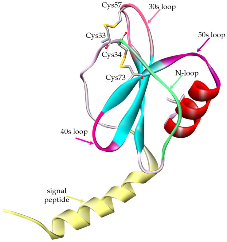
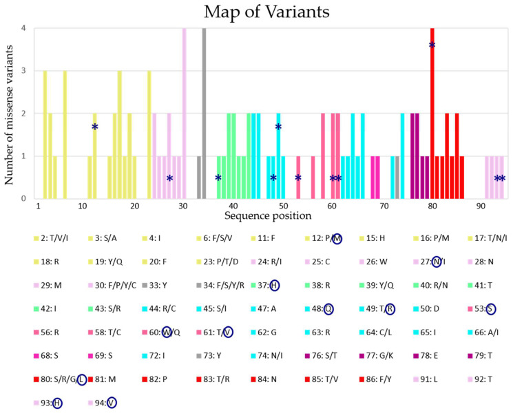
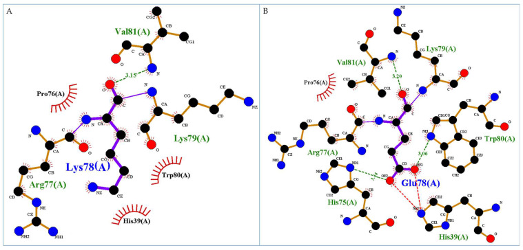
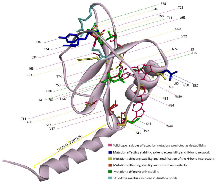
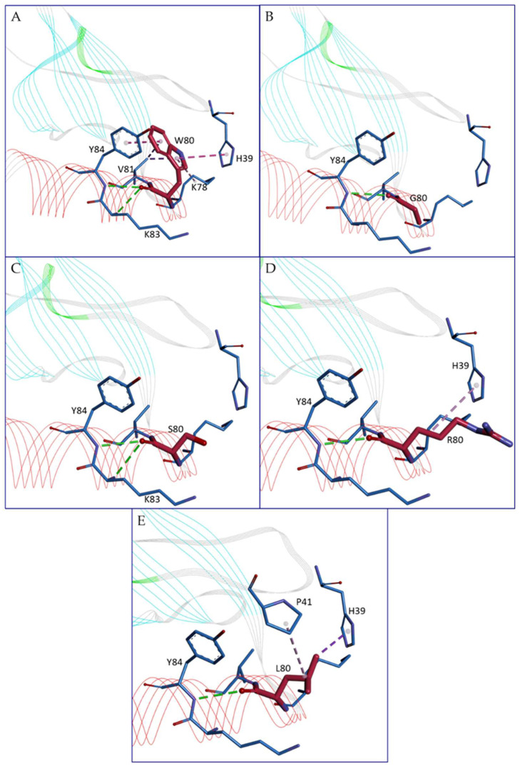
Eotaxin-3 is a key chemokine with a relevant role in eosinophilic esophagitis, a rare chronic immune/antigen-mediated inflammatory disorder. Eotaxin-3 is a potent activator of eosinophil emergence and migration, which may lead to allergic airway inflammation. We investigated, using bioinformatics tools, the protein structure and the possible effects of the known variations reported in public databases. Following a procedure already established, we created a 3D model of the whole protein and modeled the structure of 105 protein variants due to known point mutations. The effects of the amino acid substitution at the level of impact on protein structure, stability, and possibly function were detected by the bioinformatics procedure and described in detail. A web application was implemented to browse the results of the analysis and visualize the 3D models, with the opportunity of downloading the models and analyzing them using their own software. Among 105 amino acid substitutions investigated, the study evidenced in 44 cases at least one change in any of the investigated structural parameters. Other six variations are also relevant, although a structural effect was not detected by our analysis, because they affected amino acids highly conserved, which suggests a possible function role. All these variations should be the object of particular attention, as they may induce a loss of functionality in the protein.
Overview
The claims and statistics of this paper haven’t been checked yet.
Run the check — free, no account needed. It reads the open-access text and checks the sources it cites.
Integrity signals
Registry
Cites
Statistics
Figures
Verification
Claims the paper makes
The claims and statistics of this paper haven’t been checked yet.
Run the check — free, no account needed. It reads the open-access text and checks the sources it cites.
Self-check
Percentages, sums and reported differences re-computed from the paper’s own numbers.
The self-check needs the paper’s full text.
Citations
Cites
Highly accurate protein structure prediction with AlphaFold
Jumper, J., Evans, R., Pritzel, A., Green, T., Figurnov, M., Ronneberger, O., Tunyasuvunakool, K., Bates, R., Žídek, A., Potapenko, A., et al. (2021) · Nature · doi:10.1038/s41586-021-03819-2 · Cited by 43,764
Berman, H. M. (2000) · Nucleic Acids Research · doi:10.1093/nar/28.1.235 · Cited by 40,196
PROCHECK: a program to check the stereochemical quality of protein structures
Laskowski, R. A., MacArthur, M. W., Moss, D. S., & Thornton, J. M. (1993) · Journal of Applied Crystallography · doi:10.1107/s0021889892009944 · Cited by 24,581
Kabsch, W., & Sander, C. (1983) · Biopolymers · doi:10.1002/bip.360221211 · Cited by 15,320
Wiederstein, M., & Sippl, M. J. (2007) · Nucleic Acids Research · doi:10.1093/nar/gkm290 · Cited by 5,909
Comparative Protein Structure Modeling Using Modeller
Eswar, N., Webb, B., Marti‐Renom, M. A., Madhusudhan, M. S., Eramian, D., Shen, M., Pieper, U., & Sali, A. (2006) · Current Protocols in Bioinformatics · doi:10.1002/0471250953.bi0506s15 · Cited by 4,478
Comparative Protein Structure Modeling Using MODELLER
Webb, B., & Sali, A. (2016) · Current Protocols in Bioinformatics · doi:10.1002/cpbi.3 · Cited by 4,316
Toward the estimation of the absolute quality of individual protein structure models
Benkert, P., Biasini, M., & Schwede, T. F. (2010) · Bioinformatics · doi:10.1093/bioinformatics/btq662 · Cited by 2,321
Satisfying Hydrogen Bonding Potential in Proteins
McDonald, I. K., & Thornton, J. M. (1994) · Journal of Molecular Biology · doi:10.1006/jmbi.1994.1334 · Cited by 2,136
UniProt: the universal protein knowledgebase
The Uniprot Consortium (2018) · Nucleic Acids Research · doi:10.1093/nar/gky092 · Cited by 2,097
Metadata
- Authors
- Giordano, D., d’Acierno, A., Marabotti, A., Iovino, P., Iacomino, G., & Facchiano, A.
- Affiliations
- Institute of Food Science (IT)
- University of Salerno (IT)
- Corresponding author
- Iacomino, Giuseppe
- ORCID
- Venue
- Genes
- Publisher
- MDPI AG
- Published
- 2024-08-14
- Volume
- 15
- Issue
- 8
- Pages
- 1073
- ISSN
- 2073-4425 (ISSN-L)
- PMC
- PMC11354214
- OpenAlex
- W4401584344
- Cited by
- 1
- References
- 36
- Citations by year
- 2026: 1
- FWCI
- 0.42
- Type
- journal article
- Language
- en
- Open access
- gold
- doi.org
- Licence
- Locations
- Genes (publishedVersion, cc-by)
- Preprints.org (acceptedVersion, cc-by)
- PubMed Central (publishedVersion, cc-by)
- PubMed (publishedVersion)
- Funders
- PRIN-2022 PNRR project (P2022BCXTK, NUTRAGE FOE-2021 DBA.AD005.225)
- CNR project (P2022BCXTK, NUTRAGE FOE-2021 DBA.AD005.225)
- BANCA D’ITALIA (P2022BCXTK, NUTRAGE FOE-2021 DBA.AD005.225)
- Topics
- Health Sciences › Medicine › Gastroenterology › Eosinophilic Esophagitis
- Life Sciences › Biochemistry, Genetics and Molecular Biology › Molecular Biology › Protein Structure and Dynamics
- Health Sciences › Medicine › Pulmonary and Respiratory Medicine › Asthma and respiratory diseases
- Keywords
- eotaxin-3, eosinophilic esophagitis, chemokines, point mutations, protein variants, protein structure, protein structure modeling, protein stability, amino acid substitutions, web application, eosinophil migration
- MeSH terms
- Chemokine CCL26, Humans, Models, Molecular, Computational Biology, Amino Acid Substitution, Eosinophilic Esophagitis
About these checks
Registry, retraction, Cites and statistics results are computed, not judged, with one exception: a printed reference that no index lists is read once by a model before it counts as a citation error. Claims are extracted by a model and kept only when their quote appears verbatim in the text; each cited source is then checked, with a model’s help, for whether it says what the claim relies on. Those verdicts can be wrong — read the claim’s quote and the cited source before relying on one.
Statistics and arithmetic are recomputed, not judged.
Confidence bands are shown only for fields calibrated on our annotated test set. None is calibrated yet, so no band is shown.
What counts as a claim ›How we measure confidence ›How we check this ›
Are you an author, or do you see a problem with this page? Request a review
How to cite
Cite CoChat for the checks on this page, and the paper for its own findings.
- This page
- CoChat. (2026). Citation check: Bioinformatics Study on Site-Specific Variations of Eotaxin-3, a Key Chemokine in Eosinophilic Esophagitis (EoE). https://app.cochat.ai/papers/10.3390/genes15081073
- The paper
- Giordano, D., d’Acierno, A., Marabotti, A., Iovino, P., Iacomino, G., & Facchiano, A. (2024). Bioinformatics Study on Site-Specific Variations of Eotaxin-3, a Key Chemokine in Eosinophilic Esophagitis (EoE). Genes. https://doi.org/10.3390/genes15081073
Data sources
Bibliographic data: Semantic Scholar, OpenAlex, Crossref.
Contains information from the Semantic Scholar Academic Graph Datasets, made available under the ODC Attribution License (ODC-BY 1.0).
Semantic Scholar’s data is described in: Kinney, R., et al. (2023). The Semantic Scholar Open Data Platform. arXiv:2301.10140.
